Using electormyography in research and the lab
Alan Jenks
Introduction
Electromyography (EMG) allows for valuable information to be gained about the nervous system and skeletal muscle. The data recording is called an electromyogram. It can be used in a clinical setting to examine and diagnose neuropathies, skeletal muscle insufficiencies, issues with nerve conduction, etc. In a research setting, researchers use this technique to assess motor unit recruitment, fatigue, reaction time, muscle action, and the list goes on. Most commonly in the practice of Kinesiology, EMG is used to study the neurological activation of muscles within postural tasks, functional movements, work conditions, and treatment/training regimes (Konrad, 2005, pg.4). EMG is an obligatory investigation in motor neurone disease to demonstrate the widespread denervation and fasciculation required for secure diagnosis (Mills, 2022).
EMG is collected through two mechanisms:
- Indwelling needle
- A fine wire in the axis of the needle is insulated from the shaft, the end of the needle being cut at an acute angle
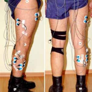
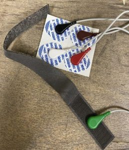
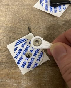
- Surface electrodes
Recall from lecture that each of these have their own pros and cons. In this lab, we will be collecting data using surface EMG.
Because surface electrodes are non-invasive, they are easy to handle but only can collect electromyographical data from surface muscles (deep muscles require indwelling needles). Silver/silver chloride pre-gelled, disposable electrodes are most used and recommended by SENIAM guidelines. Wet gel electrodes have better conduction and lower impedance, but adhesive gel electrodes have the advantage that they can be repositioned in case of error in application. This lab will use adhesive gel surface electrodes.
History
Scientists in the 17th and 18th centuries such as Jan Swammerdam, Francesco Redi, Luigi Galvani, and Alessandro Volta conducted experiments to confirm that electrical stimulation led to muscle contraction. Carlo Matteucci was the first to develop a primitive instrument related to the modern EMG to measure electrical potential in muscles. His galvanometer detected and determined the direction of small electrical currents produced by mechanical means, and then applied this to muscle contraction through studies done on frogs. Emil Du Bois-Reymond was the first to apply this to voluntary contraction of human muscle, and his work was followed by further experimentation by Guillaume Duchenne on facial muscles. Since then, EMG machines were refined and knowledge of EMG furthered at unbelievable speeds.
Motor Unit Recruitment
Reduced Recruitment
Early Recruitment
Motor Unit Potentials
Tremors
Peripheral Neuropathies
Entrapment Neuropathies
Brachial Plexus Neuropathies
Motor Neuron Diseases
Primary Muscle Disease
- Motor unit potentials are small and spiky and the recruitment pattern becomes full
- In inflammatory muscle disease where there is active degeneration of muscle fibres, fibrillations may be seen, but this is not universal or specific.
- In muscle diseases which show large variation in fibre diameter, such as the muscular dystrophies, there may be large motor units on a background of small spiky units, presumably arising from large diameter fibres.
- With just a small contraction measuring the amplitude of a compound muscle action potential evoked by nerve stimulation before and after exercise.
Validity & Reliability
Present and Future Use
EMG is currently used in the diagnosis of various of neuromuscular diseases. It has also been used to help study kinesiology as well as help map the brain for a deeper understanding of Alzheimer’s. A current use that has a prevalent future is utilizing EMG for prosthetics movement. This has been widely used in prosthetics for limbs such as arms, hands, and legs.
In addition to medical uses, use of EMG in the gaming industry is a future source for expansion.
Media Attributions
- Picture1
- Picture2
- Picture3
