Want to create or adapt books like this? Learn more about how Pressbooks supports open publishing practices.
Unit 4: Animal Structure and Function
15.1 Digestive Systems
Learning Objectives
By the end of this section, you will be able to:
Explain the processes of digestion and absorption
Compare and contrast different types of digestive systems
Explain the specialized functions of the organs involved in processing food in the body
Describe the ways in which organs work together to digest food and absorb nutrients
Animals obtain their nutrition from the consumption of other organisms. Depending on their diet, animals can be classified into the following categories: plant eaters (herbivores), meat eaters (carnivores), and those that eat both plants and animals (omnivores). The nutrients and macromolecules present in food are not immediately accessible to the cells. There are a number of processes that modify food within the animal body in order to make the nutrients and organic molecules accessible for cellular function. As animals evolved in complexity of form and function, their digestive systems have also evolved to accommodate their various dietary needs.
Herbivores, Omnivores, and Carnivores
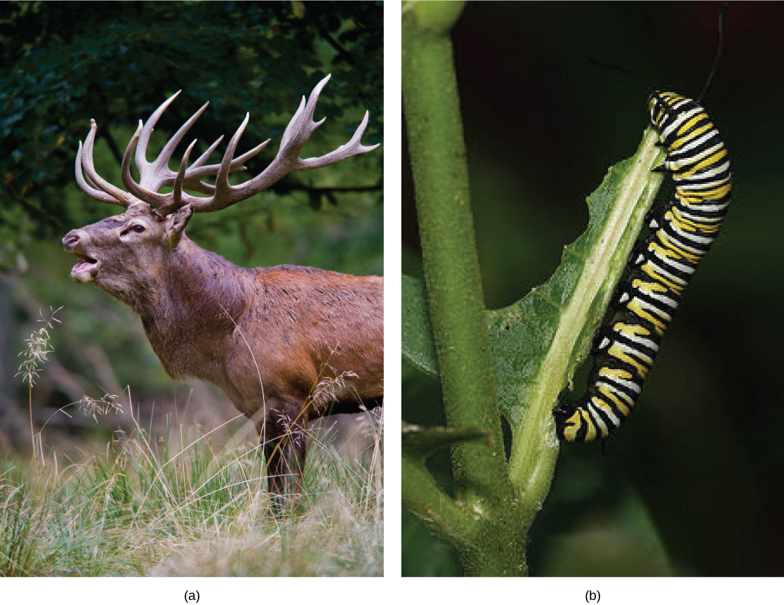
Herbivores are animals whose primary food source is plant-based. Examples of herbivores, as shown in Figure 15.2 include vertebrates like deer, koalas, and some bird species, as well as invertebrates such as crickets and caterpillars. These animals have evolved digestive systems capable of handling large amounts of plant material. Herbivores can be further classified into frugivores (fruit-eaters), granivores (seed eaters), nectivores (nectar feeders), and folivores (leaf eaters).
Figure 15.2. Herbivores, like this (a) mule deer and (b) monarch caterpillar, eat primarily plant material. (credit a: modification of work by Bill Ebbesen; credit b: modification of work by Doug Bowman)
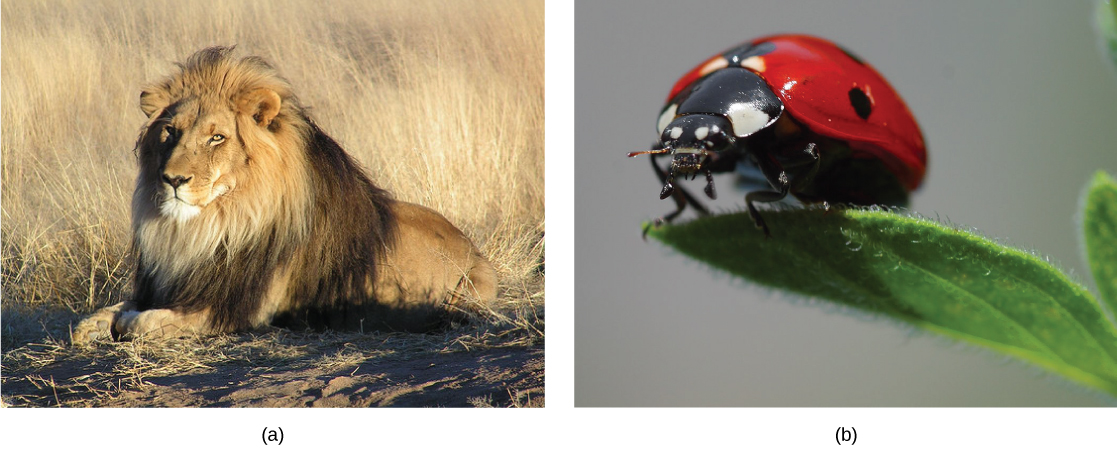
Carnivores are animals that eat other animals. The word carnivore is derived from Latin and literally means “meat eater.” Wild cats such as lions, shown in Figure 35.3 a and tigers are examples of vertebrate carnivores, as are snakes and sharks, while invertebrate carnivores include sea stars, spiders, and ladybugs, shown in Figure 15.3 b. Obligate carnivores are those that rely entirely on animal flesh to obtain their nutrients; examples of obligate carnivores are members of the cat family, such as lions and cheetahs. Facultative carnivores are those that also eat non-animal food in addition to animal food. Note that there is no clear line that differentiates facultative carnivores from omnivores; dogs would be considered facultative carnivores.
Figure 15.3. Carnivores like the (a) lion eat primarily meat. The (b) ladybug is also a carnivore that consumes small insects called aphids. (credit a: modification of work by Kevin Pluck; credit b: modification of work by Jon Sullivan)
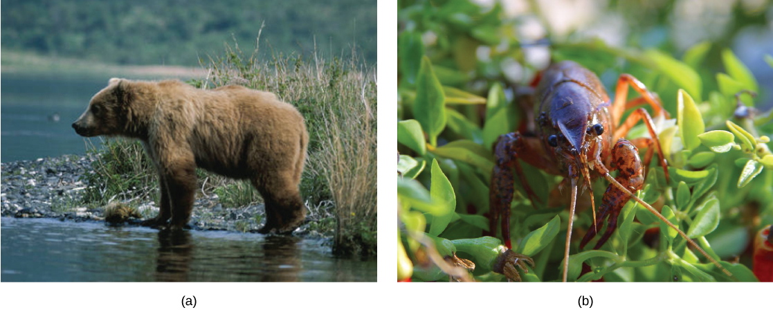
Figure 15.4. Omnivores like the (a) bear and (b) crayfish eat both plant and animal based food. (credit a: modification of work by Dave Menke; credit b: modification of work by Jon Sullivan)
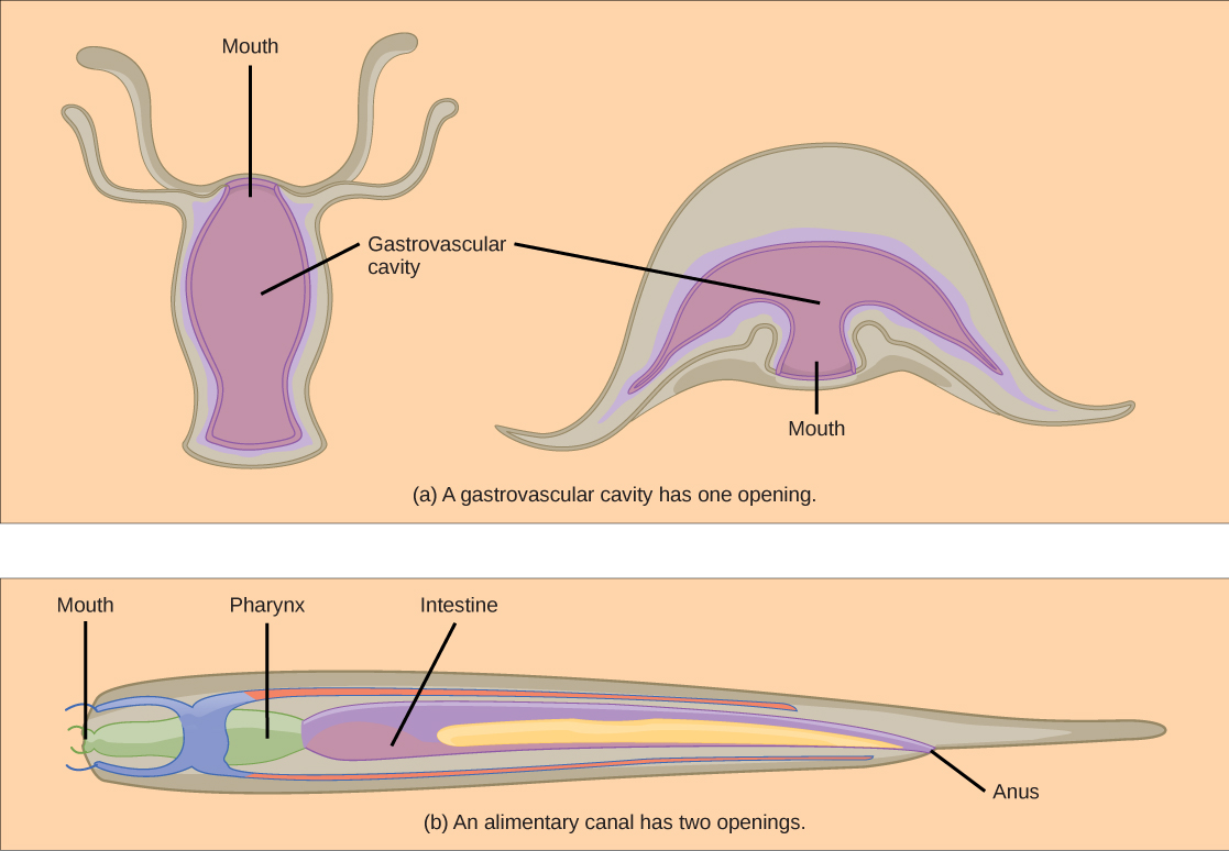
The alimentary canal, shown in Figure 15.5 b, is a more advanced system: it consists of one tube with a mouth at one end and an anus at the other. Earthworms are an example of an animal with an alimentary canal. Once the food is ingested through the mouth, it passes through the esophagus and is stored in an organ called the crop; then it passes into the gizzard where it is churned and digested. From the gizzard, the food passes through the intestine, the nutrients are absorbed, and the waste is eliminated as feces, called castings, through the anus.
Figure 15.5. (a) A gastrovascular cavity has a single opening through which food is ingested and waste is excreted, as shown in this hydra and in this jellyfish medusa. (b) An alimentary canal has two openings: a mouth for ingesting food, and an anus for eliminating waste, as shown in this nematode.
Vertebrate Digestive Systems
Vertebrates have evolved more complex digestive systems to adapt to their dietary needs. Some animals have a single stomach, while others have multi-chambered stomachs. Birds have developed a digestive system adapted to eating unmasticated food.
Monogastric: Single-chambered Stomach
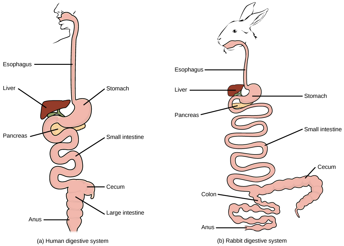
As the word monogastric suggests, this type of digestive system consists of one (“mono”) stomach chamber (“gastric”). Humans and many animals have a monogastric digestive system as illustrated in Figure 15.6 ab. The process of digestion begins with the mouth and the intake of food. The teeth play an important role in masticating (chewing) or physically breaking down food into smaller particles. The enzymes present in saliva also begin to chemically break down food. The esophagus is a long tube that connects the mouth to the stomach. Using peristalsis, or wave-like smooth muscle contractions, the muscles of the esophagus push the food towards the stomach. In order to speed up the actions of enzymes in the stomach, the stomach is an extremely acidic environment, with a pH between 1.5 and 2.5. The gastric juices, which include enzymes in the stomach, act on the food particles and continue the process of digestion. Further breakdown of food takes place in the small intestine where enzymes produced by the liver, the small intestine, and the pancreas continue the process of digestion. The nutrients are absorbed into the blood stream across the epithelial cells lining the walls of the small intestines. The waste material travels on to the large intestine where water is absorbed and the drier waste material is compacted into feces; it is stored until it is excreted through the rectum.
Figure 15.6. (a) Humans and herbivores, such as the (b) rabbit, have a monogastric digestive system. However, in the rabbit the small intestine and cecum are enlarged to allow more time to digest plant material. The enlarged organ provides more surface area for absorption of nutrients. Rabbits digest their food twice: the first time food passes through the digestive system, it collects in the cecum, and then it passes as soft feces called cecotrophes. The rabbit re-ingests these cecotrophes to further digest them.
Avian
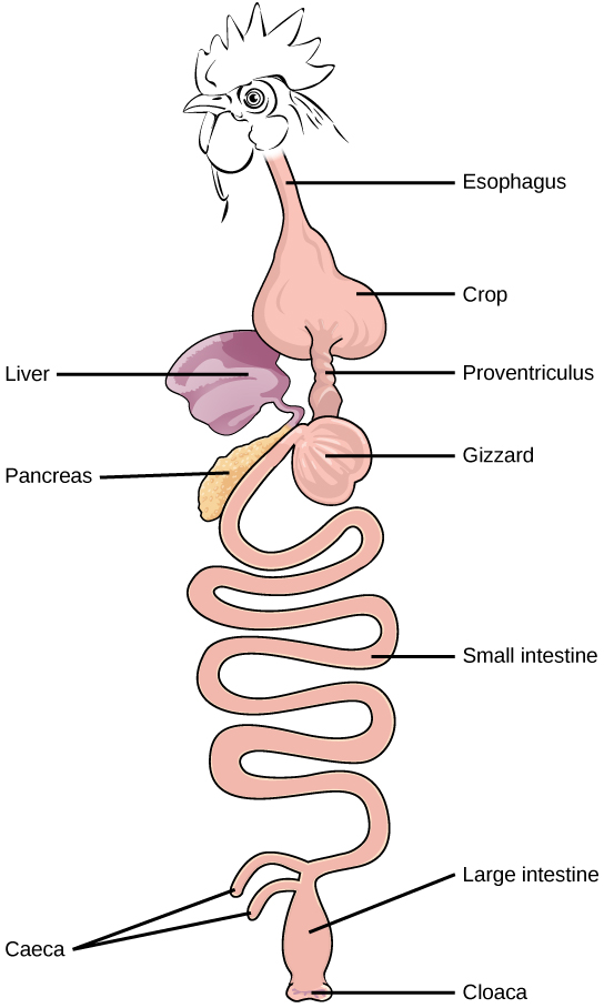
Birds face special challenges when it comes to obtaining nutrition from food. They do not have teeth and so their digestive system, shown in Figure 15.7, must be able to process un-masticated food. Birds have evolved a variety of beak types that reflect the vast variety in their diet, ranging from seeds and insects to fruits and nuts. Because most birds fly, their metabolic rates are high in order to efficiently process food and keep their body weight low. The stomach of birds has two chambers: the proventriculus, where gastric juices are produced to digest the food before it enters the stomach, and the gizzard, where the food is stored, soaked, and mechanically ground. The undigested material forms food pellets that are sometimes regurgitated. Most of the chemical digestion and absorption happens in the intestine and the waste is excreted through the cloaca.
Figure 15.7. The avian esophagus has a pouch, called a crop, which stores food. Food passes from the crop to the first of two stomachs, called the proventriculus, which contains digestive juices that break down food. From the proventriculus, the food enters the second stomach, called the gizzard, which grinds food. Some birds swallow stones or grit, which are stored in the gizzard, to aid the grinding process. Birds do not have separate openings to excrete urine and feces. Instead, uric acid from the kidneys is secreted into the large intestine and combined with waste from the digestive process. This waste is excreted through an opening called the cloaca.
Parts of the Digestive System
The vertebrate digestive system is designed to facilitate the transformation of food matter into the nutrient components that sustain organisms.
Oral Cavity
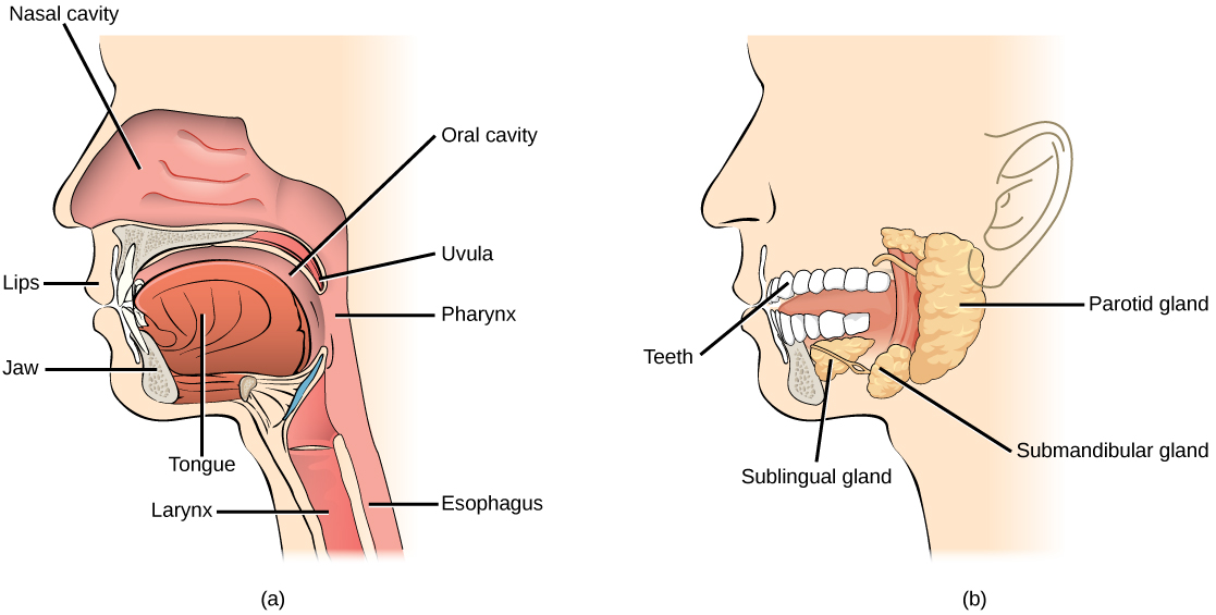
The oral cavity, or mouth, is the point of entry of food into the digestive system, illustrated in Figure 15.9. The food consumed is broken into smaller particles by mastication, the chewing action of the teeth. All mammals have teeth and can chew their food.
Figure 15.9. Digestion of food begins in the (a) oral cavity. Food is masticated by teeth and moistened by saliva secreted from the (b) salivary glands. Enzymes in the saliva begin to digest starches and fats. With the help of the tongue, the resulting bolus is moved into the esophagus by swallowing. (credit: modification of work by the National Cancer Institute)
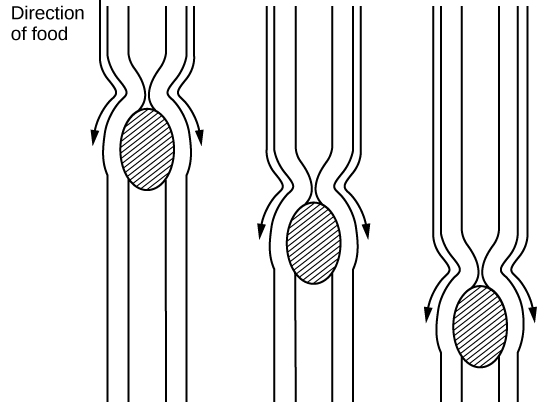
Figure 15.10. The esophagus transfers food from the mouth to the stomach through peristaltic movements.
A ring-like muscle called a sphincter forms valves in the digestive system. The gastro-esophageal sphincter is located at the stomach end of the esophagus. In response to swallowing and the pressure exerted by the bolus of food, this sphincter opens, and the bolus enters the stomach. When there is no swallowing action, this sphincter is shut and prevents the contents of the stomach from traveling up the esophagus. Many animals have a true sphincter; however, in humans, there is no true sphincter, but the esophagus remains closed when there is no swallowing action. Acid reflux or “heartburn” occurs when the acidic digestive juices escape into the esophagus.
Figure 15.11. The human stomach has an extremely acidic environment where most of the protein gets digested. (credit: modification of work by Mariana Ruiz Villareal)
Which of the following statements about the digestive system is false?
Chyme is a mixture of food and digestive juices that is produced in the stomach.
Food enters the large intestine before the small intestine.
In the small intestine, chyme mixes with bile, which emulsifies fats.
The stomach is separated from the small intestine by the pyloric sphincter.
When digesting protein and some fats, the stomach lining must be protected from getting digested by pepsin. There are two points to consider when describing how the stomach lining is protected. First, as previously mentioned, the enzyme pepsin is synthesized in the inactive form. This protects the chief cells, because pepsinogen does not have the same enzyme functionality of pepsin. Second, the stomach has a thick mucus lining that protects the underlying tissue from the action of the digestive juices. When this mucus lining is ruptured, ulcers can form in the stomach. Ulcers are open wounds in or on an organ caused by bacteria (Helicobacter pylori) when the mucus lining is ruptured and fails to reform.
The second part of the small intestine is called the jejunum, shown in Figure 15.11. Here, hydrolysis of nutrients is continued while most of the carbohydrates and amino acids are absorbed through the intestinal lining. The bulk of chemical digestion and nutrient absorption occurs in the jejunum.
The ileum, also illustrated in Figure 15.11 is the last part of the small intestine and here the bile salts and vitamins are absorbed into blood stream. The undigested food is sent to the colon from the ileum via peristaltic movements of the muscle. The ileum ends and the large intestine begins at the ileocecal valve. The vermiform, “worm-like,” appendix is located at the ileocecal valve. The appendix of humans secretes no enzymes and has an insignificant role in immunity.
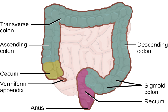
Large Intestine
The large intestine, illustrated in Figure 15.13, reabsorbs the water from the undigested food material and processes the waste material. The human large intestine is much smaller in length compared to the small intestine but larger in diameter. It has three parts: the cecum, the colon, and the rectum. The cecum joins the ileum to the colon and is the receiving pouch for the waste matter. The colon is home to many bacteria or “intestinal flora” that aid in the digestive processes. The colon can be divided into four regions, the ascending colon, the transverse colon, the descending colon and the sigmoid colon. The main functions of the colon are to extract the water and mineral salts from undigested food, and to store waste material. Carnivorous mammals have a shorter large intestine compared to herbivorous mammals due to their diet.
Figure 15.13. The large intestine reabsorbs water from undigested food and stores waste material until it is eliminated.
Rectum and Anus
The rectum is the terminal end of the large intestine, as shown in Figure 15.13. The primary role of the rectum is to store the feces until defecation. The feces are propelled using peristaltic movements during elimination. The anus is an opening at the far-end of the digestive tract and is the exit point for the waste material. Two sphincters between the rectum and anus control elimination: the inner sphincter is involuntary and the outer sphincter is voluntary.
Accessory Organs
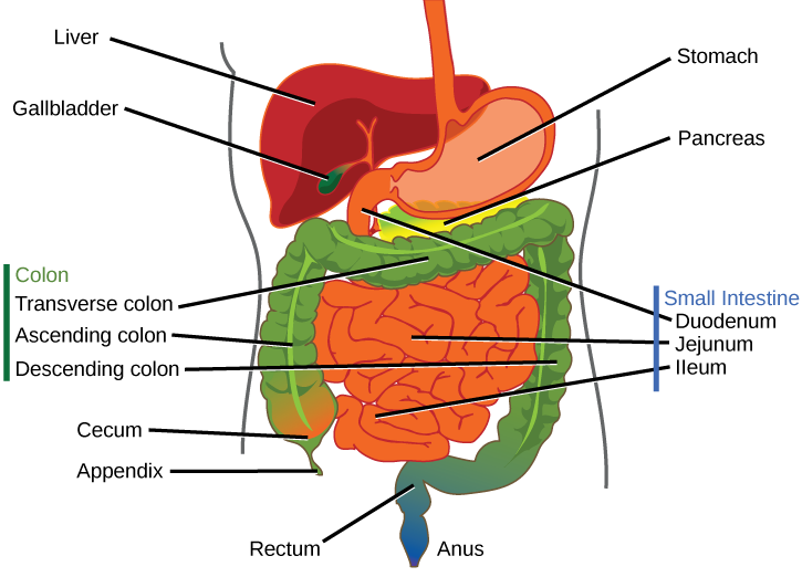
The organs discussed above are the organs of the digestive tract through which food passes. Accessory organs are organs that add secretions (enzymes) that catabolize food into nutrients. Accessory organs include salivary glands, the liver, the pancreas, and the gallbladder. The liver, pancreas, and gallbladder are regulated by hormones in response to the food consumed.
The liver is the largest internal organ in humans and it plays a very important role in digestion of fats and detoxifying blood. The liver produces bile, a digestive juice that is required for the breakdown of fatty components of the food in the duodenum. The liver also processes the vitamins and fats and synthesizes many plasma proteins.
Different animals have evolved different types of digestive systems specialized to meet their dietary needs. Humans and many other animals have monogastric digestive systems with a single-chambered stomach. Birds have evolved a digestive system that includes a gizzard where the food is crushed into smaller pieces. This compensates for their inability to masticate. Ruminants that consume large amounts of plant material have a multi-chambered stomach that digests roughage. Pseudo-ruminants have similar digestive processes as ruminants but do not have the four-compartment stomach. Processing food involves ingestion (eating), digestion (mechanical and enzymatic breakdown of large molecules), absorption (cellular uptake of nutrients), and elimination (removal of undigested waste as feces).
Many organs work together to digest food and absorb nutrients. The mouth is the point of ingestion and the location where both mechanical and chemical breakdown of food begins. Saliva contains an enzyme called amylase that breaks down carbohydrates. The food bolus travels through the esophagus by peristaltic movements to the stomach. The stomach has an extremely acidic environment. An enzyme called pepsin digests protein in the stomach. Further digestion and absorption take place in the small intestine. The large intestine reabsorbs water from the undigested food and stores waste until elimination.
Exercises
1. Which of the following statements about the digestive system is false?
A) Chyme is a mixture of food and digestive juices that is produced in the stomach.
B) Food enters the large intestine before the small intestine.
C) In the small intestine, chyme mixes with bile, which emulsifies fats.
D) The stomach is separated from the small intestine by the pyloric sphincter.
Answer: B
2. Which of the following statements about the small intestine is false?
A) Absorptive cells that line the small intestine have microvilli, small projections that increase surface area and aid in the absorption of food.
B) The inside of the small intestine has many folds, called villi.
C) Microvilli are lined with blood vessels as well as lymphatic vessels.
D) The inside of the small intestine is called the lumen.
Answer: C
Multiple Choice
3. Which of the following is a pseudo-ruminant?
A) cow
B) pig
C) crow
D) horse
Answer: D
4. Which of the following statements is untrue?
A) Roughage takes a long time to digest.
B) Birds eat large quantities at one time so that they can fly long distances.
C) Cows do not have upper teeth.
D) In pseudo-ruminants, roughage is digested in the cecum.
Answer: B
5. The acidic nature of chyme is neutralized by ________.
A) potassium hydroxide
B) sodium hydroxide
C) bicarbonates
D) vinegar
Answer: C
6. The digestive juices from the liver are delivered to the ________.
A) stomach
B) liver
C) duodenum
D) colon
Answer: C
7. How does the polygastric digestive system aid in digesting roughage?
Animals with a polygastric digestive system have a multi-chambered stomach. The four compartments of the stomach are called the rumen, reticulum, omasum, and abomasum. These chambers contain many microbes that break down the cellulose and ferment the ingested food. The abomasum is the “true” stomach and is the equivalent of a monogastric stomach chamber where gastric juices are secreted. The four-compartment gastric chamber provides larger space and the microbial support necessary for ruminants to digest plant material.
8. How do birds digest their food in the absence of teeth?
Birds have a stomach chamber called a gizzard. Here, the food is stored, soaked, and ground into finer particles, often using pebbles. Once this process is complete, the digestive juices take over in the proventriculus and continue the digestive process.
9. What is the role of the accessory organs in digestion?
Accessory organs play an important role in producing and delivering digestive juices to the intestine during digestion and absorption. Specifically, the salivary glands, liver, pancreas, and gallbladder play important roles. Malfunction of any of these organs can lead to disease states.
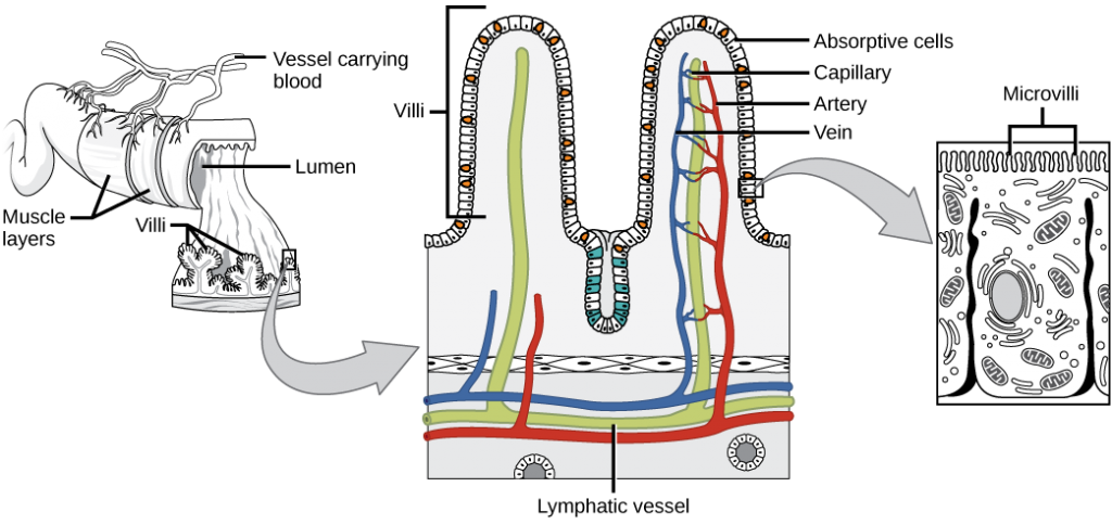
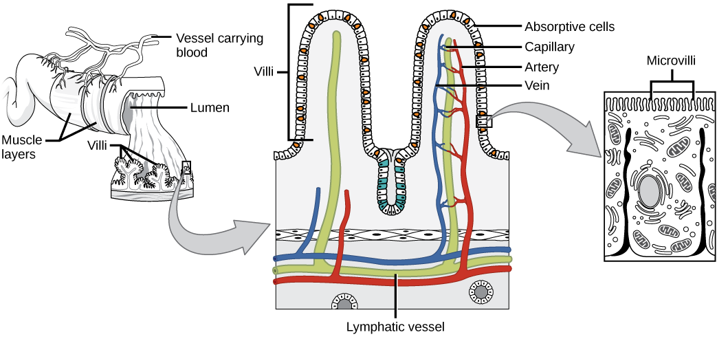
10. Explain how the villi and microvilli aid in absorption.
The villi and microvilli are folds on the surface of the small intestine. These folds increase the surface area of the intestine and provide more area for the absorption of nutrients.
Glossary
alimentary canal
tubular digestive system with a mouth and anus
anus
exit point for waste material
bile
digestive juice produced by the liver; important for digestion of lipids
bolus
mass of food resulting from chewing action and wetting by saliva
carnivore
animal that consumes animal flesh
chyme
mixture of partially digested food and stomach juices
digestion
mechanical and chemical break down of food into small organic fragments
duodenum
first part of the small intestine where a large part of digestion of carbohydrates and fats occurs
endocrine system
system that controls the response of the various glands in the body and the release of hormones at the appropriate times
esophagus
tubular organ that connects the mouth to the stomach
essential nutrient
nutrient that cannot be synthesized by the body; it must be obtained from food
gallbladder
organ that stores and concentrates bile
gastric inhibitory peptide
hormone secreted by the small intestine in the presence of fatty acids and sugars; it also inhibits acid production and peristalsis in order to slow down the rate at which food enters the small intestine
gastrin
hormone which stimulates hydrochloric acid secretion in the stomach
gastrovascular cavity
digestive system consisting of a single opening
gizzard
muscular organ that grinds food
herbivore
animal that consumes strictly plant diet
ileum
last part of the small intestine; connects the small intestine to the large intestine; important for absorption of B-12
ingestion
act of taking in food
jejunum
second part of the small intestine
lactase
enzyme that breaks down lactose into glucose and galactose
large intestine
digestive system organ that reabsorbs water from undigested material and processes waste matter
lipase
enzyme that chemically breaks down lipids
liver
organ that produces bile for digestion and processes vitamins and lipids
maltase
enzyme that breaks down maltose into glucose
mineral
inorganic, elemental molecule that carries out important roles in the body
monogastric
digestive system that consists of a single-chambered stomach
omnivore
animal that consumes both plants and animals
pancreas
gland that secretes digestive juices
pepsinogen
inactive form of pepsin
pepsin
enzyme found in the stomach whose main role is protein digestion
peristalsis
wave-like movements of muscle tissue
proventriculus
glandular part of a bird’s stomach
rectum
area of the body where feces is stored until elimination
roughage
component of food that is low in energy and high in fiber
ruminant
animal with a stomach divided into four compartments
salivary amylase
enzyme found in saliva, which converts carbohydrates to maltose
small intestine
organ where digestion of protein, fats, and carbohydrates is completed
somatostatin
hormone released to stop acid secretion when the stomach is empty
sphincter
band of muscle that controls movement of materials throughout the digestive tract
stomach
saclike organ containing acidic digestive juices
villi
folds on the inner surface of the small intestine whose role is to increase absorption area
vitamin
organic substance necessary in small amounts to sustain life