Want to create or adapt books like this? Learn more about how Pressbooks supports open publishing practices.
Unit 4: Animal Structure and Function
22.2. The Kidneys and Osmoregulatory Organs
Learning Objectives
By the end of this section, you will be able to:
Explain how the kidneys serve as the main osmoregulatory organs in mammalian systems
Describe the structure of the kidneys and the functions of the parts of the kidney
Describe how the nephron is the functional unit of the kidney and explain how it actively filters blood and generates urine
Detail the three steps in the formation of urine: glomerular filtration, tubular reabsorption, and tubular secretion
Although the kidneys are the major osmoregulatory organ, the skin and lungs also play a role in the process. Water and electrolytes are lost through sweat glands in the skin, which helps moisturize and cool the skin surface, while the lungs expel a small amount of water in the form of mucous secretions and via evaporation of water vapor.
Kidneys: The Main Osmoregulatory Organ
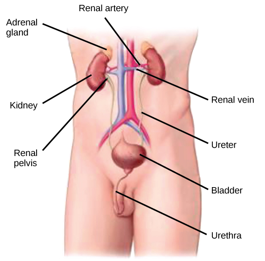
The kidneys, illustrated in Figure 22.4, are a pair of bean-shaped structures that are located just below and posterior to the liver in the peritoneal cavity. The adrenal glands sit on top of each kidney and are also called the suprarenal glands. Kidneys filter blood and purify it. All the blood in the human body is filtered many times a day by the kidneys; these organs use up almost 25 percent of the oxygen absorbed through the lungs to perform this function. Oxygen allows the kidney cells to efficiently manufacture chemical energy in the form of ATP through aerobic respiration. The filtrate coming out of the kidneys is called urine.
Figure 22.4. Kidneys filter the blood, producing urine that is stored in the bladder prior to elimination through the urethra. (credit: modification of work by NCI)
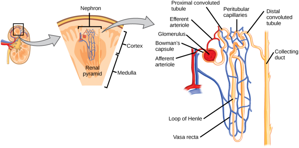
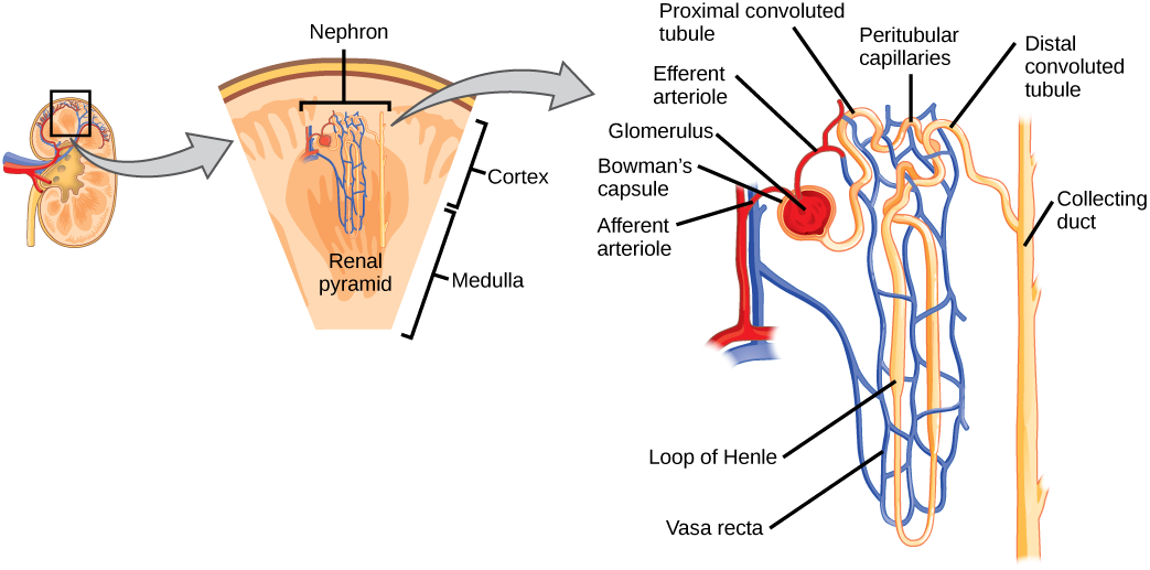
Figure 22.6. The nephron is the functional unit of the kidney. The glomerulus and convoluted tubules are located in the kidney cortex, while collecting ducts are located in the pyramids of the medulla. (credit: modification of work by NIDDK)
Which of the following statements about the nephron is false?
The collecting duct empties into the distal convoluted tubule.
The Bowman’s capsule surrounds the glomerulus.
The loop of Henle is between the proximal and distal convoluted tubules.
The loop of Henle empties into the distal convoluted tubule.
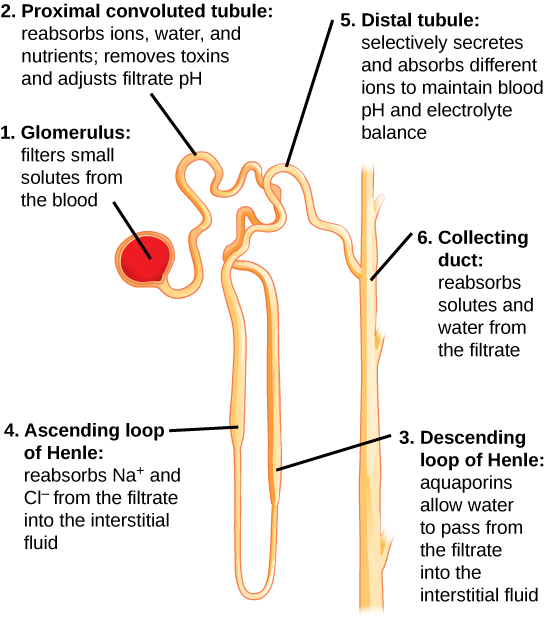
Figure 22.7. Each part of the nephron performs a different function in filtering waste and maintaining homeostatic balance. (1) The glomerulus forces small solutes out of the blood by pressure. (2) The proximal convoluted tubule reabsorbs ions, water, and nutrients from the filtrate into the interstitial fluid, and actively transports toxins and drugs from the interstitial fluid into the filtrate. The proximal convoluted tubule also adjusts blood pH by selectively secreting ammonia (NH3) into the filtrate, where it reacts with H+ to form NH4+. The more acidic the filtrate, the more ammonia is secreted. (3) The descending loop of Henle is lined with cells containing aquaporins that allow water to pass from the filtrate into the interstitial fluid. (4) In the thin part of the ascending loop of Henle, Na+ and Cl- ions diffuse into the interstitial fluid. In the thick part, these same ions are actively transported into the interstitial fluid. Because salt but not water is lost, the filtrate becomes more dilute as it travels up the limb. (5) In the distal convoluted tubule, K+ and H+ ions are selectively secreted into the filtrate, while Na+, Cl-, and HCO3- ions are reabsorbed to maintain pH and electrolyte balance in the blood. (6) The collecting duct reabsorbs solutes and water from the filtrate, forming dilute urine. (credit: modification of work by NIDDK)
Glomerular Filtration
Glomerular filtration filters out most of the solutes due to high blood pressure and specialized membranes in the afferent arteriole. The blood pressure in the glomerulus is maintained independent of factors that affect systemic blood pressure. The “leaky” connections between the endothelial cells of the glomerular capillary network allow solutes to pass through easily. All solutes in the glomerular capillaries, except for macromolecules like proteins, pass through by passive diffusion. There is no energy requirement at this stage of the filtration process. Glomerular filtration rate (GFR) is the volume of glomerular filtrate formed per minute by the kidneys. GFR is regulated by multiple mechanisms and is an important indicator of kidney function.
Concept in Action
To learn more about the vascular system of kidneys, click through
Tubular reabsorption occurs in the PCT part of the renal tubule. Almost all nutrients are reabsorbed, and this occurs either by passive or active transport. Reabsorption of water and some key electrolytes are regulated and can be influenced by hormones. Sodium (Na+) is the most abundant ion and most of it is reabsorbed by active transport and then transported to the peritubular capillaries. Because Na+ is actively transported out of the tubule, water follows it to even out the osmotic pressure. Water is also independently reabsorbed into the peritubular capillaries due to the presence of aquaporins, or water channels, in the PCT. This occurs due to the low blood pressure and high osmotic pressure in the peritubular capillaries. However, every solute has a transport maximum and the excess is not reabsorbed.
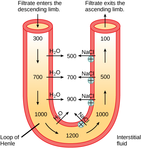
In the loop of Henle, the permeability of the membrane changes. The descending limb is permeable to water, not solutes; the opposite is true for the ascending limb. Additionally, the loop of Henle invades the renal medulla, which is naturally high in salt concentration and tends to absorb water from the renal tubule and concentrate the filtrate. The osmotic gradient increases as it moves deeper into the medulla. Because two sides of the loop of Henle perform opposing functions, as illustrated in Figure 22.8, it acts as a countercurrent multiplier. The vasa recta around it acts as the countercurrent exchanger.
Figure 22.8. The loop of Henle acts as a countercurrent multiplier that uses energy to create concentration gradients. The descending limb is water permeable. Water flows from the filtrate to the interstitial fluid, so osmolality inside the limb increases as it descends into the renal medulla. At the bottom, the osmolality is higher inside the loop than in the interstitial fluid. Thus, as filtrate enters the ascending limb, Na+ and Cl- ions exit through ion channels present in the cell membrane. Further up, Na+ is actively transported out of the filtrate and Cl- follows. Osmolarity is given in units of milliosmoles per liter (mOsm/L).
Loop diuretics are drugs sometimes used to treat hypertension. These drugs inhibit the reabsorption of Na+ and Cl– ions by the ascending limb of the loop of Henle. A side effect is that they increase urination. Why do you think this is the case?
By the time the filtrate reaches the DCT, most of the urine and solutes have been reabsorbed. If the body requires additional water, all of it can be reabsorbed at this point. Further reabsorption is controlled by hormones, which will be discussed in a later section. Excretion of wastes occurs due to lack of reabsorption combined with tubular secretion. Undesirable products like metabolic wastes, urea, uric acid, and certain drugs, are excreted by tubular secretion. Most of the tubular secretion happens in the DCT, but some occurs in the early part of the collecting duct. Kidneys also maintain an acid-base balance by secreting excess H+ ions.
A nephrologist studies and deals with diseases of the kidneys—both those that cause kidney failure (such as diabetes) and the conditions that are produced by kidney disease (such as hypertension). Blood pressure, blood volume, and changes in electrolyte balance come under the purview of a nephrologist.
Nephrologists usually work with other physicians who refer patients to them or consult with them about specific diagnoses and treatment plans. Patients are usually referred to a nephrologist for symptoms such as blood or protein in the urine, very high blood pressure, kidney stones, or renal failure.
Nephrology is a subspecialty of internal medicine. To become a nephrologist, medical school is followed by additional training to become certified in internal medicine. An additional two or more years is spent specifically studying kidney disorders and their accompanying effects on the body.
Summary
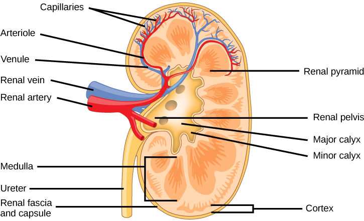
The kidneys are the main osmoregulatory organs in mammalian systems; they function to filter blood and maintain the osmolarity of body fluids at 300 mOsm. They are surrounded by three layers and are made up internally of three distinct regions—the cortex, medulla, and pelvis.
The blood vessels that transport blood into and out of the kidneys arise from and merge with the aorta and inferior vena cava, respectively. The renal arteries branch out from the aorta and enter the kidney where they further divide into segmental, interlobar, arcuate, and cortical radiate arteries.
The nephron is the functional unit of the kidney, which actively filters blood and generates urine. The nephron is made up of the renal corpuscle and renal tubule. Cortical nephrons are found in the renal cortex, while juxtamedullary nephrons are found in the renal cortex close to the renal medulla. The nephron filters and exchanges water and solutes with two sets of blood vessels and the tissue fluid in the kidneys.
There are three steps in the formation of urine: glomerular filtration, which occurs in the glomerulus; tubular reabsorption, which occurs in the renal tubules; and tubular secretion, which also occurs in the renal tubules.
Exercises
1.Which of the following statements about the kidney is false?
A) The renal pelvis drains into the ureter.
B) The renal pyramids are in the medulla.
C) The cortex covers the capsule.
D) Nephrons are in the renal cortex.
Answer: C
2. Which of the following statements about the nephron is false?
A) The collecting duct empties into the distal convoluted tubule.
B) The Bowman’s capsule surrounds the glomerulus.
C) The loop of Henle is between the proximal and distal convoluted tubules.
D) The loop of Henle empties into the distal convoluted tubule.
Answer: A
3. The macula densa is/are:
A) present in the renal medulla.
B) dense tissue present in the outer layer of the kidney.
C) cells present in the DCT and collecting tubules.
D) present in blood capillaries.
Answer: C
4. The osmolarity of body fluids is maintained at ________.
A) 100 mOsm
B) 300 mOsm
C) 1000 mOsm
D) it is not constantly maintained
Answer: B
5. The gland located at the top of the kidney is the ________ gland.
A) adrenal
B) pituitary
C) thyroid
D) thymus
Answer: A
6. Loop diuretics are drugs sometimes used to treat hypertension. These drugs inhibit the reabsorption of Na+ and Cl– ions by the ascending limb of the loop of Henle. A side effect is that they increase urination. Why do you think this is the case?
Loop diuretics decrease the excretion of salt into the renal medulla, thereby reducing its osmolality. As a result, less water is excreted into the medulla by the descending limb, and more water is excreted as urine.
7. Why are the loop of Henle and vasa recta important for the formation of concentrated urine?
The loop of Henle is part of the renal tubule that loops into the renal medulla. In the loop of Henle, the filtrate exchanges solutes and water with the renal medulla and the vasa recta (the peritubular capillary network). The vasa recta acts as the countercurrent exchanger. The kidneys maintain the osmolality of the rest of the body at a constant 300 mOsm by concentrating the filtrate as it passes through the loop of Henle.
8. Describe the structure of the kidney.
Externally, the kidneys are surrounded by three layers. The outermost layer is a tough connective tissue layer called the renal fascia. The second layer is called the perirenal fat capsule, which helps anchor the kidneys in place. The third and innermost layer is the renal capsule. Internally, the kidney has three regions—an outer cortex, a medulla in the middle, and the renal pelvis in the region called the hilum of the kidney, which is the concave part of the “bean” shape.
Glossary
afferent arteriole
arteriole that branches from the cortical radiate artery and enters the glomerulus
arcuate artery
artery that branches from the interlobar artery and arches over the base of the renal pyramids
ascending limb
part of the loop of Henle that ascends from the renal medulla to the renal cortex
Bowman’s capsule
structure that encloses the glomerulus
calyx
structure that connects the renal pelvis to the renal medulla
cortex (animal)
outer layer of an organ like the kidney or adrenal gland
cortical radiate artery
artery that radiates from the arcuate arteries into the renal cortex
cortical nephron
nephron that lies in the renal cortex
countercurrent exchanger
peritubular capillary network that allows exchange of solutes and water from the renal tubules
countercurrent multiplier
osmotic gradient in the renal medulla that is responsible for concentration of urine
descending limb
part of the loop of Henle that descends from the renal cortex into the renal medulla
distal convoluted tubule (DCT)
part of the renal tubule that is the most distant from the glomerulus
efferent arteriole
arteriole that exits from the glomerulus
glomerular filtration
filtration of blood in the glomerular capillary network into the glomerulus
glomerular filtration rate (GFR)
amount of filtrate formed by the glomerulus per minute
glomerulus (renal)
part of the renal corpuscle that contains the capillary network
hilum
region in the renal pelvis where blood vessels, nerves, and ureters bunch before entering or exiting the kidney
inferior vena cava
one of the main veins in the human body
interlobar artery
artery that branches from the segmental artery and travels in between the renal lobes
juxtaglomerular cell
cell in the afferent and efferent arterioles that responds to stimuli from the macula densa
juxtamedullary nephron
nephron that lies in the cortex but close to the renal medulla
kidney
organ that performs excretory and osmoregulatory functions
lobes of the kidney
renal pyramid along with the adjoining cortical region
loop of Henle
part of the renal tubule that loops into the renal medulla
macula densa
group of cells that senses changes in sodium ion concentration; present in parts of the renal tubule and collecting ducts
medulla
middle layer of an organ like the kidney or adrenal gland
nephron
functional unit of the kidney
perirenal fat capsule
fat layer that suspends the kidneys
peritubular capillary network
capillary network that surrounds the renal tubule after the efferent artery exits the glomerulus
proximal convoluted tubule (PCT)
part of the renal tubule that lies close to the glomerulus
renal artery
branch of the artery that enters the kidney
renal capsule
layer that encapsulates the kidneys
renal column
area of the kidney through which the interlobar arteries travel in the process of supplying blood to the renal lobes
renal corpuscle
glomerulus and the Bowman’s capsule together
renal fascia
connective tissue that supports the kidneys
renal pelvis
region in the kidney where the calyces join the ureters
renal pyramid
conical structure in the renal medulla
renal tubule
tubule of the nephron that arises from the glomerulus
renal vein
branch of a vein that exits the kidney and joins the inferior vena cava
segmental artery
artery that branches from the renal artery
transport maximum
maximum amount of solute that can be transported out of the renal tubules during reabsorption
tubular reabsorption
reclamation of water and solutes that got filtered out in the glomerulus
tubular secretion
process of secretion of wastes that do not get reabsorbed
ureter
urine-bearing tube coming out of the kidney; carries urine to the bladder
urinary bladder
structure that the ureters empty the urine into; stores urine
urine
filtrate produced by kidneys that gets excreted out of the body
vasa recta
peritubular network that surrounds the loop of Henle of the juxtamedullary nephrons