245
Athena Li
NEED AN INTRO TP THIS CHAPTER
EVERYTHING BELOW WRITTEN BY ATHENA ON SEP 14
- Cells
Part One: introduction of shapes
Lining epithelia:
Cell shape:
- Squamous (egg), think SQUAshed
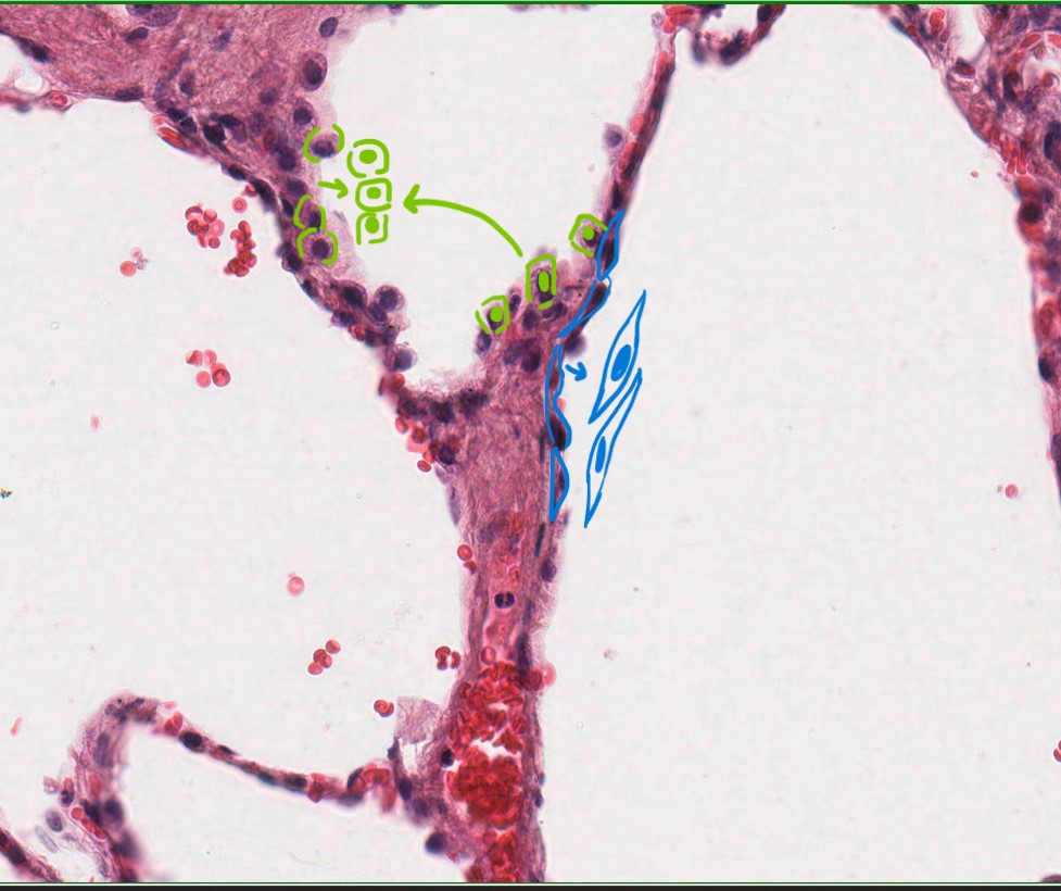
Fig 1. Slide of H&E stained lung alveoli with simple squamous type I pneumocytes marked out in blue and the cuboidal type II pneumocytes marked out in light green. (Slide ID: Path 304 030a, Image ID: 1556 – Lung)

Fig 2. H&E stained thick foot skin with stratified squamous cell lining marked out in light blue to highlight its eye-like shape. (Slide ID: Path 304 001, Image ID: 1522 – Thick skin from foot)
Above is a H&E stain of alveoli. The squamous cells are marked in blue while the cuboidal is marked in green. Squamous cells can also be distinguished based on their eye-like shape. We should know the general function of squamous epithelial linings which will inform us on the logic of its shape. There are two types of squamous epithelial linings:
- Simple – one layer (as shown in image 1 – outlined in light blue)
- Function: Generally lines blood vessels and body cavities to diffuse and regulate passage of materials between the two.
- Stratified – multiple layers (Image 2 – outlined in light blue)
- Function: protection against the environment, microbes and meant to shed off in layers.
Shape logic: So with the two functions lined out, it’s easy to see that squamous cells must be thin to allow easy diffusion between body cavities and blood vessels which gives it its “squashed” thinness. It also must be long and flexible to accommodate its stratified form where it’ll go through wear and tear to protect the body. Additionally, if we were looking from the top instead of from the side in this cross section, the squamous layer would look like a whole bunch of fried eggs crowded in a pan: so borders are irregular as is size of cells but the nucleus will be a dominant feature. The advantage of this is a lot of surface area for things like diffusion.
2. Cuboidal (cell shaped as rounded squares, containing “strings of purple beads/pearls”)
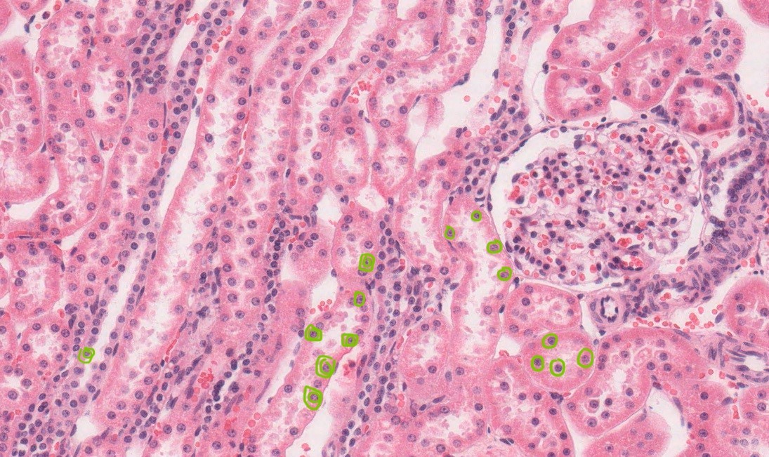
Fig. 3 Image of a H&E stained kidney slide with the simple cuboidal epithelia of the tubules marked out in light green. Specifically the uniformly round nuclei of the cuboidal cells. (Slide ID: Path 304 020, Image ID: 1546 – Kidney, Rabbit)

Fig 4. Sweat gland showing multiple layers of cuboidal epithelia. The boxed out (Blue) gland is marked as an example with three stacked cuboidal cells marked out in yellow. (Slide ID: Path 304 003, Image ID: 1521 – thin skin from scalp)
For cuboidal lining epithelia, the cells may have different shapes but the nucleus are generally very round as highlighted in green. Let’s think about function and shape logic again:
Function: generally found in glandular tissues and kidney tubules to secrete or excrete/filter substances in and from your body
Shape logic: Since secretion and selective diffusion are the main functions of cuboidal layers, it can be thought that its more voluminous cuboid shape gives room to create and secrete substances and that it gives more space for the correctly distinguish and diffuse body fluids
3. Columnar (rounded rectangles with basally located, oval nuclei)
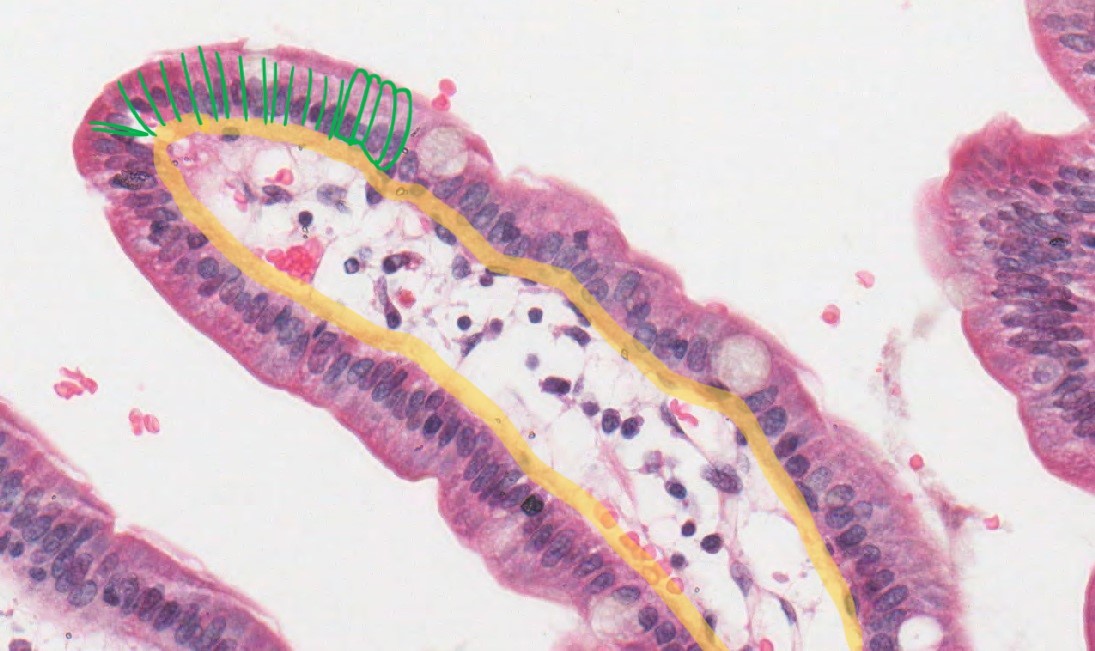
Fig. 5 Simple columnar cells in duodenum villi (stained with H&E) marked out in green. (Slide ID: Path 304 012a, Image ID: 1534 – Duodenum)
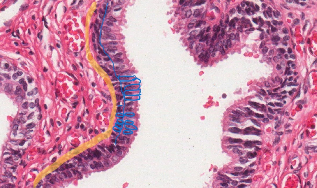
Fig. 6 stratified columnar cells marked out in blue with the basement membrane highlighted yellow found in the urethra (H&E stained). (Slide ID: Path 304 049b, Image ID: 7831 – Prostate & Urethra)
Columnar cells are recognized by their rectangular shapes and elongated, ovular nucleus which sit near the bottom of the cell (side closest to the basement membrane) all lined up in a row. But for the shape logic let’s go through the function and form thought exercise again.
Function: Absorption is one of the well known functions of columnar cells which can be found among other locations, lining the gastrointestinal tract.
Shape logic: Your body wants to be as efficient in absorption as possible to extract the most amount of nutrients and energy possible from foodstuffs. With thin columnar cells lining the GI tract, they are able to maximize absorption with their special feature – microvilli. Many columnar cells have special features like microvilli, cilia etc. because of its elongated shape which is “deep enough” to anchor these apical features.
4. Transitional – bunched rounded layers, transitions between bunched cuboidal and stretched squamous (redraw)
![]()
Fig. 7 transitional epithelium found in the bladder (H&E stained). (Slide ID: Path 304 022, Image ID: 1548 – Bladder, Guinea pig)
Transitional epithelium is easy to identify with its signature umbrella or raindrop shape cells at the top of the layer. Since this type of epithelium lines most urinary tracts and bladder it needs to be able to stretch well which is what the “umbrella” shape allows for.
- Pseudostratified

Fig. 8 Trachea pseudostratified columnar ciliated cells (H&E stained). (Slide ID: Path 304 032, Image ID: 1559 – Trachea & Esophagus, pig)
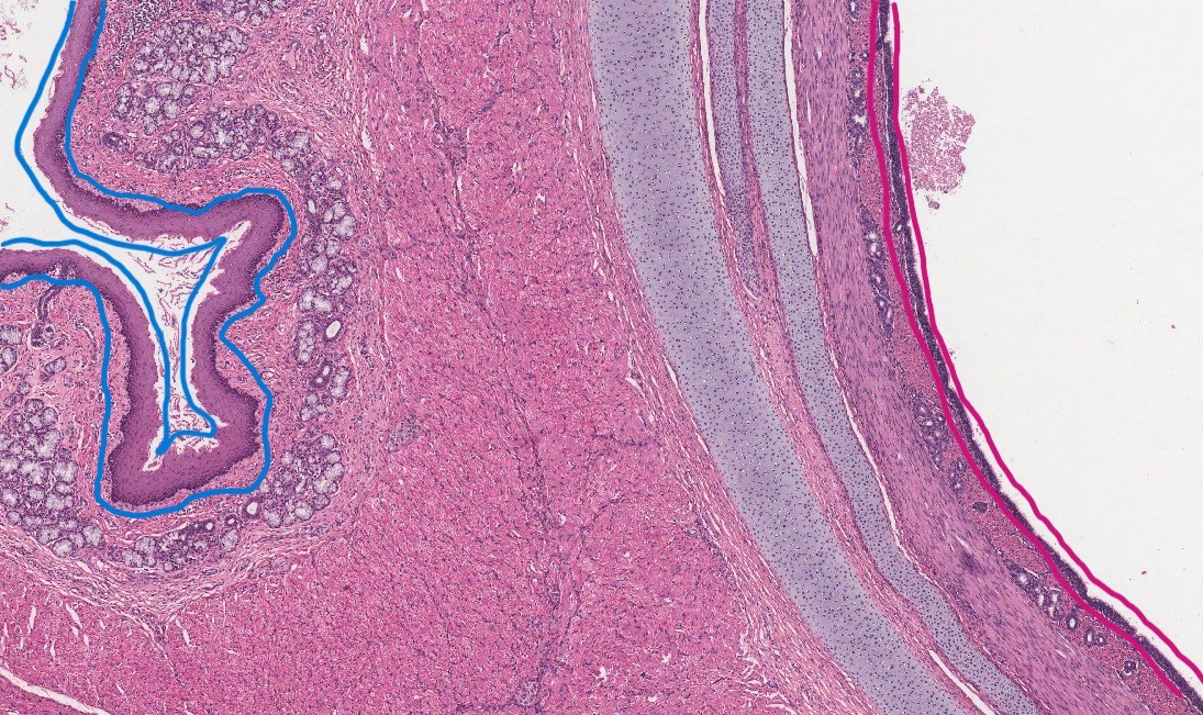
Fig 9. Comparison between pseudostratified (red, on the right) and stratified (blue on the left) thickness (Slide ID: Path 304 032, Image ID: 1559 – Trachea & Esophagus, pig)
Pseudostratified is a difficult layer type to recognize as it has virtually no visual difference from stratified epithelium up close as marked in figure 8. But if given the chance to compare layer thickness with a true stratified epithelial lining as marked in figure 8, the difference in thickness is abundantly clear. For recognition up close, pseudostratified epithelia is only present in respiratory and reproductive systems and so is generally ciliated. The nuclei in pseudostratified epithelia layers are also mismatched at different heights which separates it from the more neatly layered cuboidal and columnar stratified epithelia. Figure 8 demonstrates this mismatching of nuclei well.
