246
Athena Li
- Keratinizing – clear mesh on top of skin layers lacking nuclei

Fig. 9 The keratin layer of this thick foot skin slide is marked in yellow. (Slide ID: Path 304 001, Image ID: 1522 – Thick skin from foot)
The de-saturated mesh marked out by yellow above is the keratin layer atop of thick skin. This keratinizing layer is distinguished by its duller, less saturated staining, mesh-like, flaking texture and lack of nuclei.
- Glands
Endocrine vs Exocrine:
- – Exocrine glands: secretes products into surface epithelium or a larger organ
- – Simple tubular – individual test-tube like infoldings of the epithelium
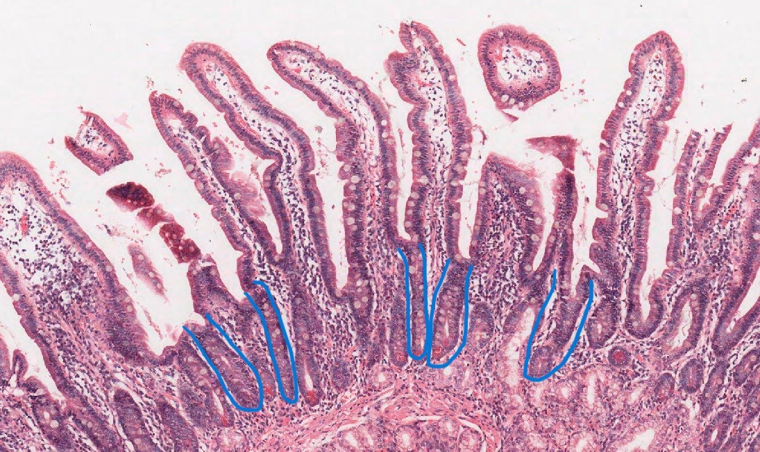
Fig. 10 Simple tubular crypts marked out in blue in the duodenum (Slide ID: Path 304 012a, Image ID: 1534 – Duodenum, stain: H&E)
- – Branched tubular – individual but branching tubular glands

Fig. 11 H&E stained stomach slide with branched tubular glands marked out in yellow and blue (Slide ID: Path 304 011b, Image ID: 1533 – stomach)
Above are branched tubular glands marked out in yellow and blue. The yellow box shows an interesting sample which could be mistaken for two simple tubular glands, but this would be due to the cut of this stomach section.
The possible cut made is visualized below:
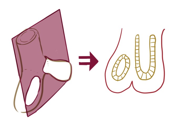
Fig. 12 Hand drawn 3-D to 2-D visualization of a branched tubular gland
- – Coiled tubular
If a coiled tubular gland was presented as a cross-section one can imagine a nest of squirmy things, each surrounding a “hole”.
