Unit 6.1: Control of Bacterial Growth
Outline
Physical Methods of Microbial Control
Learning Objectives
After reading the following, you should be able to:
- Define: sterilization, sterile, commercially sterile, disinfection, disinfectant, bactericide, bacteriostatic, sanitization, antiseptic, aseptic, degerming.
- Describe how the efficiency of anti-microbial agents (AMA) is expressed.
- Describe the factors that can affect the effectiveness of a particular antimicrobial agent.
- Explain the two general, modes of action of antimicrobial agents.
- Describe and explain specific physical and chemical methods used to control microbial growth based on application.
- Be able to identify which methods are more effective for specific types of microbes.
How clean is clean? People wash their cars and vacuum the carpets, but most would not want to eat from these surfaces. Similarly, we might eat with silverware cleaned in a dishwasher, but we could not use the same dishwasher to clean surgical instruments. As these examples illustrate, “clean” is a relative term. Car washing, vacuuming, and dishwashing all reduce the microbial load on the items treated, thus making them “cleaner.” But whether they are “clean enough” depends on their intended use. Because people do not normally eat from cars or carpets, these items do not require the same level of cleanliness that silverware does. Likewise, because silverware is not used for invasive surgery, these utensils do not require the same level of cleanliness as surgical equipment, which requires sterilization to prevent infection.
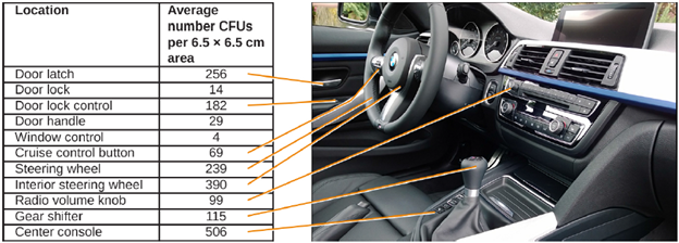
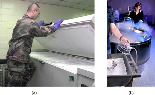
Why not play it safe and sterilize everything? Sterilizing everything we come in contact with is impractical, as well as potentially dangerous. As this chapter will demonstrate, sterilization protocols often require time- and labor-intensive treatments that may degrade the quality of the item being treated or have toxic effects on users. Therefore, the user must consider the item’s intended application when choosing a cleaning method to ensure that it is “clean enough” (Figure 6.1)
To prevent the spread of human disease, it is necessary to control the growth and abundance of microbes in or on various items frequently used by humans. Inanimate items, such as doorknobs, toys, or towels, which may harbor microbes and aid in disease transmission, are called fomites. Two factors heavily influence the level of cleanliness required for a particular fomite and, hence, the protocol chosen to achieve this level. The first factor is the application for which the item will be used. For example, invasive applications that require insertion into the human body require a much higher level of cleanliness than applications that do not. The second factor is the level of resistance to antimicrobial treatment by potential pathogens. For example, foods preserved by canning often become contaminated with the bacterium Clostridium botulinum, which produces the neurotoxin that causes botulism. Because C. botulinum can produce endospores that can survive harsh conditions, extreme temperatures and pressures must be used to eliminate the endospores. Other organisms may not require such extreme measures and can be controlled by a procedure such as washing clothes in a laundry machine.
Sterilization: The most extreme protocols for microbial control aim to achieve sterilization: the complete removal or killing of all vegetative cells, endospores, and viruses from the targeted item or environment. Sterilization protocols are generally reserved for laboratory, medical, manufacturing, and food industry settings, where it may be imperative for certain items to be completely free of potentially infectious agents. Sterilization can be accomplished through either physical means, such as exposure to high heat, pressure, or filtration through an appropriate filter, or by chemical means. Chemicals that can be used to achieve sterilization are called sterilants. Sterilants effectively kill all microbes and viruses, and, with appropriate exposure time, can also kill endospores.
For many clinical purposes, aseptic technique is necessary to prevent contamination of sterile surfaces. Aseptic technique involves a combination of protocols that collectively maintain sterility, or asepsis, thus preventing contamination of the patient with microbes and infectious agents. Failure to practice aseptic technique during many types of clinical procedures may introduce microbes to the patient’s body and put the patient at risk for sepsis, a systemic inflammatory response to an infection that results in high fever, increased heart and respiratory rates, shock, and, possibly, death. Medical procedures that carry risk of contamination must be performed in a sterile field, a designated area that is kept free of all vegetative microbes, endospores, and viruses. Sterile fields are created according to protocols requiring the use of sterilized materials, such as packaging and drapings, and strict procedures for washing and application of sterilants.
One food sterilization protocol, commercial sterilization, uses heat at a temperature low enough to preserve food quality but high enough to destroy common pathogens responsible for food poisoning, such as C. botulinum. Because C. botulinum and its endospores are commonly found in soil, they may easily contaminate crops during harvesting, and these endospores can later germinate within the anaerobic environment once foods are canned. Metal cans of food contaminated with C. botulinum will bulge due to the microbe’s production of gases; contaminated jars of food typically bulge at the metal lid. To eliminate the risk for C. botulinum contamination, commercial food-canning protocols are designed with a large margin of error. They assume an impossibly large population of endospores (1012 per can) and aim to reduce this population to 1 endospore per can to ensure the safety of canned foods.
Even so, commercial sterilization does not eliminate the presence of all microbes; rather, it targets those pathogens that cause spoilage and foodborne diseases, while allowing many nonpathogenic organisms to survive. Therefore, “sterilization” is somewhat of a misnomer in this context, and commercial sterilization may be more accurately described as “quasi-sterilization.”
Other Methods of Control: Sterilization protocols require procedures that are not practical, or necessary, in many settings. Various other methods are used in clinical and nonclinical settings to reduce the microbial load on items. Although the terms for these methods are often used interchangeably, there are important distinctions.
The process of disinfection inactivates most microbes on the surface of a fomite by using antimicrobial chemicals or heat. Because some microbes remain, the disinfected item is not considered sterile. Ideally, disinfectants should be fast acting, stable, easy to prepare, inexpensive, and easy to use. An example of a natural disinfectant is vinegar; its acidity can kill certain microbes. Chemical disinfectants, such as chlorine bleach or products containing chlorine, are used to clean nonliving surfaces such as laboratory benches, clinical surfaces, and bathroom sinks. Typical disinfection does not lead to sterilization because endospores tend to survive even when all vegetative cells have been killed.
Unlike disinfectants, antiseptics are antimicrobial chemicals safe for use on living skin or tissues. Examples of antiseptics include hydrogen peroxide and isopropyl alcohol. The process of applying an antiseptic is called antisepsis. In addition to the characteristics of a good disinfectant, antiseptics must also be selectively effective against microorganisms and able to penetrate tissue deeply without causing tissue damage.
The type of protocol required to achieve the desired level of cleanliness depends on the particular item to be cleaned. For example, those used clinically are categorized as critical, semicritical, and noncritical. Critical items must be sterile because they will be used inside the body, often penetrating sterile tissues or the bloodstream; examples of critical items include surgical instruments, catheters, and intravenous fluids. Gastrointestinal endoscopes and various types of equipment for respiratory therapies are examples of semicritical items; they may contact mucous membranes or nonintact skin but do not penetrate tissues. Semicritical items do not typically need to be sterilized but do require a high level of disinfection. Items that may contact but not penetrate intact skin are noncritical items; examples are bed linens, furniture, crutches, stethoscopes, and blood pressure cuffs. These articles need to be clean but not highly disinfected.
The act of handwashing is an example of degerming, in which microbial numbers are significantly reduced by gently scrubbing living tissue, most commonly skin, with a mild chemical (e.g., soap) to avoid the transmission of pathogenic microbes. Wiping the skin with an alcohol swab at an injection site is another example of degerming. These degerming methods remove most (but not all) microbes from the skin’s surface.
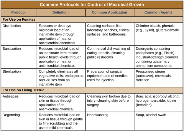
The term sanitization refers to the cleansing of fomites to remove enough microbes to achieve levels deemed safe for public health. For example, commercial dishwashers used in the food service industry typically use very hot water and air for washing and drying; the high temperatures kill most microbes, sanitizing the dishes. Surfaces in hospital rooms are commonly sanitized using a chemical disinfectant to prevent disease transmission between patients. Figure 6.2 summarizes common protocols, definitions, applications, and agents used to control microbial growth.
Measuring Microbial Control: Physical and chemical methods of microbial control that kill the targeted microorganism are identified by the suffix-cide (or -cidal). The prefix indicates the type of microbe or infectious agent killed by the treatment method: bactericides kill bacteria, viricides kill or inactivate viruses, and fungicides kill fungi. Other methods do not kill organisms but, instead, stop their growth, making their population static; such methods are identified by the suffix – stat (or -static). For example, bacteriostatic treatments inhibit the growth of bacteria, whereas fungistatic treatments inhibit the growth of fungi. Factors that determine whether a particular treatment is -cidal or -static include the types of microorganisms targeted, the concentration of the chemical used, and the nature of the treatment applied.
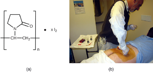
Although -static treatments do not actually kill infectious agents, they are often less toxic to humans and other animals, and may also better preserve the integrity of the item treated. Such treatments are typically sufficient to keep the microbial population of an item in check. The reduced toxicity of some of these -static chemicals also allows them to be impregnated safely into plastics to prevent the growth of microbes on these surfaces. Such plastics are used in products such as toys for children and cutting boards for food preparation. When used to treat an infection, -static treatments are typically sufficient in an otherwise healthy individual, preventing the pathogen from multiplying, thus allowing the individual’s immune system to clear the infection.
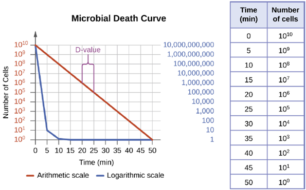
The degree of microbial control can be evaluated using a microbial death curve to describe the progress and effectiveness of a particular protocol. When exposed to a particular microbial control protocol, a fixed percentage of the microbes within the population will die. Because the rate of killing remains constant even when the population size varies, the percentage killed is more useful information than the absolute number of microbes killed. Death curves are often plotted as semilog plots just like microbial growth curves because the reduction in microorganisms is typically logarithmic (Figure 6.3). The amount of time it takes for a specific protocol to produce a one order of- magnitude decrease in the number of organisms, or the death of 90% of the population, is called the decimal reduction time (DRT) or D-value. The smaller the D-value, the stronger the antimicrobial agent.
Several factors contribute to the effectiveness of a disinfecting agent or microbial control protocol. First, as demonstrated in Figure 6.3, the length of time of exposure is important. Longer exposure times kill more microbes. Because microbial death of a population exposed to a specific protocol is logarithmic, it takes longer to kill a high-population load than a low-population load exposed to the same protocol. A shorter treatment time (measured in multiples of the D-value) is needed when starting with a smaller number of organisms. Effectiveness also depends on the susceptibility of the agent to that disinfecting agent or protocol. The concentration of disinfecting agent or intensity of exposure is also important. For example, higher temperatures and higher concentrations of disinfectants kill microbes more quickly and effectively. Conditions that limit contact between the agent and the targeted cells cells—for example, the presence of bodily fluids, tissue, organic debris (e.g., mud or feces), or biofilms on surfaces—increase the cleaning time or intensity of the microbial control protocol required to reach the desired level of cleanliness. All these factors must be considered when choosing the appropriate protocol to control microbial growth in a given situation.
Physical methods of Microbial Control: For thousands of years, humans have used various physical methods of microbial control for food preservation. Common control methods include the application of high temperatures, radiation, filtration, and desiccation (drying), among others. Most of these methods nonspecifically kill cells by disrupting membranes, changing membrane permeability and causing cellular contents to leak out, or by damaging cell’s proteins and nucleic acids by denaturation, degradation, or chemical modification, preventing metabolism. Various physical methods used for microbial control are described in this section.
- Heat: Heating is one of the most common—and oldest—forms of microbial control. It is used in simple techniques like cooking and canning. Heat can kill microbes by altering their membranes and denaturing proteins. The thermal death point (TDP) of a microorganism is the lowest temperature at which all microbes are killed in a 10-minute exposure. Different microorganisms will respond differently to high temperatures, with some (e.g., endospore-formers such as C. botulinum) being more heat tolerant. A similar parameter, the thermal death time (TDT), is the length of time needed to kill all microorganisms in a sample at a given temperature. These parameters are often used to describe sterilization procedures that use high heat, such as autoclaving. Boiling is one of the oldest methods of moist-heat control of microbes, and it is typically quite effective at killing vegetative cells and some viruses. However, boiling is less effective at killing endospores; some endospores are able to survive up to 20 hours of boiling. Additionally, boiling may be less effective at higher altitudes, where the boiling point of water is lower and the boiling time needed to kill microbes is therefore longer. For these reasons, boiling is not considered a useful sterilization technique in the laboratory or clinical setting.
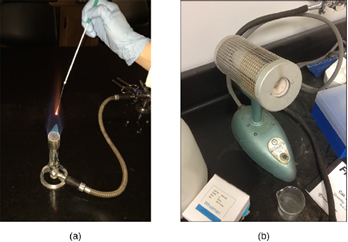
Many different heating protocols can be used for sterilization in the laboratory or clinic, and these protocols can be broken down into two main categories: dry-heat sterilization and moist-heat sterilization. Aseptic technique in the laboratory typically involves some dry-heat sterilization protocols using direct application of high heat, such as sterilizing inoculating loops (Figure 6.4). Incineration at very high temperatures destroys all microorganisms. Dry heat can also be applied for relatively long periods of time (at least 2 hours) at temperatures up to 170 °C by using a dry-heat sterilizer, such as an oven. However, moist-heat sterilization is typically the more effective protocol because it penetrates cells better than dry heat does.
1.2 Autoclaves: Autoclaves rely on moist-heat sterilization. They are used to raise temperatures above the boiling point of water to sterilize items such as surgical equipment from vegetative cells, viruses, and especially endospores, which are known to survive boiling temperatures, without damaging the items. Charles Chamberland (1851–1908) designed the modern autoclave in 1879 while working in the laboratory of Louis Pasteur. The autoclave is still considered the most effective method of sterilization. Outside laboratory and clinical settings, large industrial autoclaves called retorts allow for moist-heat sterilization on a large scale.
In general, the air in the chamber of an autoclave is removed and replaced with increasing amounts of steam trapped within the enclosed chamber, resulting in increased interior pressure and temperatures above the boiling point of water. Standard operating temperatures for autoclaves are 121 °C or, in some cases, 132 °C, typically at a pressure of 15 to 20 pounds per square inch (psi). The length of exposure depends on the volume and nature of material being sterilized, but it is typically 20 minutes or more, with larger volumes requiring longer exposure times to ensure sufficient heat transfer to the materials being sterilized. The steam must directly contact the liquids or dry materials being sterilized, so containers are left loosely closed and instruments are loosely wrapped in paper or foil. The key to autoclaving is that the temperature must be high enough to kill endospores to achieve complete sterilization.
1.3 Pasteurization: Although complete sterilization is ideal for many medical applications, it is not always practical for other applications and may also alter the quality of the product. Boiling and autoclaving are not ideal ways to control microbial growth in many foods because these methods may ruin the consistency and other organoleptic (sensory) qualities of the food. Pasteurization is a form of microbial control for food that uses heat but does not render the food sterile. Traditional pasteurization kills pathogens and reduces the number of spoilage-causing microbes while maintaining food quality. The process of pasteurization was first developed by Louis Pasteur in the 1860s as a method for preventing the spoilage of beer and wine. Today, pasteurization is most commonly used to kill heat-sensitive pathogens in milk and other food products (e.g., apple juice and honey). However, because pasteurized food products are not sterile, they will eventually spoil.
- Refrigeration and Freezing: Just as high temperatures are effective for controlling microbial growth, exposing microbes to low temperatures can also be an easy and effective method of microbial control, with the exception of psychrophiles, which prefer cold temperatures. Refrigerators used in home kitchens or in the laboratory maintain temperatures between 0 °C and 7 °C. This temperature range inhibits microbial metabolism, slowing the growth of microorganisms significantly and helping preserve refrigerated products such as foods or medical supplies. Certain types of laboratory cultures can be preserved by refrigeration for later use. In most cases refrigeration or freezing is a bacteriostatic treatment.
Freezing below −2 °C may stop microbial growth and even kill susceptible organisms. Bacterial cultures and medical specimens requiring long-term storage or transport are often frozen at ultra-low temperatures of −70 °C or lower. These ultra-low temperatures can be achieved by storing specimens on dry ice in an ultra-low freezer or in special liquid nitrogen tanks, which maintain temperatures lower than −196 °C (Figure 6.5).
2.2 Lyophilization: Freeze-drying, or lyophilization, is a method in which an item is rapidly frozen (“snap-frozen”) and placed under vacuum so that water is lost by sublimation. Lyophilization combines both exposure to cold temperatures and desiccation, making it quite effective for controlling microbial growth. In addition, lyophilization causes less damage to an item than conventional desiccation and better preserves the item’s original qualities. Lyophilized items may be stored at room temperature if packaged appropriately to prevent moisture acquisition. Lyophilization is used for preservation in the food industry and is also used in the laboratory for the long-term storage and transportation of microbial cultures.
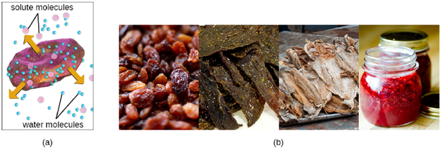
- Osmotic Pressure: All cells, including microbes, require water for their metabolism and survival and by limiting water access, we inhibit growth. While these treatments can kill microbes, they might not kill all microbes or their endospores, which may start to regrow when conditions are more favorable and water content is restored. At very high concentrations of salts or sugars, the amount of available water in microbial cells is reduced dramatically because water will be drawn from an area of low solute concentration (inside the cell) to an area of high solute concentration (outside the cell) (Figure 6.6). Many microorganisms do not survive these conditions of high osmotic pressure. Honey, for example, is 80% sucrose, an environment in which very few microorganisms are capable of growing, thereby eliminating the need for refrigeration. Salted meats and fish, like ham and cod, respectively, were critically important foods before the age of refrigeration. Fruits were preserved by adding sugar, making jams and jellies. However, certain microbes, such as molds and yeasts, tend to be more tolerant of desiccation and high osmotic pressures, and, thus, may still contaminate these types of foods.
- Radiation: Radiation in various forms, from high-energy radiation to sunlight, can be used to kill microbes or inhibit their growth. Ionizing radiation includes X-rays, gamma rays, and high-energy electron beams. Ionizing radiation is strong enough to pass into the cell, where it alters molecular structures and damages cell components. For example, ionizing radiation introduces double-strand breaks in DNA molecules. This may directly cause DNA mutations to occur, or mutations may be introduced when the cell attempts to repair the DNA damage. As these mutations accumulate, they eventually lead to cell death.
Both X-rays and gamma rays easily penetrate paper and plastic and can therefore be used to sterilize many packaged materials. In the laboratory, ionizing radiation is commonly used to sterilize materials that cannot be autoclaved, such as plastic Petri dishes and disposable plastic inoculating loops. For clinical use, ionizing radiation is used to sterilize gloves, intravenous tubing, and other latex and plastic items used for patient care. Ionizing radiation is also used for the sterilization of other types of delicate, heat-sensitive materials used clinically, including tissues for transplantation, pharmaceutical drugs, and medical equipment Packaged dried spices are also often gamma-irradiated. Because of their ability to penetrate paper, plastic, thin sheets of wood and metal, and tissue, great care must be taken when using X-rays and gamma irradiation. These types of ionizing irradiation cannot penetrate thick layers of iron or lead, so these metals are commonly used to protect humans who may be potentially exposed.
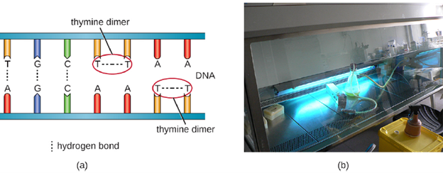
Another type of radiation, nonionizing radiation, is commonly used for sterilization and uses less energy than ionizing radiation. It does not penetrate cells or packaging. Ultraviolet (UV) light is one example; it causes thymine dimers to form between adjacent thymines within a single strand of DNA (Figure 6.7). When DNA polymerase encounters the thymine dimer, it does not always incorporate the appropriate complementary nucleotides (two adenines), and this leads to formation of mutations that can ultimately kill microorganisms. UV light can be used effectively by both consumers and laboratory personnel to control microbial growth. UV lamps are now commonly incorporated into water purification systems for use in homes. In addition, small portable
UV lights are commonly used by campers to purify water from natural environments before drinking. Germicidal lamps are also used in surgical suites, biological safety cabinets, and transfer hoods, typically emitting UV light at a wavelength of 260 nm. Because UV light does not penetrate surfaces and will not pass through plastics or glass, cells must be exposed directly to the light source.
Sunlight has a very broad spectrum that includes UV and visible light. In some cases, sunlight can be effective against certain bacteria because of both the formation of thymine dimers by UV light and by the production of reactive oxygen products induced in low amounts by exposure to visible light.
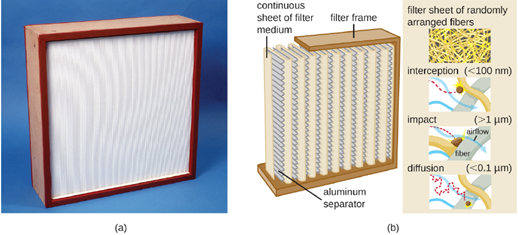
- Filtration: Filtration is a method of physically separating microbes from samples. Air is commonly filtered through high efficiency particulate air (HEPA) filters (Figure 6.8). HEPA filters have effective pore sizes of 0.3 μm, small enough to capture bacterial cells, endospores, and many viruses, as air passes through these filters, nearly sterilizing the air on the other side of the filter. HEPA filters have a variety of applications and are used widely in clinical settings, in cars and airplanes, and even in the home. For example, they may be found in vacuum cleaners, heating and air-conditioning systems, and air purifiers.
HEPA filters are also commonly used in hospitals and surgical suites to prevent contamination and the spread of airborne microbes through ventilation systems. HEPA filtration systems may be designed for entire buildings or for individual rooms. For example, burn units, operating rooms, or isolation units may require special HEPA-filtration systems to remove opportunistic pathogens from the environment because patients in these rooms are particularly vulnerable to infection.
5.2 Membrane Filtration: Filtration can also be used to remove microbes from liquid samples using membrane filtration. Membrane filters for liquids function similarly to HEPA filters for air. Typically, membrane filters that are used to remove bacteria have an effective pore size of 0.2 μm, smaller than the average size of a bacterium (1 μm), but filters with smaller pore sizes are available for more specific needs. Membrane filtration is useful for removing bacteria from various types of heat-sensitive solutions used in the laboratory, such as antibiotic solutions and vitamin solutions. Large volumes of culture media may also be filter sterilized rather than autoclaved to protect heat-sensitive components. Often when filtering small volumes, syringe filters are used, but vacuum filters are typically used for filtering larger volumes (Figure 6.9).
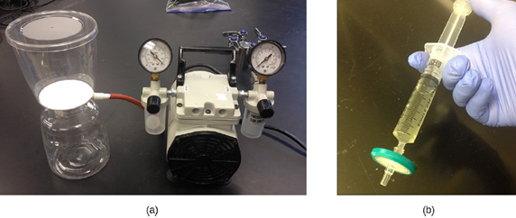
Larger volumes are filtered in units like these. The solution is drawn through the filter by connecting the unit to a vacuum. (b) Smaller volumes are often filtered using syringe filters, which are units that fit on the end of a syringe. In this case, the solution is pushed through by depressing the syringe’s plunger. (credit a, b: modification of work by Brian Forster)
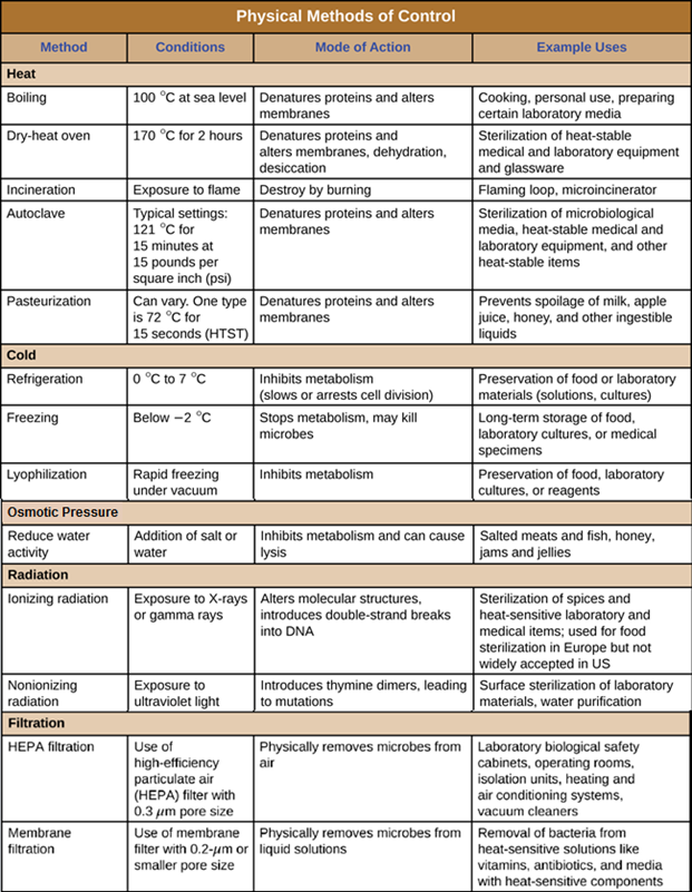
Physical methods of microbial control are summarized in Figure 6.10.
Chemical Methods of Microbial Control: In addition to physical methods of microbial control, chemicals are also used to control microbial growth. A wide variety of chemicals can be used as disinfectants or antiseptics. When choosing which to use, it is important to consider the type of microbe targeted; how clean the item needs to be; the disinfectant’s effect on the item’s integrity; its safety to animals, humans, and the environment; its expense; and its ease of use. This section describes the variety of chemicals used as disinfectants and antiseptics, including their mechanisms of action and common uses.
- Phenolics: In the 1800s, scientists began experimenting with a variety of chemicals for disinfection. In the 1860s, British surgeon Joseph Lister (1827–1912) began using carbolic acid, known as phenol, as a disinfectant for the treatment of surgical wounds. In 1879, Lister’s work inspired the American chemist Joseph Lawrence (1836–1909) to develop Listerine, an alcohol-based mixture of several related compounds that is still used today as an oral antiseptic. Today, carbolic acid is no longer used as a surgical disinfectant because it is a skin irritant, but the chemical compounds found in antiseptic mouthwashes and throat lozenges are called phenolics.
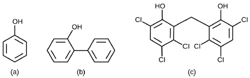
Chemically, phenol consists of a benzene ring with an –OH group, and phenolics are compounds that have this group as part of their chemical structure (Figure 6.11). Phenolics such as thymol and eucalyptol occur naturally in plants. Other phenolics can be derived from creosote, a component of coal tar. Phenolics tend to be stable, persistent on surfaces, and less toxic than phenol. They inhibit microbial growth by denaturing proteins and disrupting membranes.
Since Lister’s time, several phenolic compounds have been used to control microbial growth. Phenolics like cresols (methylated phenols) and o-phenylphenol were active ingredients in various formulations of Lysol since its invention in 1889. The bisphenol hexachlorophene, a disinfectant, is the active ingredient in pHisoHex, a topical cleansing detergent widely used for handwashing in hospital settings. pHisoHex is particularly effective against gram-positive bacteria, including those causing staphylococcal and streptococcal skin infections. pHisoHex was formerly used for bathing infants, but this practice has been discontinued because it has been shown that exposure to hexachlorophene can lead to neurological problems.
Triclosan is another bisphenol compound that has seen widespread application in antibacterial products over the last several decades. Initially used in toothpastes, triclosan is now commonly used in hand soaps and is frequently impregnated into a wide variety of other products, including cutting boards, knives, shower curtains, clothing, and concrete, to make them antimicrobial. It is particularly effective against gram-positive bacteria on the skin, as well as certain gram-negative bacteria and yeasts.
- Heavy Metals: Some of the first chemical disinfectants and antiseptics to be used were heavy metals. Heavy metals kill microbes by binding to proteins, thus inhibiting enzymatic activity (Figure 6.12). Heavy metals are oligodynamic, meaning that very small concentrations show significant antimicrobial activity. Ions of heavy metals bind to sulfur-containing amino acids strongly and bioaccumulate within cells, allowing these metals to reach high localized concentrations. This causes proteins to denature.
Heavy metals are not selectively toxic to microbial cells. They may bioaccumulate in human or animal cells, as well, and excessive concentrations can have toxic effects on humans. If too much silver accumulates in the body, for example, it can result in a condition called argyria, in which the skin turns irreversibly blue-gray. One way to reduce the potential toxicity of heavy metals is by carefully controlling the duration of exposure and concentration of the heavy metal.
One example of a commonly used heavy metal is silver. Silver has long been used as an antiseptic. In ancient times, drinking water was stored in silver jugs. Silvadene cream is commonly used to treat topical wounds and is particularly helpful in preventing infection in burn wounds. Silver nitrate drops were once routinely applied to the eyes of newborns to protect against ophthalmia neonatorum, eye infections that can occur due to exposure to pathogens in the birth canal, but antibiotic creams are more now commonly used. Silver is often combined with antibiotics, making the antibiotics thousands of times more effective. Silver is also commonly incorporated into catheters and bandages, rendering them antimicrobial; however, there is evidence that heavy metals may also enhance selection for antibiotic resistance.
- Halogens: Other chemicals commonly used for disinfection are the halogens iodine, chlorine, and fluorine. Iodine works by oxidizing cellular components, including sulfur-containing amino acids, nucleotides, and fatty acids, and destabilizing the macromolecules that contain these molecules. It is often used as a topical tincture, but it may cause staining or skin irritation. An iodophor is a compound of iodine complexed with an organic molecule, thereby increasing iodine’s stability and, in turn, its efficacy. One common iodophor is povidone-iodine, which includes a wetting agent that releases iodine relatively slowly. Betadine is a brand of povidone-iodine commonly used as a hand scrub by medical personnel before surgery and for topical antisepsis of a patient’s skin before incision (Figure 6.13).
Chlorine is another halogen commonly used for disinfection. When chlorine gas is mixed with water, it produces a strong oxidant called hypochlorous acid, which is uncharged and enters cells easily, damaging cellular components. Chlorine gas is commonly used in municipal drinking water and wastewater treatment plants, with the resulting hypochlorous acid producing the actual antimicrobial effect. Sodium hypochlorite is the chemical component of common household bleach, and it is also used for a wide variety of disinfecting purposes. Although chlorinated compounds are relatively effective disinfectants, they have their disadvantages. Some may irritate the skin, nose, or eyes of some individuals, and they may not completely eliminate certain hardy organisms from contaminated drinking water.
The halogen fluorine is also known to have antimicrobial properties that contribute to the prevention of dental caries (cavities). Fluoride is the main active ingredient of toothpaste and is also commonly added to tap water to help communities maintain oral health. Chemically, fluoride can become incorporated into the hydroxyapatite of tooth enamel, making it more resistant to corrosive acids produced by the fermentation of oral microbes. Fluoride also enhances the uptake of calcium and phosphate ions in tooth enamel, promoting remineralization. In addition to strengthening enamel, fluoride also seems to be bacteriostatic. It accumulates in plaque-forming bacteria, interfering with their metabolism and reducing their production of the acids that contribute to tooth decay.
- Alcohols: Alcohols make up another group of chemicals commonly used as disinfectants and antiseptics. They work by rapidly denaturing proteins, which inhibits cell metabolism, and by disrupting membranes, which leads to cell lysis. Once denatured, the proteins may potentially refold if enough water is present in the solution. Alcohols are typically used at concentrations of about 70% aqueous solution and, in fact, work better in aqueous solutions than 100% alcohol solutions. This is because alcohols coagulate proteins. In higher alcohol concentrations, rapid coagulation of surface proteins prevents effective penetration of cells. The most commonly used alcohols for disinfection are ethyl alcohol (ethanol) and isopropyl alcohol (isopropanol, rubbing alcohol).
Alcohols tend to be bactericidal and fungicidal, but may also be viricidal for enveloped viruses only. Although alcohols are not sporicidal, they do inhibit the processes of sporulation and germination. Alcohols are volatile and dry quickly, but they may also cause skin irritation because they dehydrate the skin at the site of application. One common clinical use of alcohols is swabbing the skin for degerming before needle injection. Alcohols also are the active ingredients in instant hand sanitizers, which have gained popularity in recent years. The alcohol in these hand sanitizers works both by denaturing proteins and by disrupting the microbial cell membrane, but will not work effectively in the presence of visible dirt. All in all, alcohols are inexpensive and quite effective for the disinfection of a broad range of vegetative microbes. However, one disadvantage of alcohols is their high volatility, limiting their effectiveness to immediately after application.
- Surfactants: Surface-active agents, or surfactants, are a group of chemical compounds that lower the surface tension of water. Surfactants are the major ingredients in soaps and detergents. Soaps are salts of long-chain fatty acids and have both polar and nonpolar regions, allowing them to interact with polar and nonpolar regions in other molecules. They can interact with nonpolar oils and grease to create emulsions in water, loosening and lifting away dirt and microbes from surfaces and skin. Soaps do not kill or inhibit microbial growth and so are not considered antiseptics or disinfectants. However, proper use of soaps mechanically carries away microorganisms, effectively degerming a surface. Some soaps contain added bacteriostatic agents such as triclocarban or cloflucarban, compounds structurally related to triclosan, that introduce antiseptic or disinfectant properties to the soaps.
Soaps, however, often form films that are difficult to rinse away, especially in hard water, which contains high concentrations of calcium and magnesium mineral salts. Detergents contain synthetic surfactant molecules with both polar and nonpolar regions that have strong cleansing activity but are more soluble, even in hard water, and, therefore, leave behind no soapy deposits. Anionic detergents, such as those used for laundry, have a negatively charged anion at one end attached to a long hydrophobic chain, whereas cationic detergents have a positively charged cation instead.
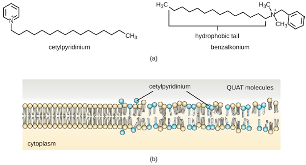
Cationic detergents include an important class of disinfectants and antiseptics called the quaternary ammonium salts (quats), named for the characteristic quaternary nitrogen atom that confers the positive charge (Figure 6.14). Overall, quats have properties similar to phospholipids, having hydrophilic and hydrophobic ends. As such, quats have the ability to insert into the bacterial phospholipid bilayer and disrupt membrane integrity. The cationic charge of quats appears to confer their antimicrobial properties, which are diminished when neutralized. Quats have several useful properties. They are stable, nontoxic, inexpensive, colorless, odorless, and tasteless. They tend to be bactericidal by disrupting membranes. They are also active against fungi, protozoans, and enveloped viruses, but endospores are unaffected. In clinical settings, they may be used as antiseptics or to disinfect surfaces. Mixtures of quats are also commonly found in household cleaners and disinfectants, including many current formulations of Lysol brand products, which contain benzalkonium chlorides as the active ingredients. Benzalkonium chlorides, along with the quat cetylpyrimidine chloride, are also found in products such as skin antiseptics, oral rinses, and mouthwashes.
- Alkylating Agents: The alkylating agents are a group of strong disinfecting chemicals that act by replacing a hydrogen atom within a molecule with an alkyl group, thereby inactivating enzymes and nucleic acids. The alkylating agent formaldehyde is commonly used in solution at a concentration of 37% (known as formalin) or as a gaseous disinfectant and biocide. It is a strong, broad-spectrum disinfectant and biocide that has the ability to kill bacteria, viruses, fungi, and endospores, leading to sterilization at low temperatures, which is sometimes a convenient alternative to the more labor-intensive heat sterilization methods. It also cross-links proteins and has been widely used as a chemical fixative. Because of this, it is used for the storage of tissue specimens and as an embalming fluid. It also has been used to inactivate infectious agents in vaccine preparation. Formaldehyde is very irritating to living tissues and is also carcinogenic; therefore, it is not used as an antiseptic.
Glutaraldehyde is structurally similar to formaldehyde but has two reactive aldehyde groups, allowing it to act more quickly than formaldehyde. It is commonly used as a 2% solution for sterilization and is marketed under the brand name Cidex. It is used to disinfect a variety of surfaces and surgical and medical equipment. However, similar to formaldehyde, glutaraldehyde irritates the skin and is not used as an antiseptic.
Ethylene oxide is a type of alkylating agent that is used for gaseous sterilization. It is highly penetrating and can sterilize items within plastic bags such as catheters, disposable items in laboratories and clinical settings (like packaged Petri dishes), and other pieces of equipment. Ethylene oxide exposure is a form of cold sterilization, making it useful for the sterilization of heat-sensitive items, or items that it would be impractical to use liquid disinfectants on, such as mattresses. Great care needs to be taken with the use of ethylene oxide, however; it is carcinogenic, like the other alkylating agents, and is also highly explosive. With careful use and proper aeration of the products after treatment, ethylene oxide is highly effective, and ethylene oxide sterilizers are commonly found in medical settings for sterilizing packaged materials.
- Peroxygens: Peroxygens are strong oxidizing agents that can be used as disinfectants or antiseptics. The most widely used peroxygen is hydrogen peroxide (H2O2), which is often used in solution to disinfect surfaces and may also be used as a gaseous agent. Hydrogen peroxide solutions are inexpensive skin antiseptics that break down into water and oxygen gas, both of which are environmentally safe. This decomposition is accelerated in the presence of light, so hydrogen peroxide solutions typically are sold in brown or opaque bottles. One disadvantage of using hydrogen peroxide as an antiseptic is that it also causes damage to skin that may delay healing or lead to scarring. Contact lens cleaners often include hydrogen peroxide as a disinfectant.
Hydrogen peroxide works by producing free radicals that damage cellular macromolecules. Hydrogen peroxide has broad-spectrum activity, working against gram-positive and gram-negative bacteria (with slightly greater efficacy against gram-positive bacteria), fungi, viruses, and endospores. However, bacteria that produce the oxygen detoxifying enzymes catalase or peroxidase may have inherent tolerance to low hydrogen peroxide concentrations. To kill endospores, the length of exposure or concentration of solutions of hydrogen peroxide must be increased. Gaseous hydrogen peroxide has greater efficacy and can be used as a sterilant for rooms or equipment.
Other examples of peroxygens include benzoyl peroxide and carbamide peroxide. Benzoyl peroxide is a peroxygen that used in acne medication solutions. It kills the bacterium Cutibacterium acnes, which is associated with acne. Carbamide peroxide, an ingredient used in toothpaste, is a peroxygen that combats oral biofilms that cause tooth discoloration and halitosis (bad breath). Last, ozone gas is a peroxygen with disinfectant qualities and is used to clean air or water supplies. Overall, peroxygens are highly effective and commonly used, with no associated environmental hazard.
Testing the effectiveness of chemical methods of control: The effectiveness of various chemical disinfectants is reflected in the terms used to describe them. Chemical disinfectants are grouped by the power of their activity, with each category reflecting the types of microbes and viruses its component disinfectants are effective against. High-level germicides have the ability to kill vegetative cells, fungi, viruses, and endospores, leading to sterilization, with extended use. Intermediate-level germicides, as their name suggests, are less effective against endospores and certain viruses, and low-level germicides kill only vegetative cells and certain enveloped viruses, and are ineffective against endospores.
However, several environmental conditions influence the potency of an antimicrobial agent and its effectiveness. For example, length of exposure is particularly important, with longer exposure increasing efficacy. Similarly, the concentration of the chemical agent is also important, with higher concentrations being more effective than lower ones. Temperature, pH, and other factors can also affect the potency of a disinfecting agent.
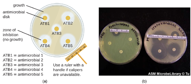
One approach that can be used to compare the relative effectiveness of antimicrobial agents is the disk-diffusion method. The disk-diffusion method involves applying different chemicals to separate, sterile filter paper disks (Figure 6.15). The disks are then placed on an agar plate that has been inoculated with the targeted bacterium and the chemicals diffuse out of the disks into the agar where the bacteria have been inoculated. As the “lawn” of bacteria grows, zones of inhibition of microbial growth are observed as clear areas around the disks. Although there are other factors that contribute to the sizes of zones of inhibition (e.g., whether the agent is water soluble and able to diffuse in the agar), larger zones typically correlate to increased inhibition effectiveness of the chemical agent. The diameter across each zone is measured in millimeters.
Unit 6.2: Antimicrobial Drugs.
