120
Jennifer Kong
Learning Objectives
By the end of this chapter, the reader will be able to:
- Describe the major surface markings of the brain and brainstem
- Identify the motor cortex and subcortical fibers
- Describe the gross anatomy of the spinal cord and peripheral nerve plexus
Visualization of the entire neuromuscular control is such a large topic that it can encompass years of post-secondary education and only just scratch the proverbial surface. Thus, this chapter relies on Open Education resources created by our Open Education collaborators at UBC, the Hackspace for Innovation and Visualization in Education (The HIVE). However, images of brain specimens with anatomical labels can be viewed directly in their Open education resources at neuroanatomy.ca and clinicalanatomy.ca
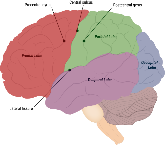
As mentioned in an earlier sections, voluntary motor control starts at the motor cortex in the grey matter of the cerebral cortex. Anatomically, the motor cortex is located in the frontal lobe, immediately anterior (‘front of’) a deep groove known as the central sulcus, called the precentral gyrus.
The outgoing motor message travels from the grey matter in the cerebral cortex down through the myelinated fibers in the white matter, heading down towards the brain stem and spinal cord.
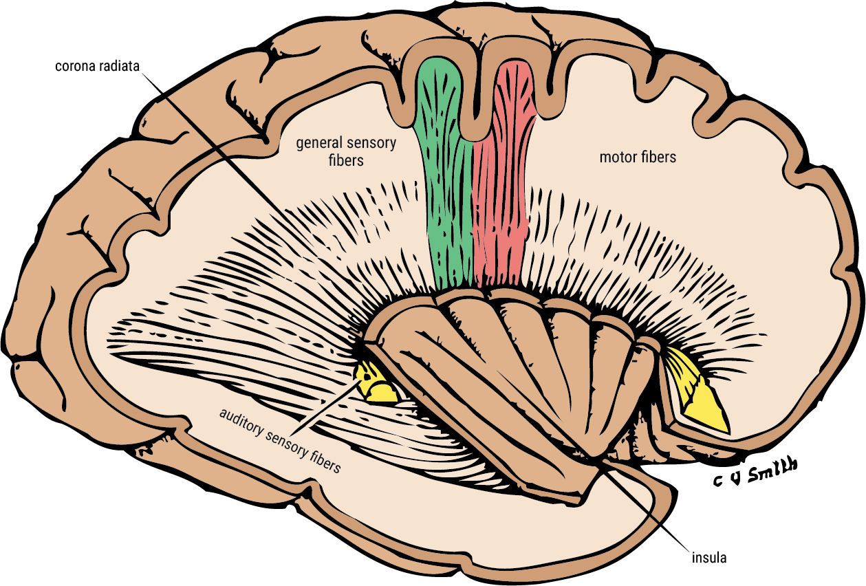
The motor message descends from the cortex, into the brainstem (midbrain, pons, & medulla) and into specific areas of the spinal cord which is a continuation of the brainstem, at the approximate level of the upper neck. Recall that the spinal cord is protected within the bony vertebrae of the backbone.
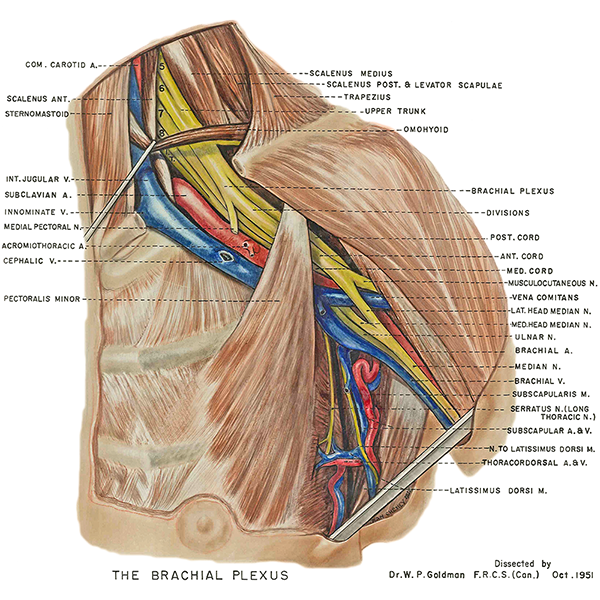
The motor message travels to its desired target such as the arm, finger, legs, etc. The motor message leaves the central nervous system of the spinal cord and transfers it to the peripheral nervous system of the desired target. To demonstrate this, below is an illustration of the peripheral nerves (in yellow) which have left the spinal cord and are traversing the shoulder girdle on the way to the arms and fingers.
The motor message from the peripheral nerve will eventually end at the neuromuscular junction which is a microscopic structure. However, thousands of neuromuscular junctions are situated within skeletal muscles where the relay the motor message for voluntary movement.
.