Renal Module
Kidney Functions
Learning Objectives
By the end of this section, you will be able to:
- Describe the characteristics of a normal urine sample, including normal range of pH, osmolarity, and volume
- Explain the normal physiological role and the five (5) functions of kidneys:
- urine formation
- regulation of fluid and electrolyte balance
- excretion of metabolic waste products (urea and creatinine)
- regulation of acid-base balance
- production of hormones (calcitriol, erythropoietin, renin)
- Describe the utility of a selection of clinical tests associated with renal function
Urine Formation
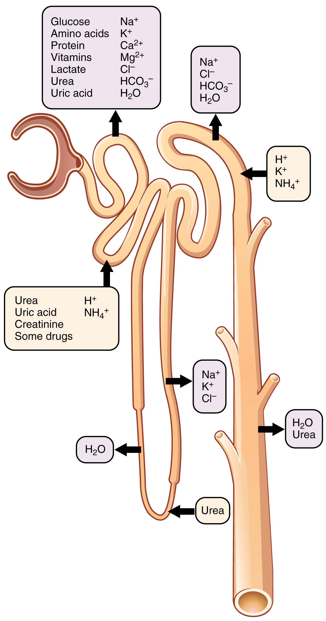
The physiologic goal is to modify the composition of the plasma and, in doing so, produce the waste product urine. Different parts of the nephron utilize specific processes to produce urine: filtration, reabsorption, and secretion.
- Glomerular Filtration: Water and solutes move from the capillary lumen to Bowman’s space across 3 layers of the glomerular filter.
The outermost part of Bowman’s capsule, the parietal layer, is a simple squamous epithelium. It transitions onto the glomerular capillaries in an intimate embrace to form the visceral layer of the capsule. Here, the cells are not squamous, but uniquely shaped cells (podocytes) extending finger-like arms (pedicels) to cover the glomerular capillaries (Figure 1). These projections interdigitate to form filtration slits, leaving small gaps between the digits to form a sieve. Where the fenestrae (windows) in the glomerular capillaries match the spaces between the podocyte “fingers,” the only thing separating the capillary lumen and the lumen of Bowman’s capsule is their shared basement membrane (Figure 2). These three features comprise what is known as the filtration membrane. Overall, filtration is regulated by fenestrations in capillary endothelial cells, podocytes with filtration slits, membrane charge, and the basement membrane between capillary cells. The result is the creation of a filtrate that does not contain cells or large proteins, and has a slight predominance of positively charged substances.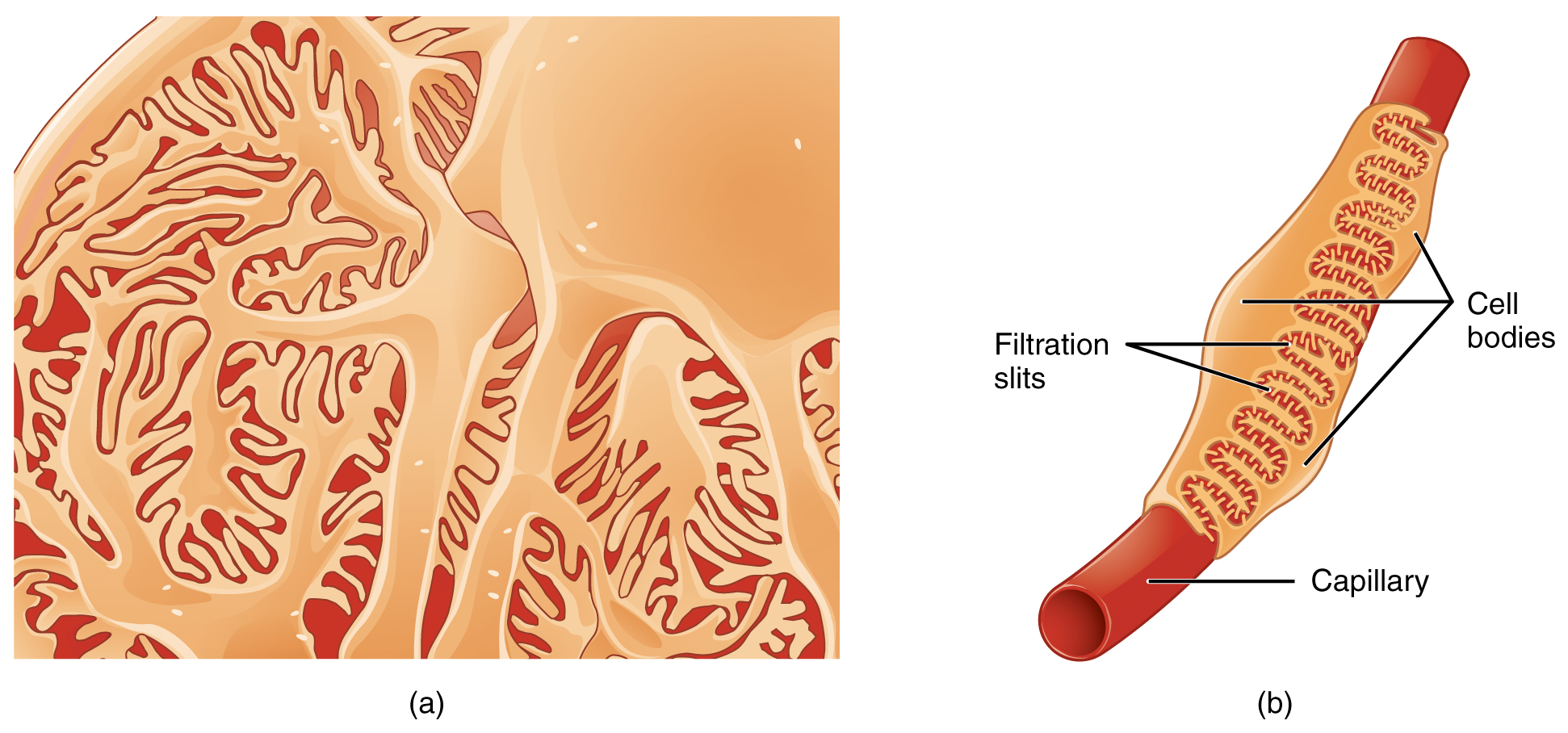
Figure 1. Podocytes. Podocytes interdigitate with structures called pedicels and filter substances in a way similar to fenestrations. In (a), the large cell body can be seen at the top right corner, with branches extending from the cell body. The smallest finger-like extensions are the pedicels. Pedicels on one podocyte always interdigitate with the pedicels of another podocyte. (b) This capillary has three podocytes wrapped around it. 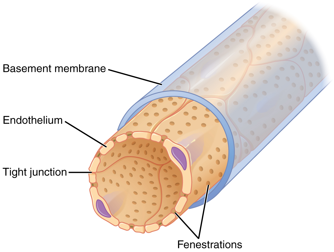
Figure 2. Fenestrated Capillary. Fenestrations allow many substances to diffuse from the blood based primarily on size. - Reabsorption back into the blood: lots in the proximal convoluted tubules (PCT) and some in Loop of Henle; fine-tuning after that [distal convoluted tubules (DCT) and collecting duct]
The filtrate enters the PCT where absorption and secretion of several substances occur. The descending and ascending limbs of the loop of Henle consist of thick and thin segments. Absorption and secretion continue in the DCT but to a lesser extent than in the PCT. - Secretion from blood into tubule lumen: in the PCT, DCT & collecting duct. What remains in the lumen from the collecting duct on is urine which will be excreted.
Various portions of the nephron differ in their capacity to reabsorb water and specific solutes. While much of the reabsorption and secretion occur passively based on concentration gradients, the amount of water that is reabsorbed or lost is tightly regulated (Figure 3). This control is exerted directly by ADH and aldosterone, and indirectly by renin. Most water is recovered in the PCT, loop of Henle, and DCT. About 10 percent (about 18 L) reaches the collecting ducts. The collecting ducts, under the influence of ADH, can recover almost all of the water passing through them, in cases of dehydration, or almost none of the water, in cases of over-hydration.
Physical Characteristics of Urine
The urinary system’s ability to filter the blood resides in about 2 to 3 million tufts of specialized capillaries—the glomeruli—distributed more or less equally between the two kidneys. Because the glomeruli filter the blood based mostly on particle size, large elements like blood cells, platelets, antibodies, and albumen are excluded. The glomerulus is the first part of the nephron, which then continues as a highly specialized tubular structure responsible for creating the final urine composition. All other solutes, such as ions, amino acids, vitamins, and wastes, are filtered to create a filtrate composition very similar to plasma. The glomeruli create about 200 liters (189 quarts) of this filtrate every day, yet you excrete less than two liters of waste you call urine.
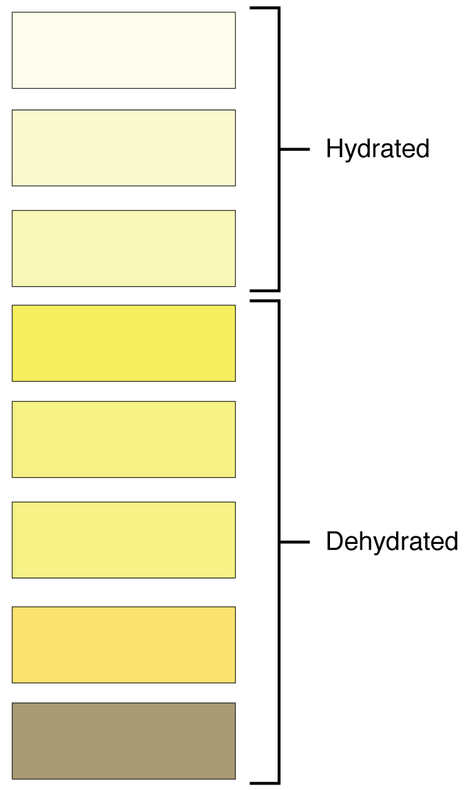
Characteristics of the urine change, depending on influences such as water intake, exercise, environmental temperature, nutrient intake, and other factors (Table 1). Some of the characteristics such as color and odor are rough descriptors of your state of hydration. For example, if you exercise or work outside, and sweat a great deal, your urine will turn darker and produce a slight odor, even if you drink plenty of water. Athletes are often advised to consume water until their urine is clear. This is good advice; however, it takes time for the kidneys to process body fluids and store it in the bladder. Another way of looking at this is that the quality of the urine produced is an average over the time it takes to make that urine. Producing clear urine may take only a few minutes if you are drinking a lot of water or several hours if you are working outside and not drinking much.
| Normal Urine Characteristics (Table 1) | |
|---|---|
| Characteristic | Normal values |
| Color | Pale yellow to deep amber |
| Odor | Odorless |
| Volume | 1500 mL/24 hour |
| pH | 4.5 – 8.0 |
| Osmolarity | 50 – 1200 mOsm/kg |
| Urobilinogen |
5 – 10 mg/L
Negative (less sensitive test)
|
| Protein | None or trace |
| Ketones | None |
| Blood | None |
| Glucose | None |
Urinalysis (urine analysis) often provides clues to renal disease. Normally, only traces of protein are found in urine, and when higher amounts are found, damage to the glomeruli is the likely basis. The color of urine is determined mostly by the breakdown products of red blood cell destruction (Figure 4). The “heme” of hemoglobin is converted by the liver into water-soluble forms that can be excreted into the bile and indirectly into the urine. Urine color may also be affected by certain foods like beets, berries, and fava beans. A kidney stone or a cancer of the urinary system may produce sufficient bleeding to manifest as pink or even bright red urine. Diseases of the liver or obstructions of bile drainage from the liver impart a dark “tea” or “cola” hue to the urine. Dehydration produces darker, concentrated urine that may also possess the slight odor of ammonia. Most of the ammonia produced from protein breakdown is converted into urea by the liver, so ammonia is rarely detected in fresh urine. The strong ammonia odor you may detect in bathrooms or alleys is due to the breakdown of urea into ammonia by bacteria in the environment. About one in five people detect a distinctive odor in their urine after consuming asparagus; other foods such as onions, garlic, and fish can impart their own aromas! These food-caused odors are harmless.
Urine volume varies considerably. The normal range is one to two liters per day (Table 2). The kidneys must produce a minimum urine volume of about 500 mL/day to rid the body of wastes. Output below this level may be caused by severe dehydration or renal disease and is termed oliguria. The virtual absence of urine production is termed anuria. Excessive urine production is polyuria, which may be due to diabetes mellitus or diabetes insipidus. In diabetes mellitus, blood glucose levels exceed the number of available sodium-glucose transporters in the kidney, and glucose appears in the urine. The osmotic nature of glucose attracts water, leading to its loss in the urine. In the case of diabetes insipidus, insufficient pituitary antidiuretic hormone (ADH) release or insufficient numbers of ADH receptors in the collecting ducts means that too few water channels are inserted into the cell membranes that line the collecting ducts of the kidney. Insufficient numbers of water channels (aquaporins) reduce water absorption, resulting in high volumes of very dilute urine.
| Urine Volumes (Table 2) | ||
|---|---|---|
| Volume condition | Volume | Causes |
| Normal | 1–2 L/day | |
| Polyuria | >2.5 L/day | Diabetes mellitus; diabetes insipidus; excess caffeine or alcohol; kidney disease; certain drugs, such as diuretics; sickle cell anemia; excessive water intake |
| Oliguria | 300–500 mL/day | Dehydration; blood loss; diarrhea; cardiogenic shock; kidney disease; enlarged prostate |
| Anuria | <50 mL/day | Kidney failure; obstruction, such as kidney stone or tumor; enlarged prostate |
The pH (hydrogen ion concentration) of the urine can vary more than 1000-fold, from a normal low of 4.5 to a maximum of 8.0. Diet can influence pH; meats lower the pH, whereas citrus fruits, vegetables, and dairy products raise the pH. Chronically high or low pH can lead to disorders, such as the development of kidney stones or osteomalacia.
Protein does not normally leave the glomerular capillaries, so only trace amounts of protein should be found in the urine, approximately 10 mg/100 mL in a random sample. If excessive protein is detected in the urine, it usually means that the glomerulus is damaged and is allowing protein to “leak” into the filtrate.
Ketones are byproducts of fat metabolism. Finding ketones in the urine suggests that the body is using fat as an energy source in preference to glucose. In diabetes mellitus when there is not enough insulin (type I diabetes mellitus) or because of insulin resistance (type II diabetes mellitus), there is plenty of glucose, but without the action of insulin, the cells cannot take it up, so it remains in the bloodstream. Instead, the cells are forced to use fat as their energy source, and fat consumed at such a level produces excessive ketones as byproducts. These excess ketones will appear in the urine. Ketones may also appear if there is a severe deficiency of proteins or carbohydrates in the diet.
There should be no blood found in the urine. It may sometimes appear in urine samples as a result of menstrual contamination, but this is not an abnormal condition. Now that you understand what the normal characteristics of urine are, the next section will introduce you to how you store and dispose of this waste product and how you make it.
Fluid and Electrolyte Balance
Related to the process of filtration, reabsoprtion, and secretion of urine formation.
Proximal tubule
- reabsorption including:
- ~80% salt(NaCl) and water reabsorbed from filtrate
- 100% of filtered glucose(normally)
- ions: HCO3–, PO4– ,Cl–, K+, Mg2+, Ca2+
- secretion, including:
- H+, K+
Loop of Henle reabsorbs relatively large quantities of Na+& Cl–and to a lesser extent water.
Distal tubule and collecting tubule then do the fine tuning for most substances, determining the final amounts excreted in the urine by adjusting their rates of reabsorption (water,Na+, Cl–) and secretion (K+, H+) back into the lumen and hence into urine.
See a video of animated nephron anatomy and functions by Handwritten Tutorials. Nephron Anatomy and Function. YouTube.
Excretion of waste products of protein metabolism
- Urea:
- amino acid breakdown requires deamination, producing ammonia, NH3, which is toxic.
- NH3 in blood is converted to urea in LIVER (urea cycle).
- Much (but not all) of the urea is excreted in the urine.
- Blood Urea Nitrogen measures the amount of nitrogen in the blood that comes from urea.
- Creatinine
- waste product from the breakdown of muscle tissue, specifically of the creatine-phosphate in muscle
- Creatinine Clearance Rate (CCr) provides a good estimate of the glomerular filtration rate (GFR) and is a standard test for determining kidney function. Creatinine is a nearly ideal substance for measurement of clearance because it is small and therefore readily filtered in glomerulus. Moreover, almost none of it is reabsorbed. (Any that was reabsorbed gets secreted back into the filtrate).
Acid-Base Balance
see Homeostasis Section.
Hormonal Functions
- In order for vitamin D to become active, it must undergo a hydroxylation reaction in the kidney, that is, an –OH group must be added to calcidiol to make calcitriol (1,25-dihydroxycholecalciferol). Activated vitamin D is important for absorption of Ca++ in the digestive tract, its reabsorption in the kidney, and the maintenance of normal serum concentrations of Ca++ and phosphate. Calcium is vitally important in bone health, muscle contraction, hormone secretion, and neurotransmitter release.
- Production & release of renin, which can influence urine volume & urine [Na+]. Renin release in response to low ECF volume. Renin stimulates the Renin-Angiotensin-Aldosterone system by enzymatically converts angiotensinogen into angiotensin I. When it is then converted to angiotensin II, it triggers the release of aldosterone. Aldosterone, often called the “salt-retaining hormone,” promotes Na+ reabsorption by the nephron, promoting the retention of water. It is also important in regulating K+, promoting its excretion. As a result, renin has an immediate effect on blood pressure due to angiotensin II–stimulated vasoconstriction and a prolonged effect through Na+ recovery due to aldosterone.
- Erythropoetin, or EPO, is a 193-amino acid protein that stimulates the formation of red blood cells in the bone marrow. The kidney produces 85 percent of circulating EPO; the liver, the remainder. Renal failure (loss of EPO production) is associated with anemia, which makes it difficult for the body to cope with increased oxygen demands or to supply oxygen adequately even under normal conditions. Anemia diminishes performance and can be life threatening.
