Levels of Organization
Unit 4: Cell Structure and Function
Unit Outline
Part 1. Characteristics of life
- Organization
- Metabolism
- Exchange of material
- Responsiveness
- Movement
- Development, growth and reproduction
Part 2. Structural organization of the body
Part 3. Cell structure, cellular organelles and functions
- General cell structure: plasma membrane, cytoplasm and nucleus
- Organelles of the endomembrane system
- Organelles for energy processing
Part 4. Cellular processes involved in growth
Practice Questions
Learning Objectives
At the end of this unit, you should be able to:
I. Specify the characteristics associated with life and explain why the cell is the basic unit of life.
II. Describe the levels of structural organization in the body.
III. Describe the structure and the functions of major components of a cell.
IV. Define metabolism, and distinguish between anabolism and catabolism.
V. Describe the cellular processes involved in the growth of the human body from a fertilized egg to an adult.
VI. Describe the importance of cell differentiation to an organism.
VII. Describe the general characteristics of each of the following cell types and relate their characteristics to their functions: nerve cell, muscle cell, red blood cell (erythrocyte), white blood cell (leukocyte).
Learning Objectives and Guiding Questions
At the end of this unit, you should be able to complete all the following tasks, including answering the guiding questions associated with each task.
I. Specify the characteristics associated with life and explain why the cell is the basic unit of life.
- List, explain and provide examples of each of the characteristics of life.
- In reference to your answer to question #1, above, explain in one sentence why the cell is considered to be the basic unit of life.
II. Describe the levels of structural organization in the body.
- Define each of the following levels of organization in the body:
- Chemical level
- Cellular level
- Tissue level
- Organ level
- Organ system level
- Organismal level
- Write a clear description of the relationships between the chemical, cellular, tissue, organ, organ system, and organismal levels of organization in the body.
III. Describe the structure and the functions of major components of a cell.
- Describe the structure (be able to identify them in a diagram) and describe the functions of the following cytoplasmic components:
- Cell membrane (plasma membrane)
- Endoplasmic reticulum
- Ribosomes
- Golgi apparatus (Golgi complex)
- Lysosomes
- Mitochondria
- Vesicle
- Describe the structure (be able to identify them in a diagram) and describe the functions of the following nuclear components:
- Nuclear envelope
- Chromosomes
- Nucleolus
- Use a full page to draw (by hand, and neatly!) an annotated diagram of a cell showing all of the following structures and briefly describing the function of each (a simple, flattened diagram is fine, no need to show 3D structures, but the defining characteristics of each named structure should be clear):
- Plasma membrane
- Nuclear envelope
- Nucleus
- Nucleolus
- Smooth endoplasmic reticulum
- Rough endoplasmic reticulum
- Bound ribosomes
- Free ribosomes
- Golgi apparatus (or Golgi complex)
- Lysosomes
- Mitochondria
- Vesicles
- Describe the structure (name all the components and describe their relationships to each other) and list the general functions of the “endomembrane system”.
IV. Define metabolism, and distinguish between anabolism and catabolism.
- Define the term “metabolism”.
- Write a single sentence that clearly differentiates between “anabolism” and “catabolism”.
V. Describe the cellular processes involved in the growth of the human body from a fertilized egg to an adult.
- Distinguish between cell division, cell growth and cell differentiation.
- Provide two examples of cell types in the human body that do not undergo cell division.
- Define the term “stem cell”.
VI. Describe the importance of cell differentiation to an organism.
- Describe how cell differentiation allows cells to serve specialized functions.
- Explain why it is important in the human body to have cells specialized in performing particular functions, rather than having all cells be identical to each other.
VII. Describe the general characteristics of each of the following cell types and relate their characteristics to their functions: nerve cell, muscle cell, red blood cell (erythrocyte), white blood cell (leukocyte).
- Describe the general structural characteristics (morphology) and function of each of the following cell types:
- Neuron
- Muscle fiber
- Erythrocyte
- Leukocyte
Part 1: Characteristics of Life
The different organ systems each have different functions and therefore unique roles to perform in the body. These many functions can be summarized in terms of a few that we might consider definitive of human life: organization, metabolism, exchange of materials, responsiveness, movement, development, growth and reproduction.
Organization
A human body consists of trillions of cells organized in a way that maintains distinct internal compartments. These compartments keep body cells separated from external environmental threats and keep the cells moist and nourished. They also separate internal body fluids from the countless microorganisms that grow on body surfaces, including the lining of certain tracts, or passageways. The intestinal tract, for example, is home to even more bacteria cells than the total of all human cells in the body, yet these bacteria are outside the body and cannot circulate freely inside the body.
Cells, for example, have a membrane (also referred to as the plasma membrane) that keeps the intracellular environment—the fluids and organelles—separate from the environment outside the cell (the extracellular environment). Blood vessels keep blood inside a closed system, and nerves and muscles are wrapped in tissue sheaths that separate them from surrounding structures. In the chest and abdomen, a variety of internal membranes keep major organs such as the lungs, heart, and kidneys separate from others.
The body’s largest organ system is the integumentary system, which includes the skin and its associated structures, such as hair and nails. The surface tissue of skin is a barrier that protects internal structures and fluids from potentially harmful microorganisms, toxins and the external environment.
Metabolism
The first law of thermodynamics holds that energy can neither be created nor destroyed—it can only change form. Your basic function as an organism is to consume (ingest) energy and molecules in the foods you eat, convert some of it into fuel for movement, sustain your body functions, and build and maintain your body structures. There are two types of reactions that accomplish this: anabolism and catabolism.
- Anabolism is the process whereby smaller, simpler molecules are combined into larger, more complex substances. For example, amino acids can be combined together to make proteins. Your body can assemble, by utilizing energy, the complex chemicals it needs by combining small molecules derived from the foods you eat.
- Catabolism is the process by which larger more complex substances are broken down into smaller simpler molecules. For example, sugars are broken down to carbon dioxide and water. Catabolism releases energy. The complex molecules found in foods are broken down so the body can use their parts to assemble the structures and substances needed for life.
Taken together, these two processes are called metabolism. Metabolism is the sum of all anabolic and catabolic reactions that take place in the body. Both anabolism and catabolism occur simultaneously and continuously to keep you alive.
Every cell in your body makes use of a chemical compound, adenosine triphosphate (ATP), to store and release energy. The cell stores energy in the molecule of ATP, then moves the ATP molecules to the location where energy is needed to fuel cellular activities. Then the ATP is broken down and a controlled amount of energy is released, which is used by the cell to perform a particular job.

Exchange of Material
Organisms do not exist solely within their own boundaries, but interact with the external environment that surrounds them. One of the ways in which they do this is by exchanging materials with their external environment: taking in materials from their external environment and by expelling waste products out into their external environment. These materials and waste products may be anything from very small, relatively simple molecules (e.g. glucose, carbon dioxide) that must cross an individual cell’s plasma membrane to whole cells or foods that were ingested but not fully digested and/or absorbed and so must be excreted from the organism.
Responsiveness
Responsiveness is the ability of an organism to adjust to changes in its internal and external environments. An example of responsiveness to external stimuli could include moving toward sources of food and water and away from perceived dangers. Changes in an organism’s internal environment, such as increased body temperature, can cause the responses of sweating and the dilation of blood vessels in the skin in order to decrease body temperature.
Movement
Human movement includes not only actions at the joints of the body, but also the motion of individual organs and even individual cells. As you read these words, red and white blood cells are moving throughout your body, muscle cells are contracting and relaxing to maintain your posture and to focus your vision, and glands are secreting chemicals to regulate body functions. Your body is coordinating the action of entire muscle groups to enable you to move air into and out of your lungs, to push blood throughout your body, and to propel the food you have eaten through your digestive tract. Consciously, of course, you contract your skeletal muscles to move the bones of your skeleton to get from one place to another, and to carry out all of the activities of your daily life.
Development, growth and reproduction
- Development is all of the changes the body goes through in life. Development includes the process of cell differentiation, in which unspecialized cells become specialized in structure and function to perform certain tasks in the body. Development also includes the processes of growth and repair, both of which involve cell differentiation.
- Growth is the increase in body size. Humans, like all multicellular organisms, grow by increasing the number of existing cells, increasing the amount of non-cellular material around cells (such as mineral deposits in bone), and, within very narrow limits, increasing the size of existing cells.
- Reproduction is the formation of a new organism from parent organisms. In humans, reproduction is carried out by the male and female reproductive systems. Because death will come to all complex organisms, without reproduction, the line of organisms would end.
Part 2: Structural Organization of the Human Body
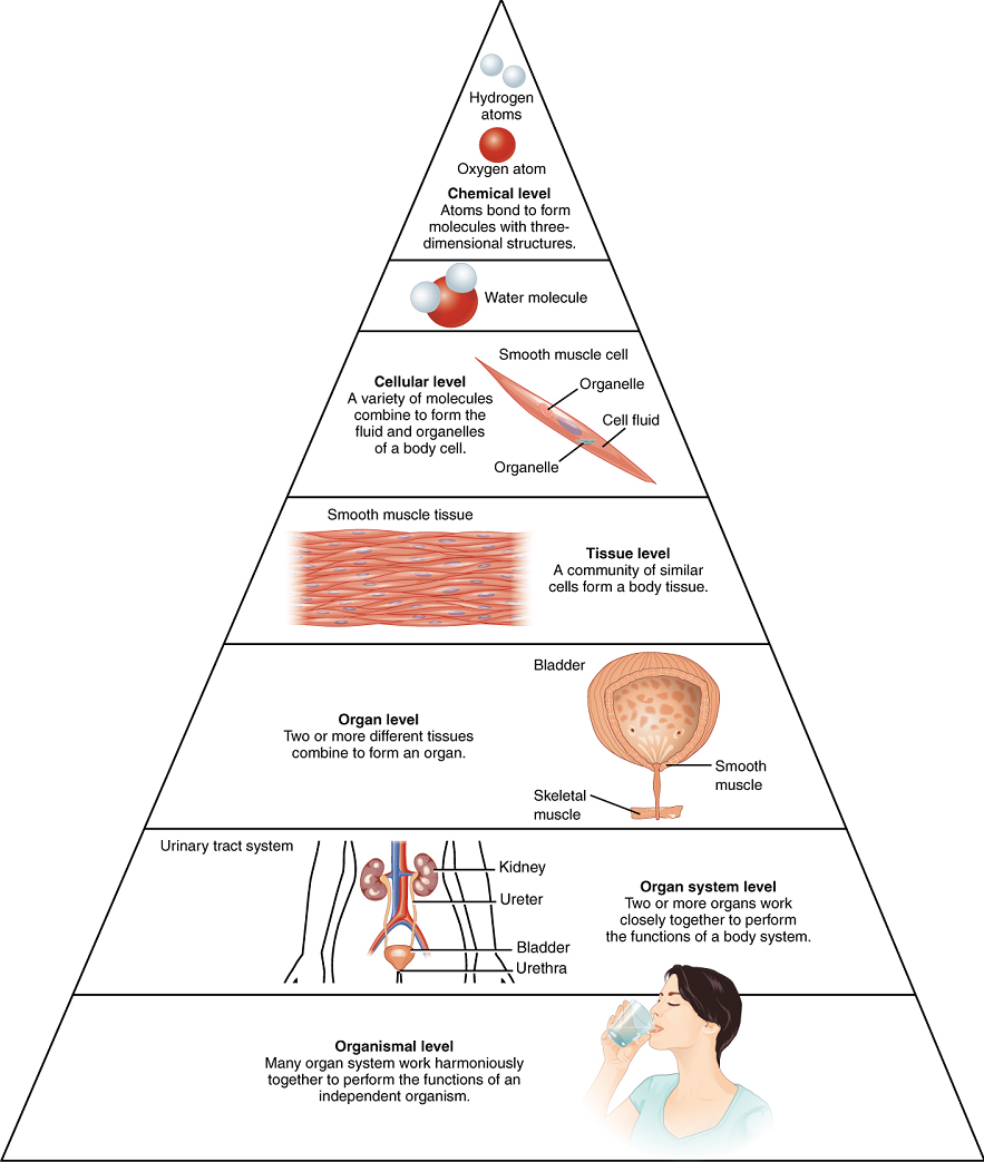
Before you begin to study the different structures and functions of the human body, it is helpful to consider its basic architecture; that is, how its smallest parts are assembled into larger structures. It is convenient to consider the structures of the body in terms of fundamental levels of organization that increase in complexity: subatomic particles, atoms, molecules, organelles, cells, tissues, organs, organ systems and organisms (Figure 1).
The Levels of Organization
To study the chemical level of organization, scientists consider the simplest building blocks of matter: subatomic particles, atoms and molecules. All matter in the universe is composed of one or more unique pure substances called elements, familiar examples of which are hydrogen, oxygen, carbon, nitrogen, calcium, and iron. The smallest unit of any of these pure substances (elements) is an atom. Atoms are made up of subatomic particles such as the proton, electron and neutron. Two or more atoms combine to form a molecule, such as the water molecules, proteins, and sugars found in living things. Molecules are the chemical building blocks of all body structures.
A cell is the smallest independently functioning unit of a living organism. All living structures of human anatomy contain cells, and almost all functions of human physiology are performed in cells or are initiated by cells. Even bacteria, which are extremely small single celled, independently-living organisms, have a cellular structure.
A human cell typically consists of flexible membranes that enclose cytoplasm, a water-based fluid together with a variety of tiny functioning units called organelles. In humans, as in all organisms, cells perform all functions of life. A tissue is a group of many similar cells (though sometimes composed of a few related types) that work together to perform a specific function. An organ is an anatomically distinct structure of the body composed of two or more tissue types. Each organ performs one or more specific physiological functions. An organ system is a group of organs that work together to perform major functions or meet physiological needs of the body. Assigning organs to organ systems can be imprecise since organs that “belong” to one system can also have functions integral to another system. In fact, most organs contribute to more than one system.
The organism level is the highest level of organization. An organism is a living being that has a cellular structure and that can independently perform all physiologic functions necessary for life. In multicellular organisms, including humans, all cells, tissues, organs, and organ systems of the body work together to maintain the life and health of the organism.
The Cellular Level of Organization
You developed from a single fertilized egg cell into the complex organism containing trillions of cells that you see when you look in a mirror. Early during this developmental process, cells differentiate and become specialized in their structure and function. These different cell types form specialized tissues that work in concert to perform all of the functions necessary for the living organism. Cellular and developmental biologists study how the continued division of a single cell leads to such complexity.
Consider the difference between a cell in the skin and a nerve cell. A skin cell may be shaped like a flat plate (squamous) and live only for a short time before it is shed and replaced. Packed tightly into rows and sheets, the squamous skin cells provide a protective barrier for the cells and tissues that lie beneath. A nerve cell, on the other hand, may be shaped something like a star, sending out long processes up to a meter in length and may live for the entire lifetime of the organism. With their long winding processes, nerve cells can communicate with one another and with other types of body cells and send rapid signals that inform the organism about its environment and allow it to interact with that environment. These differences illustrate one very important theme that is consistent at all organizational levels of biology: the form of a structure is optimally suited to perform particular functions assigned to that structure. Keep this theme in mind as you tour the inside of a cell and are introduced to the various types of cells in the body.
The concept of a cell started with microscopic observations of dead cork tissue by scientist Robert Hooke in 1665. Without realizing their function or importance, Hook coined the term “cell” based on the resemblance of the small subdivisions in the cork to the rooms that monks inhabited, called cells. About ten years later, Antonie van Leeuwenhoek became the first person to observe living and moving cells under a microscope. In the century that followed, the theory that cells represented the basic unit of life would develop. These tiny fluid-filled sacs house components responsible for the thousands of biochemical reactions necessary for an organism to grow and survive. In this chapter, you will learn about the major components and functions of a generalized cell and discover some of the different types of cells in the human body.

Part 3: Cell structure, cellular organelles and functions
General cell structure: Plasma Membrane, Cytoplasm and Nucleus
The cell membrane (also known as the plasma membrane) separates the inner contents of a cell from its external environment. This membrane provides a protective barrier around the cell and regulates which materials can pass in or out. It is primarily composed of phospholipids arranged in a two layers but also contains cholesterol and a mosaic of different proteins. You will learn more about the structure and function of the plasma membrane in Unit 5. All living cells in multicellular organisms contain an internal cytoplasmic compartment, composed of cytosol and organelles. Cytosol, the jelly-like substance within the cell, provides the fluid medium necessary for biochemical reactions and is mostly composted of water. Eukaryotic cells, including all animal cells, also contain various cellular organelles. An organelle (“little organ”) is one of several different types of membrane-enclosed bodies in the cell, each performing a unique function.
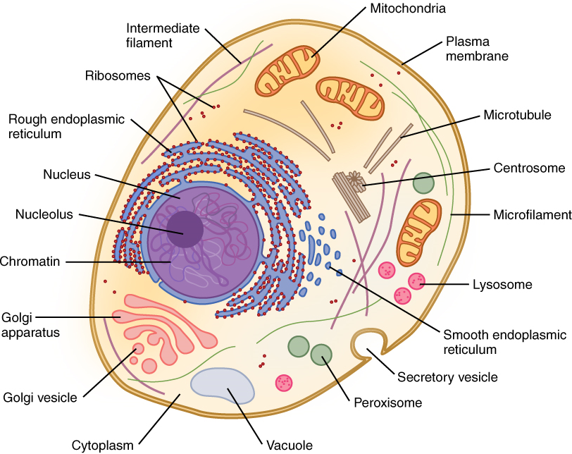
Just as the various bodily organs work together in harmony to perform all of a human’s functions, the many different cellular organelles work together to keep the cell healthy and performing all of its important functions. The organelles and cytosol, taken together, compose the cell’s cytoplasm. The nucleus is a cell’s central organelle, which contains the cell’s DNA (Figure 2).
Organelles of the Endomembrane System
Most membranous organelles found in a human cell together form a system within the cell called the endomembrane system. These organelles work together to perform various cellular jobs, including the task of producing, packaging, and exporting certain cellular products. The components of the endomembrane system include the nuclear envelope, endoplasmic reticulum, Golgi apparatus, vesicles, and plasma membrane.
Endoplasmic Reticulum: The endoplasmic reticulum (ER) is a system of channels that is continuous with the nuclear membrane (or “envelope”) covering the nucleus (see Part 7) and composed of the same lipid bilayer material. The ER can be thought of as a series of winding thoroughfares similar to the waterway canals in Venice. The ER provides passages throughout much of the cell that function in transporting, synthesizing, and storing materials. The winding structure of the ER results in a large membranous surface area that supports its many functions (Figure 3).
Endoplasmic reticulum can exist in two forms: rough ER and smooth ER. These two types of ER perform some very different functions and can be found in different amounts depending on the type of cell. Rough ER (RER) is so-called because its membrane is dotted with embedded granules—organelles called ribosomes, giving the RER a bumpy appearance. A ribosome is an organelle that serves as the site of protein synthesis and it is composed of two subunits. Ribosomes can either be bound (attached to ER) or free (floating in the cytosol). Smooth ER (SER) lacks ribosomes.
One of the main functions of the smooth ER is in the synthesis of lipids. The smooth ER synthesizes phospholipids, the main component of biological membranes, as well as steroid hormones.
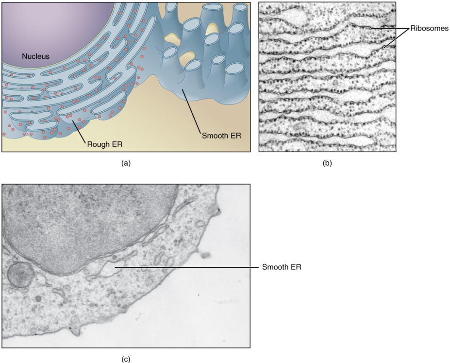
For this reason, cells that produce large quantities of such hormones, such as those of the female ovaries and male testes, contain large amounts of smooth ER. In addition to lipid synthesis, the smooth ER also sequesters (i.e., stores) and regulates the concentration of cellular calcium (Ca2+) which is extremely important in cells of the nervous system where Ca2+ is the trigger for neurotransmitter release. Additionally, the smooth ER, especially in the liver, performs a detoxification role, breaking down certain toxins.
In contrast with the smooth ER, the primary job of the rough ER is the synthesis and modification of proteins destined for the cell membrane or for export from the cell. For this protein synthesis, many ribosomes attach to the ER (giving it the studded appearance of rough ER). Typically, a protein is synthesized within the ribosome and released inside the channel of the rough ER, where sugars can be added to it (by a process called glycosylation) before it is transported within a vesicle (a small fluid filled sac) to the next stage in the packaging and shipping process: the Golgi apparatus.
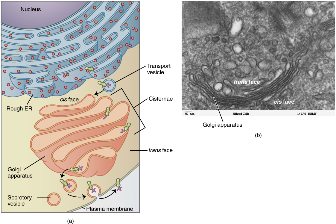
The Golgi Apparatus: The Golgi apparatus is responsible for sorting, modifying, and shipping off the products that come from the rough ER, much like a post-office. The Golgi apparatus looks like stacked flattened discs, almost like stacks of oddly shaped pancakes. Like the ER, these discs are membranous. The Golgi apparatus has two distinct sides, each with a different role. One side (the cis face) of the apparatus receives products in vesicles. These products are sorted through the apparatus, and then they are released from the opposite side (the trans face) after being repackaged into new vesicles. If the product is to be exported from the cell, the vesicle migrates to the cell surface and fuses to the cell membrane, and the cargo is secreted (Figure 4).
Lysosomes: Some of the protein products from the Golgi include digestive enzymes that are meant to remain inside the cell for use in breaking down certain materials. These enzymes are packaged into to vesicles called lysosomes. A lysosome is an organelle that contains enzymes that break down and digest unneeded cellular components, such as a damaged organelle in a process called autophagy (“self-eating”).
Lysosomes are also important for breaking down foreign material. For example, when certain immune defense cells, like white blood cells, phagocytize (engulf) bacteria, the bacterial cell is transported to a lysosome and digested by the enzymes inside. Under certain circumstances, lysosomes perform a more grand and dire function. In the case of damaged or unhealthy cells, lysosomes can be triggered to open up and release their digestive enzymes into the cytoplasm of the cell, killing the cell. This “self-destruct” mechanism is called autolysis, and makes the process of cell death controlled (a mechanism called “apoptosis”).
Organelles for Energy Processing
In addition to the jobs performed by the endomembrane system, the cell has many other important functions. Just as you must consume nutrients to provide yourself with energy, so must each of your cells take in nutrients, some of which convert to chemical energy that can be used to power biochemical reactions.
Mitochondrion: A mitochondrion (plural = mitochondria) is a membranous, bean-shaped organelle that is the “energy transformer” of the cell. Mitochondria consist of an outer lipid bilayer membrane as well as an additional inner lipid bilayer membrane (Figure 5). The inner membrane is highly folded into winding structures with a great deal of surface area, called cristae. It is along this inner membrane that a series of proteins, enzymes, and other molecules perform the biochemical reactions of cellular respiration.
These reactions harvest the energy stored in nutrient molecules (such as glucose) to power the synthesis of ATP, which provides usable energy to the cell. Cells use ATP constantly, and so the mitochondria are constantly at work. Oxygen molecules are required during cellular respiration, which is why you must constantly breathe it in. One of the organ systems in the body that uses huge amounts of ATP is the muscular system because ATP is required to sustain muscle contraction. As a result, muscle cells are packed full of mitochondria.
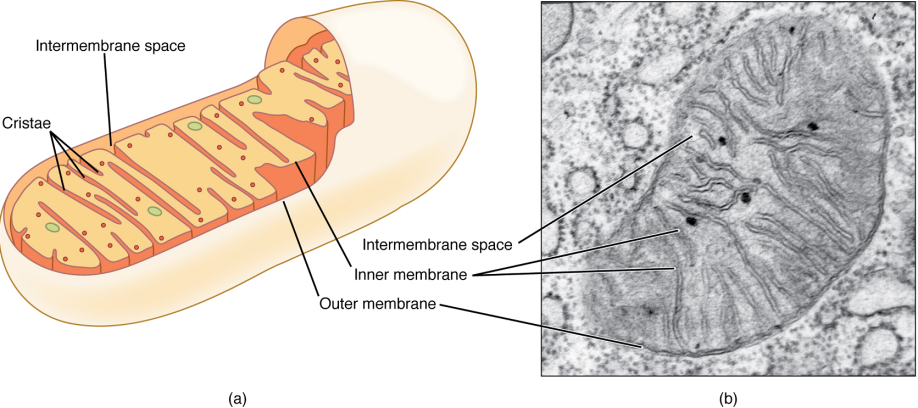
Nerve cells also need large quantities of ATP to run their sodium-potassium pumps which are used to generate an action potential. Therefore, an individual neuron will be loaded with over a thousand mitochondria. On the other hand, a bone cell, which is not nearly as metabolically-active, might only have a couple hundred mitochondria.
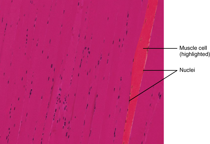
The Nucleus: The nucleus is the largest and most prominent of a cell’s organelles (Figure 6). The nucleus is generally considered the control center of the cell because it stores all of the genetic instructions for manufacturing proteins. Interestingly, some cells in the body, such as muscle cells, contain more than one nucleus (Figure 7), which is known as multinucleated. Other cells, such as mammalian red blood cells (RBCs), do not contain nuclei at all. RBCs eject their nuclei as they mature, making space for the large numbers of hemoglobin molecules that carry oxygen throughout the body.
Inside the nucleus lies the blueprint that dictates everything a cell will do and all of the products it will make. This information is stored within DNA. The nucleus sends “commands” to the cell via molecular messengers that translate the information from DNA. Each cell in your body (with the exception of the cells that produce eggs and sperm) contains the complete set of your DNA. When a cell divides, the DNA must be duplicated so that the each new cell receives a full complement of DNA.
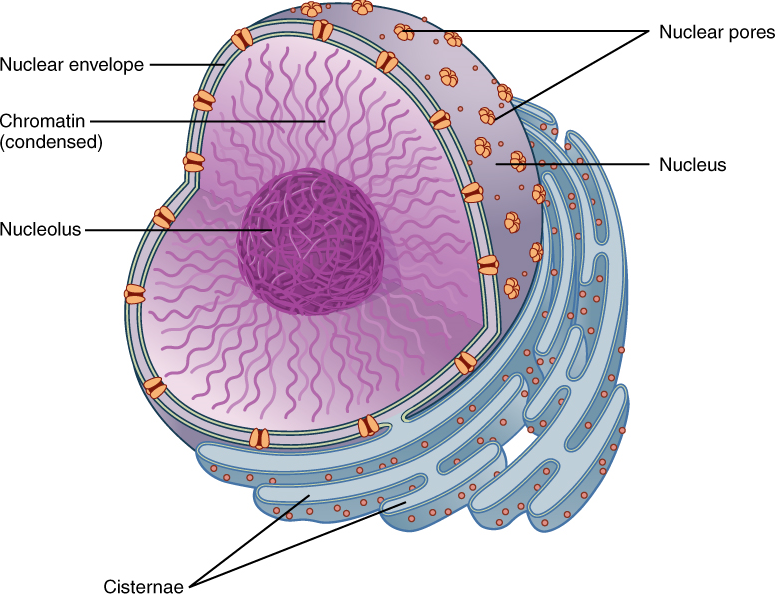
Organization of the Nucleus and Its DNA: Like most other cellular organelles, the nucleus is surrounded by a membrane called the nuclear envelope. This membranous covering consists of two adjacent lipid bilayers with a thin fluid space in between them. Spanning these two bilayers are nuclear pores. A nuclear pore is a tiny passageway for the passage of proteins, RNA, and solutes between the nucleus and the cytoplasm.
Inside the nuclear envelope is a gel-like nucleoplasm with solutes that include the building blocks of nucleic acids. There also can be a dark-staining mass often visible under a simple light microscope, called a nucleolus (plural = nucleoli). The nucleolus is a region of the nucleus that is responsible for manufacturing the RNAs necessary for construction of ribosomes. Once synthesized, newly made ribosomal subunits exit the cell’s nucleus through the nuclear pores.
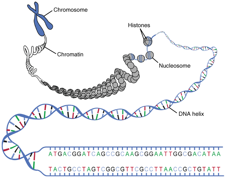
The genetic instructions that are used to build and maintain an organism are arranged in an orderly manner in strands of DNA. Within the nucleus are threads of chromatin composed of DNA and associated proteins (Figure 8). Along the chromatin threads, the DNA is wrapped around a set of histone proteins. When a cell is in the process of division, the chromatin condenses into chromosomes, so that the DNA can be safely transported to the “daughter cells.” The chromosome is composed of DNA and proteins; it is the condensed form of chromatin. It is estimated that humans have almost 22,000 genes distributed on 46 chromosomes.
Part 4: Cellular processes involved in growth
Cell Division, Growth, and Differentiation
Cell Division: cells in the body must replace themselves over the lifetime of a person. For example, the cells lining the gastrointestinal tract must be frequently replaced when constantly “worn off” by the movement of food through the gut. But what triggers a cell to divide, and how does it prepare for and complete cell division? The cell cycle is the sequence of events in the life of the cell from the moment it is created at the end of a previous cycle of cell division until it then divides itself, generating two new cells.
While there are a few cells in the body that do not undergo cell division (such as gametes, red blood cells, most neurons, and some muscle cells), most somatic cells divide regularly. A somatic cell is a general term for a body cell, and all human cells, except for the cells that produce eggs and sperm (which are referred to as germ cells), are somatic cells.
Cell Growth: Once cells divide, they grow and increase in size. For example, nerve cells first appear as relatively small cells but then they elongate to become extremely long cells. Similarly, muscle cells grow to become extremely long cells as muscles are formed.
Cell Differentiation: How does a complex organism such as a human develop from a single cell—a fertilized egg—into the vast array of cell types such as nerve cells, muscle cells, and epithelial cells that characterize the adult? Throughout development and adulthood, the process of cellular differentiation leads cells to assume their final morphology and physiology. Differentiation is the process by which unspecialized cells become specialized to carry out distinct functions.
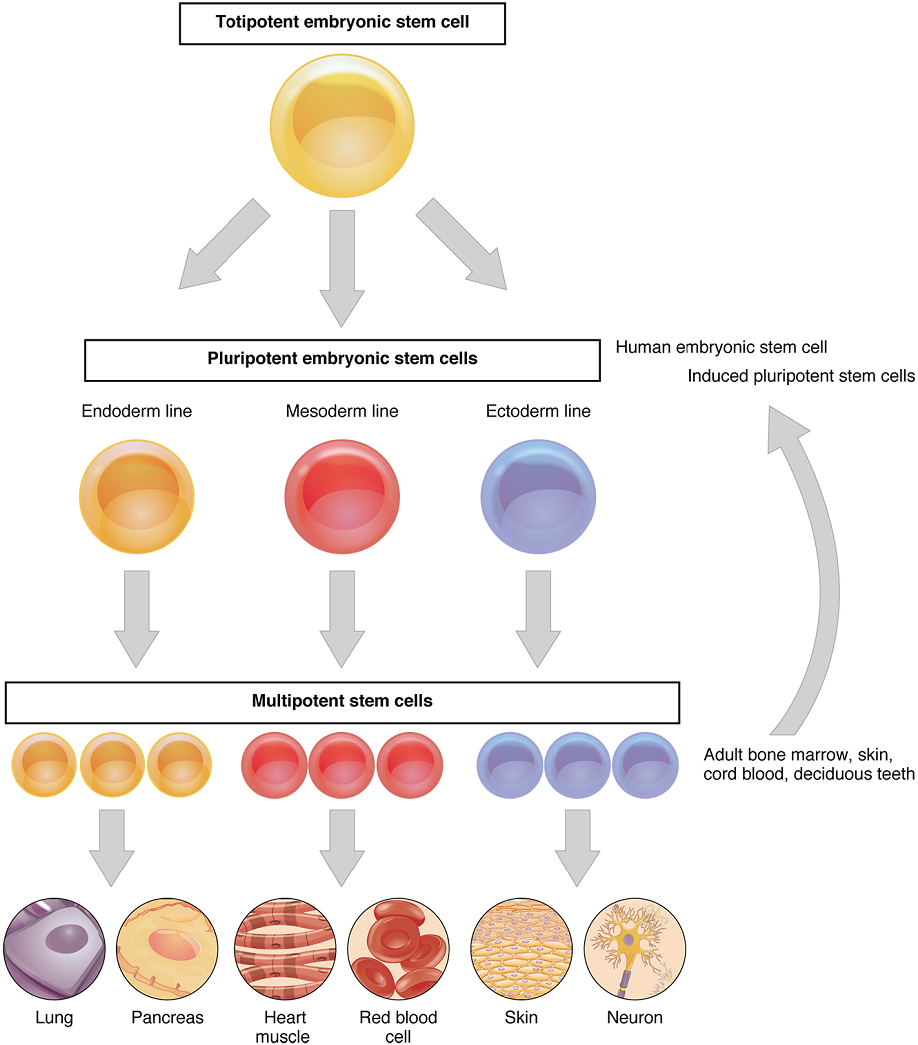
A stem cell is an unspecialized cell that can divide without limit as needed and can, under specific conditions, differentiate into specialized cells. Stem cells are unique in that they can also continually divide and regenerate new stem cells instead of further specializing. There are different stem cells present at different stages of a human’s life. They include the embryonic stem cells of the embryo, fetal stem cells of the fetus, and adult stem cells in the adult. One type of adult stem cell is the epithelial stem cell, which gives rise to the keratinocytes in the multiple layers of epithelial cells in the epidermis of skin.
When a cell differentiates they becomes specialized; yet all cells in the body, beginning with the fertilized egg, contain the same DNA, how do the different cell types come to be so different? The answer is analogous to a movie script. The different actors in a movie all read from the same script, however, they are each only reading their own part of the script. Similarly, all cells contain the same full complement of DNA, but each type of cell only “reads” the portions of DNA that are relevant to its own function. In biology, this is referred to as the unique genetic expression of each cell.
Cell specialization
As cells specialize they may undertake major changes in its size, shape, metabolic activity, and overall function. The morphology (structure) of a mature cell is closely related to the function it is specialized to serve (Figure 9). Muscle fibres for example are far removed in structure and function from the zygote that they ultimately arose from: they are long, slender structures that are well-suited to contracting to produce macroscopic movements over relatively long distances. Some neurons (nerve cells) are exceptionally long and slender in shape, again to act over relatively long distances, although in this case their function is to transmit information rather than move body structures directly. Erythrocytes (red blood cells) are used to transport oxygen in the blood; their tiny size and lack of a nucleus make them well-suited to squeezing through the smallest of capillaries, and their lack of mitochondria mean they do not themselves use up the oxygen they are supposed to be delivering to other cells. Leukocytes (white blood cells) on the other hand are noticeably larger than erythrocytes, and do have mitochondria. The large size of macrophages, for example, means they are capable of physically engulfing relatively large particles or whole cells such as bacteria by phagocytosis, and their mitochondria allow them access to the chemical energy required to move themselves through body tissues towards invading pathogens.


Part 1. Characteristics of life
Part 2. Structural organization of the body
Part 3. Cell structure, cellular organelles and functions
Part 4. Cellular processes involved in growth
Sum of all catabolic and anabolic reactions that take place in the body.
Any of several different types of membrane-enclosed specialized structures in the cell that perform specific functions for the cell.
Skin and its accessory structures.
Reactions that build smaller molecules into larger molecules.
Chemical reaction that breaks down more complex organic molecules.
Building block of proteins; characterized by an amino and carboxyl functional groups and a variable side-chain.
Nucleotide containing ribose and an adenine base that is essential in energy transfer.
Becoming wider, larger or more open.
Process by which unspecialized cells become more specialized in structure and function.
Consisting of more than one cell (as opposed organisms such as bacteria which are unicellular).
The smallest unit of an element that retains the unique properties of that element.
Two or more atoms covalently bonded together.
Group of many similar cells (though sometimes composed of a few related types) that work together to perform a specific function.
An anatomically distinct structure of the body composed of two or more tissue types.
Internal material between the cell membrane and nucleus of a cell, mainly consisting of a water-based fluid called cytosol, within which are all the other organelles and cellular solute and suspended materials.
Clear, semi-fluid medium of the cytoplasm, made up mostly of water.
One of two major divisions of living things (or their cells) that have membrane-bound nuclei and other organelles, and can form large complex organisms (including all animals, plants, fungi). By contrast, bacteria are prokaryotic.
Cell’s central organelle; contains the cell’s DNA.
Set of cellular organelles that often work together to produce, package and export certain products.
Cellular organelle that consists of interconnected membrane-bound tubules, which may or may not be associated with ribosomes (rough type or smooth type, respectively).
Cellular organelle formed by a series of flattened, membrane-bound sacs that functions in protein modification, tagging, packaging, and transport.
Cellular organelle that functions in protein synthesis.
Class of organic compounds that are composed of many amino acids linked together by peptide bonds.
Class of nonpolar organic compounds built from hydrocarbons and distinguished by the fact that they are not soluble in water.
An amphipathic lipid molecule containing a phosphate head (polar) and two fatty acid tails (non-polar). The major molecule comprising plasma membranes.
(Also, sterol) lipid compound composed of four hydrocarbon rings bonded to a variety of other atoms and molecules; not to be confused with anabolic steroids, a synthetic supplement
Secretion of an endocrine organ that travels via the bloodstream or lymphatics to induce a response in target cells or tissues in another part of the body.
Chemical signal that is released from the synaptic end bulb of a neuron to cause a change in the target cell.
Membrane-bound structure that contains materials within or outside of the cell.
Membrane-bound cellular organelle originating from the Golgi apparatus and containing digestive enzymes.
Cell process (a form of endocytosis) in which a cell engulfs and ingests another large particle or cell.
Programmed cell death.
One of the cellular organelles bound by a double lipid bilayer that function primarily in the production of cellular energy (ATP).
Molecule (usually a protein) that catalyzes chemical reactions.
Production of ATP from glucose oxidation via glycolysis, the Krebs cycle, and oxidative phosphorylation.
Oxygen-carrying protein in erythrocytes (red blood cells).
Deoxyribose-containing nucleic acid that stores genetic information.
Membrane that surrounds the nucleus; consisting of a double lipid-bilayer.
One of the small, protein-lined openings found scattered throughout the nuclear envelope.
Ribose-containing nucleic acid that helps manifest the genetic code as protein.
Small region of the nucleus that functions in ribosome synthesis.
Substance consisting of DNA and associated proteins.
A long DNA molecule, combined with proteins that contains a number of genes. The normal chromosome compliment is 23 pairs of homologous chromosomes, one each from mother and father.
Life cycle of a single cell, from its birth until its division into two new daughter cells.
Haploid reproductive cell (egg or sperm in humans) that contributes genetic material to form an offspring.
Excitable neural cell that transfer nerve impulses.
A body cell, excluding germ cells. Normally diploid, each cell containing a complete set of genes.
Cell that gives rise to a gamete.
Type of tissue that serves primarily as a covering or lining of body parts, protecting the body; it also functions in absorption, transport, and secretion.
Cell that is oligo-, multi-, or pleuripotent that has the ability to produce additional stem cells rather than becoming further specialized.
Developing human during weeks 3–8.
Developing human during the time from the end of the embryonic period (week 9) to birth.
Cell that produces keratin and is the most predominant type of cell found in the epidermis.
outermost tissue layer of the skin
Red blood cell.
White blood cell.
