Levels of Organization
Unit 7: Body Structure
Unit Outline
Part 2: Anatomical Terminology
- The anatomical position
- Regional terms
- Directional terms
- Body planes
- Body cavities and serous membranes
- Subdivisions of the posterior and anterior cavities
- Abdominopelvic regions and quadrants
- Membranes of the anterior body cavity
Practice Questions
Learning Objectives
At the end of this unit, you should be able to:
I. Define the terms organ, organ system and organism.
II. Name the eleven organ systems of the human body, identify the major organs, and give a major function of each system.
III. Define and demonstrate the anatomical position.
IV. Locate the anterior (ventral) and posterior (dorsal) surfaces for the body, hands and feet.
V. Define the directional terms used in human anatomy.
VI. Define sagittal, frontal, and transverse planes, and distinguish between midsagittal (median) and parasagittal planes.
VII. Specify and describe the limits of the body cavities.
VIII. Describe how the abdominopelvic region is divided into either nine regions or four quadrants.
Learning Objectives and Guiding Questions
At the end of this unit, you should be able to complete all the following tasks, including answering the guiding questions associated with each task.
I. Define the terms organ, organ system and organism.
II. Name the eleven organ systems of the human body, identify the major organs, and give a major function of each system.
- Create a table specifying the eleven organ systems of the human body, the major organs found in each organ system, and at least one major function of each organ system.
III. Define and demonstrate the anatomical position.
- Sketch a human body in standard anatomical position.
- Use complete sentences to clearly describe the position of each of the following components of the human body when in standard anatomical position:
- Feet
- Upper limbs
- Trunk
- Head
IV. Locate the anterior (ventral) and posterior (dorsal) surfaces of the human body, hands, and feet.
- Clearly define each of the following terms:
- Anterior
- Posterior
- Ventral
- Dorsal
- Explain why ‘anterior’ and ‘ventral’ can be used interchangeably to describe relative locations in the human body.
- Explain why ‘posterior’ and ‘dorsal’ can be used interchangeably to describe relative locations in the human body.
- Identify the direction (ventral or dorsal) in which the palms face when in standard anatomical position, and thus identify which side of the hand (palm or back) of the hand is the ventral side of the hand, and which is the dorsal side.
- Compare the anatomy of the foot to the anatomy of the hand to identify which side of the foot (sole or top) is the ventral side of the foot, and which is the dorsal side.
- Sketch each of the following, and on each diagram clearly indicate the anterior (ventral) and posterior (dorsal) surfaces:
- A human body
- A human hand
- A human foot
- Use complete sentences to describe how to identify the anterior (ventral) and posterior (dorsal) surfaces of each of the following:
- The human body
- The human hand
- The human foot
V. Define the directional terms used in human anatomy.
- Define each of the following terms and provide one complete sentence that correctly uses each term to describe the relative position of two or three body structures (as appropriate).
- Superior
- Inferior
- Medial
- Lateral
- Intermediate
- Peripheral
- Central
- Proximal
- Distal
- Deep
- Superficial
- Anterior
- Posterior
- Cranial
- Caudal
- Distinguish between the terms prone and supine.
VI. Define sagittal, frontal, and transverse planes, and distinguish between midsagittal (median) and parasagittal planes.
- Explain why a body structure is often cut into thin sections before viewing.
- Define each of the following:
- Sagittal plane
- Midsagittal (median) plane
- Parasagittal plane
- Frontal (coronal) plane
- Transverse plane
VII. Specify and describe the limits of the body cavities.
- Sketch a diagram of the human body showing the relative locations of all the following body cavities:
- Dorsal body cavity
- Ventral body cavity
- Cranial cavity
- Vertebral cavity
- Thoracic cavity
- Abdominal cavity
- Pelvic cavity
- Abdominopelvic cavity
- For each of the following cavities, specify whether there is a physical body structure separating them or not. If there is a major body structure separating them, name that structure.
- Dorsal and ventral body cavities
- Cranial and vertebral cavities
- Thoracic and abdominal cavities
- Abdominal and pelvic cavities
- Abdominal and vertebral cavities
- For each of the following cavities, name all the organs found within that cavity:
- Cranial cavity
- Vertebral cavity
- Thoracic cavity
- Abdominal cavity
- Pelvic cavity
- For each of the following cavities, name any organ systems that are (mostly) contained within that cavity:
- Cranial cavity
- Vertebral cavity
- Thoracic cavity
- Abdominal cavity
- Pelvic cavity
- Define each of the following terms, using only a single short sentence for each:
- Parietal serosa
- Visceral serosa
- Serous fluid
- Parietal pericardium
- Visceral pericardium
- Parietal peritoneum
- Visceral peritoneum
- Parietal pleurae (NB: “pleurae” means “more than one pleura”; why are there multiple pleurae?)
- Visceral pleurae
- Briefly describe the general function of serosa and the serous fluid in the human body.
VIII. Describe how the abdominopelvic region is divided into either nine regions or four quadrants.
- Specify the locations of each of the following nine abdominopelvic regions:
- Right hypochondriac
- Epigastric
- Left hypochondriac
- Right lumbar
- Umbilical
- Left lumbar
- Right iliac
- Hypogastric
- Left iliac
- Describe the location of each of the following abdominopelvic quadrants:
- Right upper quadrant
- Left upper quadrant
- Right lower quadrant
- Left lower quadrant
- Specify the location(s), within the nine abdominopelvic regions, of each of the following organs:
- Liver
- Gall bladder
- Spleen
- Stomach
- Small intestine
- Caecum
- Appendix
- Ascending colon
- Transverse colon
- Descending colon
- Urinary bladder
Part 1: Body Systems
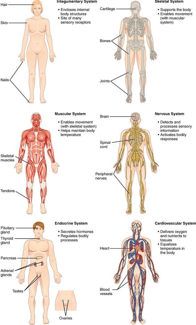
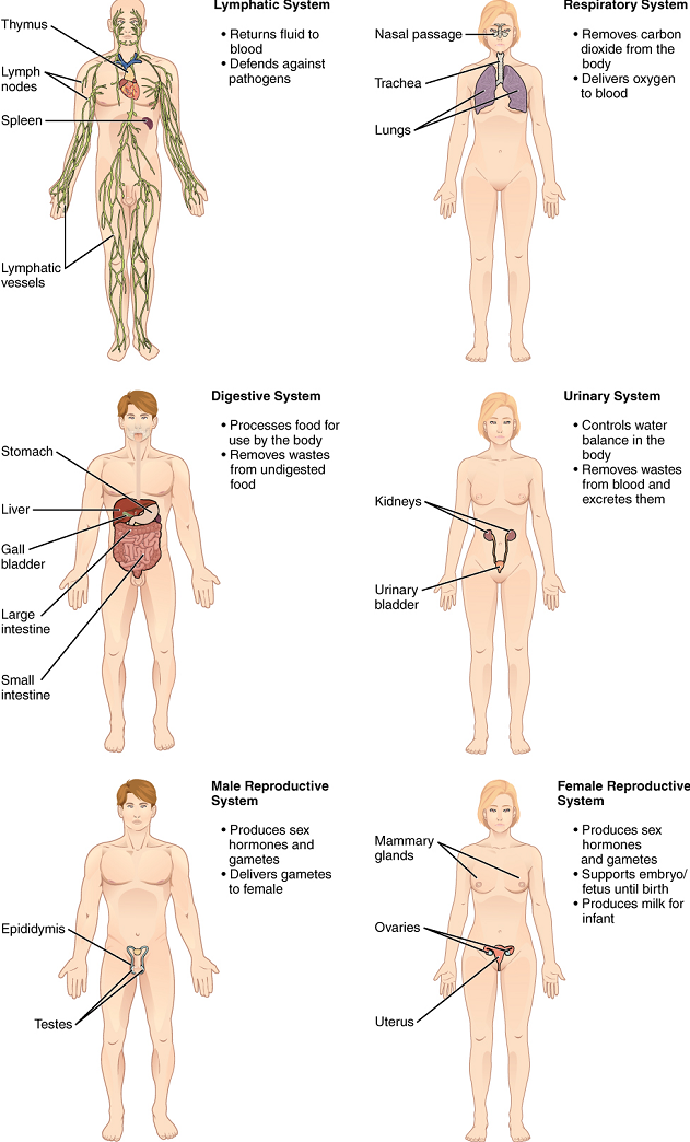
An organ is an anatomically distinct structure of the body composed of two or more tissue types. Each organ performs one or more specific physiological functions. An organ system is a group of organs that work together to perform major functions or meet physiological needs of the body.
The human body contains eleven distinct organ systems (Figure 1 and Figure 2). Assigning organs to organ systems can be imprecise since organs that “belong” to one system can also have functions integral to another system. In fact, most organs contribute to more than one system.
The organism level is the highest level of anatomical organization. An organism is a living being that has a cellular structure and that can independently perform all physiologic functions necessary for life. In multicellular organisms, including humans, all cells, tissues, organs, and organ systems of the body work together to maintain the life and health of the organism.
Part 2: Anatomical Terminology
Anatomists and health care providers use terminology that can be bewildering to the uninitiated. However, the purpose of this language is not to confuse, but rather to increase precision and reduce medical errors. For example, is a scar “above the wrist” located on the forearm two or three inches away from the hand? Or is it at the base of the hand? Is it on the palm-side or back-side? By using precise anatomical terminology, we eliminate ambiguity. Anatomical terms derive from Ancient Greek and Latin words. Because these languages are no longer used in everyday conversation, the meaning of their words does not change.
Anatomical terms are made up of roots, prefixes, and suffixes (Appendix II). The root of a term often refers to an organ, tissue, or condition, whereas the prefix or suffix often describes the root. For example, in the disorder hypertension, the prefix “hyper-” means “high” or “over,” and the root word “tension” refers to pressure, so the word “hypertension” refers to abnormally high blood pressure.
The anatomical position
To further increase precision, anatomists standardize the way in which they view the body. Just as maps are normally oriented with north at the top, the standard body “map,” or anatomical position, is that of the body standing upright, with the feet parallel, at shoulder width apart and with toes forward. The upper limbs are held out to each side, and the palms of the hands face forward (Figure 3). Using this standard position reduces confusion. It does not matter how the body being described is oriented, the terms are used as if it is in anatomical position. For example, a scar in the “anterior (front) carpal (wrist) region” would be present on the palm side of the wrist. The term “anterior” would be used even if the hand were palm down on a table.
A body that is lying down is described as either prone or supine. Prone describes a face-down orientation, and supine describes a face up orientation. These terms are sometimes used in describing the position of the body during specific physical examinations or surgical procedures.
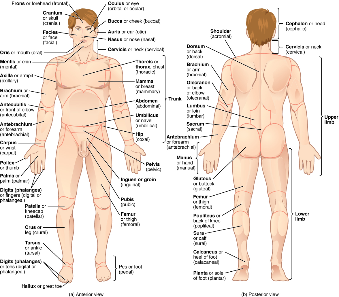
Regional Terms
The human body’s numerous regions have specific terms to help increase precision (Figure 3). Notice that the term “brachium” or “arm” is reserved for the “upper arm” and “antebrachium” or “forearm” is used rather than “lower arm.” Similarly, “femur” or “thigh” is correct, and “leg” or “crus” is reserved for the portion of the lower limb between the knee and the ankle. You will be able to describe the body’s regions using the terms from the figure.
Directional Terms
Certain directional anatomical terms appear throughout this and any other anatomy textbook (Figure 4). These terms are essential for describing the relative locations of different body structures. For instance, an anatomist might describe one band of tissue as “inferior to” another or a physician might describe a tumor as “superficial to” a deeper body structure. Commit these terms to memory to avoid confusion when you are studying or describing the locations of particular body parts.
- Anterior (or ventral) describes the front or direction toward the front of the body. The toes are anterior to the foot.
- Posterior (or dorsal) describes the back or direction toward the back of the body. The popliteus is posterior to the patella.
- Superior (or cranial) describes a position above or higher than another part of the body proper. The orbits are superior to the oris.
- Inferior (or caudal) describes a position below or lower than another part of the body proper; near or toward the tail (in humans, the coccyx, or lowest part of the spinal column). The pelvis is inferior to the abdomen.
- Lateral describes the side or direction toward the side of the body. The thumb (pollex) is lateral to the digits.
- Medial describes the middle or direction toward the middle of the body. The hallux is the medial toe.
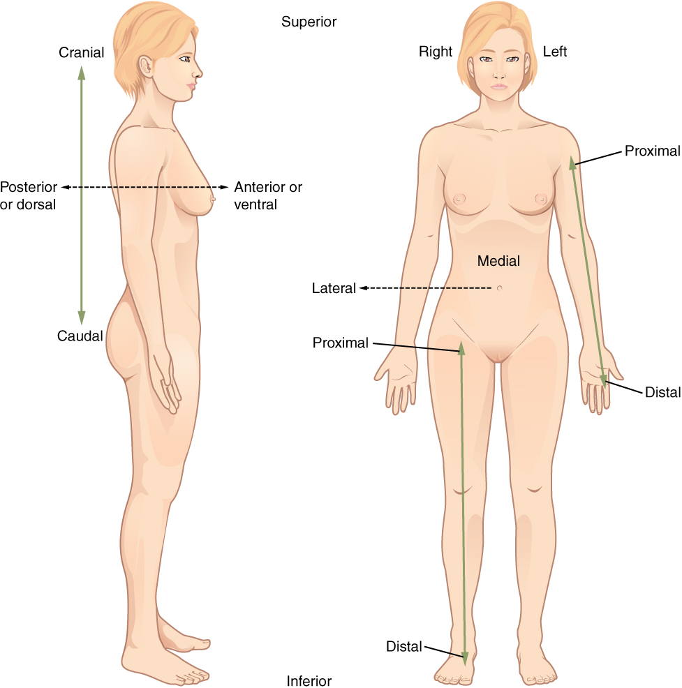
- Intermediate describes a position between a more medial and a more lateral structure. The middle finger is intermediate between the ring and index fingers.
- Proximal describes a position in a limb that is nearer to the point of attachment or the trunk of the body. The brachium is proximal to the antebrachium.
- Distal describes a position in a limb that is farther from the point of attachment or the trunk of the body. The crus is distal to the femur.
- Central describes a position towards the middle (centre) of a structure or organ system. The central nervous system is contained within the skull and vertebral column.
- Peripheral describes a position towards the outer edge (periphery) of a structure or organ system. The peripheral nervous system is found outside the skull and vertebral column.
- Superficial describes a position closer to the surface of the body. The skin is superficial to the bones.
- Deep describes a position farther from the surface of the body. The brain is deep to the skull.
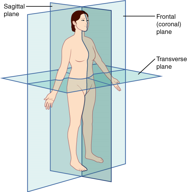
Body Planes
Sectioning, or cutting, is frequently used in the study of Anatomy. The body can be sectioned in various ways to produce a plane, this is a two-dimensional surface of a three-dimensional structure that has been cut. A body structure is often cut into thin sections before macroscopic viewing to allow visualization of the structure’s interior and assist with identification of local disease or infiltration as these pathologies may not be obvious when observing the surface anatomy alone. Modern medical imaging devices enable clinicians to obtain “virtual sections” of living bodies. We call these scans. Body sections and scans can be correctly interpreted, however, only if the viewer understands the plane along which the section was made. A plane is an imaginary two-dimensional surface that passes through the body. There are three planes commonly referred to in anatomy and medicine (Figure 5).
- A sagittal plane is a plane that divides the body or an organ vertically into right and left sides. If this vertical plane runs directly down the middle of the body, it is called the midsagittal or median plane. If it divides the body into unequal right and left sides, it is called a parasagittal plane or (less commonly) a longitudinal section.
- A frontal plane is a plane that divides the body or an organ into an anterior (front) portion and a posterior (rear) portion. A frontal plane is often referred to as a coronal plane (“corona” is Latin for “crown”).
- A transverse plane is a plane that divides the body or organ horizontally into upper and lower portions. Transverse planes produce images referred to as cross sections.
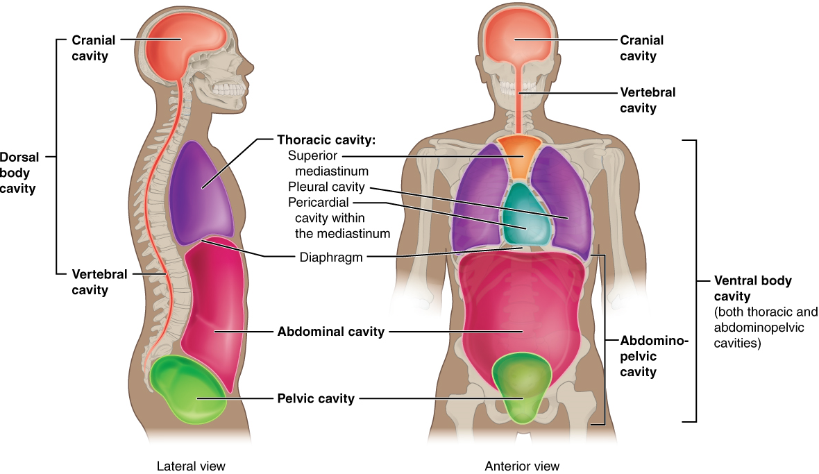
Body Cavities and Serous Membranes
The body maintains its internal organization by means of membranes, sheaths, and other structures that separate compartments. The dorsal (posterior) cavity and the ventral (anterior) cavity are the largest body compartments (Figure 6). These cavities contain and protect delicate internal organs, and the ventral cavity allows for significant changes in the size and shape of the organs as they perform their functions. The lungs, heart, stomach, and intestines, for example, can expand and contract without distorting other tissues or disrupting the activity of nearby organs.
Subdivisions of the Posterior (Dorsal) and Anterior (Ventral) Cavities: The posterior (dorsal) and anterior (ventral) cavities are each subdivided into smaller cavities. In the posterior (dorsal) cavity, the cranial cavity houses the brain, and the spinal cavity (or vertebral cavity) encloses the spinal cord. Just as the brain and spinal cord make up a continuous, uninterrupted structure, the cranial and spinal cavities that house them are also continuous. The brain and spinal cord are protected by the bones of the skull and vertebral column and by cerebrospinal fluid, a colorless fluid produced by the brain, which cushions the brain and spinal cord within the posterior (dorsal) cavity.
The anterior (ventral) cavity has two main subdivisions: the thoracic cavity and the abdominopelvic cavity (Figure 6). The thoracic cavity is the more superior subdivision of the anterior cavity, and it is enclosed by the rib cage. The thoracic cavity contains the lungs and the heart, which is located in the mediastinum. The diaphragm forms the floor of the thoracic cavity and separates it from the more inferior abdominopelvic cavity. The abdominopelvic cavity is the largest cavity in the body. Although no membrane physically divides the abdominopelvic cavity, it can be useful to distinguish between the abdominal cavity, the division that houses the digestive organs, and the pelvic cavity, the division that houses the organs of reproduction.
Abdominopelvic Regions and Quadrants: To promote clear communication, for instance about the location of a patient’s abdominal pain or a suspicious mass, health care providers typically divide up the abdominopelvic cavity into either nine regions or four quadrants (Figure 7).
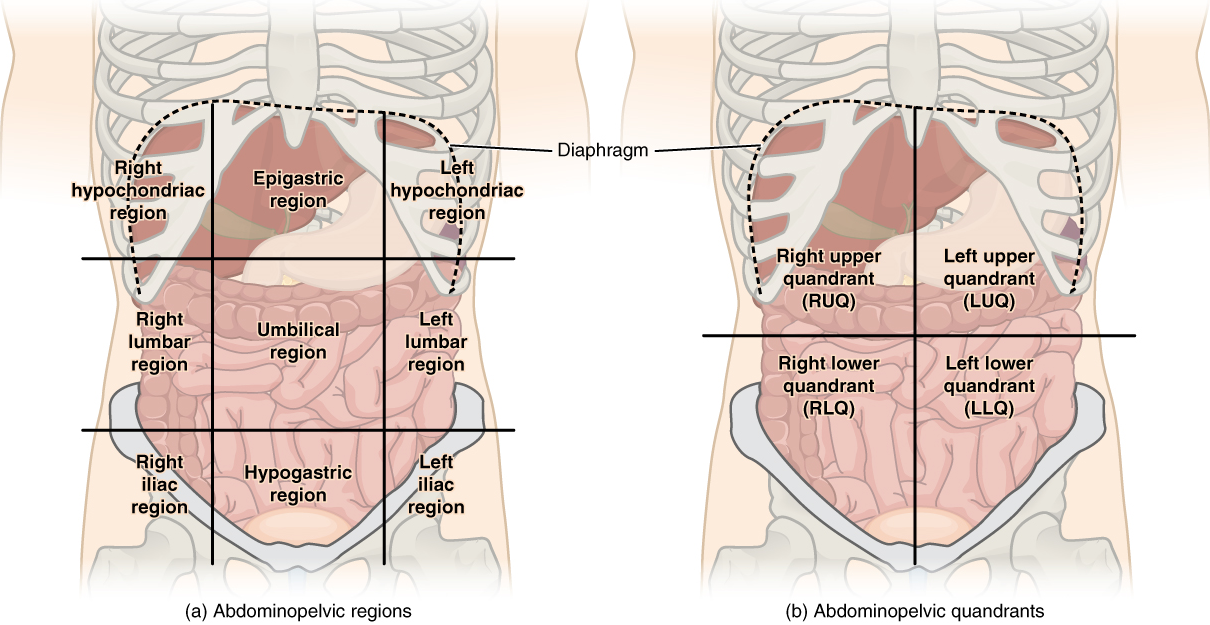
The more detailed regional approach subdivides the cavity with one horizontal line immediately inferior to the ribs and one immediately superior to the pelvis, and two vertical lines drawn as if dropped from the midpoint of each clavicle (collarbone). There are nine resulting regions. The simpler quadrants approach, which is also commonly used in medicine, subdivides the cavity with one horizontal and one vertical line that intersect at the patient’s umbilicus (navel).
These regions can be used to identify the location of abdominal organs more precisely. For example:
- The right hypochondriac region contains the gall bladder and part of the liver, and the right kidney
- The epigastric region contains part of the liver and part of the stomach
- The left hypochondriac region contains part of the spleen and part of the stomach, and the left kidney
- The right lumbar region contains most of the ascending colon
- The umbilical region contains the transverse colon and part of the small intestine
- The left lumbar region contains most of the descending colon
- The right iliac region contains the appendix and caecum
- The hypogastric region contains the lower small intestine, the distal sigmoid colon and anus, and the urinary bladder, as well as the uterus and ovaries in females and the prostate in males
- The left iliac region contains the proximal sigmoid colon
Membranes of the Anterior (Ventral) Body Cavity: A serous membrane (also referred to as a serosa) is one of the thin membranes that cover the walls and organs in the thoracic and abdominopelvic cavities. The parietal layers of the membranes line the walls of the body cavity (pariet- refers to a cavity wall). The visceral layer of the membrane covers the organs (the viscera). Between the parietal and visceral layers is a very thin, fluid-filled serous space, or cavity (Figure 8).
There are three serous cavities and their associated membranes. The pleura is the serous membrane that surrounds the lungs in the pleural cavity; the pericardium is the serous membrane that surrounds the heart in the pericardial cavity; and the peritoneum is the serous membrane that surrounds several organs in the abdominopelvic cavity.
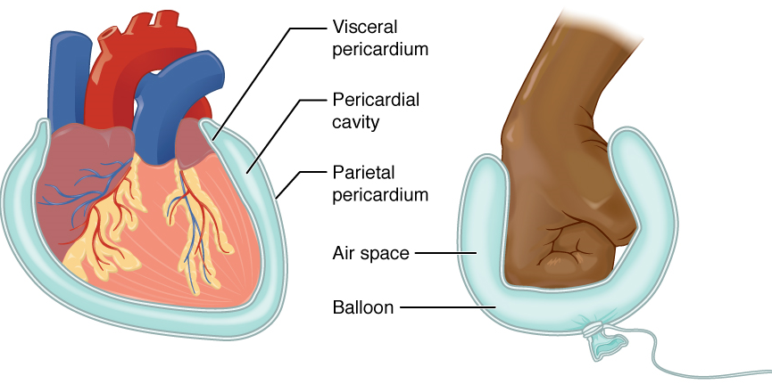
The serous membranes form fluid-filled sacs, or cavities, that cushion and reduce friction on internal organs when they move, such as when the lungs inflate or the heart beats. Both the parietal and visceral serosa secrete the thin, slippery serous fluid located within the serous cavities.
The pleural cavity reduces friction between the lungs and the body wall. Likewise, the pericardial cavity reduces friction between the heart and the wall of the pericardium. The peritoneal cavity reduces friction between the abdominal and pelvic organs and the body wall. Therefore, serous membranes provide additional protection to the viscera they enclose by reducing friction that could lead to inflammation of the organs.
Group of organs that work together to carry out a particular function.
Living being that has a cellular structure and that can independently perform all physiologic functions necessary for life.
Group of many similar cells (though sometimes composed of a few related types) that work together to perform a specific function.
An anatomically distinct structure of the body composed of two or more tissue types.
Face down position.
Face up.
Describes the front or direction toward the front of the body; also referred to as ventral.
Describes the back or direction toward the back of the body; also referred to as dorsal.
Back of the knee.
Knee cap.
Describes a position above or higher than another part of the body proper; also referred to as cranial.
Mouth
Describes a position below or lower than another part of the body proper; near or toward the tail (in humans, the coccyx, or lowest part of the spinal column); also referred to as caudal.
Lowest part of the vertebral column; 'tailbone'
Describes the side or direction toward the side of the body.
Thumb
Describes the middle or direction toward the middle of the body.
Big toe
Describes a position between a more medial and a more lateral structure.
Describes a position in a limb that is nearer to the point of attachment or the trunk of the body.
Upper arm, between shoulder and elbow.
Lower arm, between elbow and wrist.
Describes a position in a limb that is farther from the point of attachment or the trunk of the body.
Thigh bone; the single bone of the thigh.
(In anatomy) describes a position towards the middle (centre) of a structure or organ system.
Describes a position towards the outer edge (periphery) of a structure or organ system.
Describes a position closer to the surface of the body.
(In anatomy) describes a position farther from the surface of the body.
(In anatomy) imaginary two-dimensional surface that passes through the body.
Two-dimensional, vertical plane that divides the body or organ into right and left sides.
A sagittal plane on the midline, dividing the body into equal left and right halves (also medial plane).
A sagittal plane that does not divide the body into equal left and right halves (also longitudinal section).
Two-dimensional, vertical plane that divides the body or organ into anterior and posterior portions.
Two-dimensional, horizontal plane that divides the body or organ into superior and inferior portions.
Posterior body cavity that houses the brain and spinal cord; also referred to the posterior body cavity.
Larger body cavity located anterior to the posterior (dorsal) body cavity; includes the serous membrane-lined pleural cavities for the lungs, pericardial cavity for the heart, and peritoneal cavity for the abdominal and pelvic organs; also referred to as anterior body cavity.
Division of the posterior (dorsal) cavity that houses the brain.
Division of the dorsal cavity that houses the spinal cord; also referred to as vertebral cavity.
Circulatory medium within the CNS that is produced by ependymal cells in the choroid plexus filtering the blood.
Division of the anterior (ventral) cavity that houses the heart, lungs, esophagus, and trachea.
A central compartment in the thoracic cavity located intermediate to the left and right pleural cavities.
Division of the anterior (ventral) cavity that houses the abdominal and pelvic viscera.
Abdominopelvic region (left or right) located under the lowest ribs in the superior corners of the abdominopelvic cavity.
Abdominopelvic region located in the central superior area below the xiphoid process.
Left or right central abdominopelvic region.
Portion of the large intestine.
central abdominopelvic region including the belly button.
Abdominopelvic region in the left or right inferior corners of the abdomen, below the hip bone.
Beginning of the large intestine, forming a small pouch.
Central inferior abdominopelvic region.
Membrane that covers organs and reduces friction; also referred to as serosa.
Facing the body wall.
Facing the organs (opposite of parietal).
Serous membrane that lines the pleural cavity and covers the lungs.
Cavity surrounding the heart filled with a lubricating serous fluid that reduces friction as the heart contracts (also, pericardial cavity or cardiac sac).
Serous membrane that lines the abdominopelvic cavity and covers the organs found there.
