Chapter 4 Selected Musculoskeletal Disease and Disorders, including Trauma and Rheumatic Disorders
Bone Fracture – Complications
Pictures coming soon!
Zoë Soon
Bone Fracture Complications
At times, complications can occur as a result of bone fractures. For example:
Muscle spasms can be triggered by different irritating chemicals released by damaged cells and blood vessels. Activated nociceptors and feelings of pain can cause muscle spasms. Unfortunately, muscle spasms can make the injury worse, as the sharp bone ends can scrape more tissue causing further damage as well as worsen the alignment between bone ends.
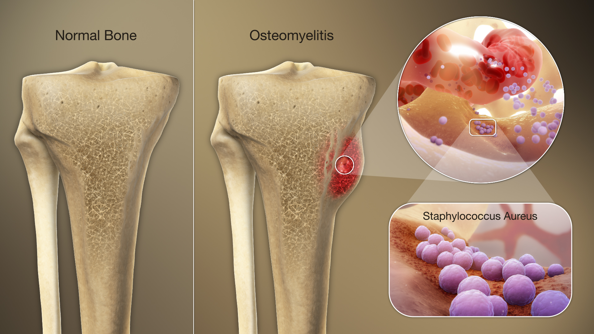
Infection of the wound or bone itself in open fractures can lead to delayed healing. Bone infections (osteomyelitis) are frequently treated with antibiotics as bacteria such as Clostridium tetani can release deadly toxins that can lead to lockjaw and paralysis of respiratory muscle. Vaccinations are available against tetanus are available and recommended at 2, 4, 6, and 18 months, with a booster dose at 4-6 years of age. Teenagers and adults should receive a booster every 10 years as a preventative measure. Other precautions include sterile cleaning of the wound, wound debridement, and prophylactic antibiotics.
Ischemia of surrounding tissue can occur, due to a few problems:
Firstly, if the amount of edema is excessive it may limit gas diffusion within the injured site.
Secondly if there is an increase in edema in the first 48 hours, the cast may become too tight, compressing blood vessels, blocking blood flow, and causing ischemia within the site. If the cast becomes too tight it will need to be replaced.
- If left too long, the affected tissues and organs become deprived of adequate oxygen and nutrients to be fully functional and these tissues and organs become less operable.
- The affected cells will resort to anaerobic cellular respiration (to produce ATP for enzymatic functioning and survival), which means that the cells, will be producing more lactate (lactic acid). The increase in lactate creates a more acidic environment which further contributes to the problem.
- The affected cell enzymes are less able to sustain the cell, as they now are affected by inadequate levels of oxygen, nutrient, pH and excessive waste buildup. Depending on the cell type (which determines their activity level and amount of stored resources), cells will start dying.
- Neurons and heart cells can survive for only minutes when deprived of oxygen.
- Skeletal muscle cells are at risk as they are most often surrounding the broken bone. Skeletal muscle cells can survive approximately 1-2 hours.
- Death of tissue due to ischemia is termed an infarction. A skeletal muscle infarction results in necrosis of the skeletal muscle, causing acute pain and inflammation. Although skeletal muscle does contain myosatellite cells that are capable of proliferation to help regenerate and repair muscle, their self-renewal capacity is limited, which means that complete regeneration of lost muscle cells is not usually possible. As such, permanent muscular atrophy and loss of strength may occur as a result of muscle infarction.
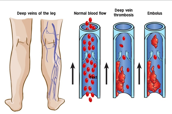
Thirdly, ischemia can be caused by blood clots (thrombi) or fat emboli that have developed in the injured site. Fat emboli comprised of lipid (mainly triglyceride) globules can enter the blood stream from the yellow bone marrow of femur breaks. When either thrombi or fat emboli break off and travel the bloodstream, they are termed emboli. Unfortunately, emboli can lodge in downstream blood vessels (either near or far) causing ischemia. Here are four examples:
- Emboli that travel into the lungs are termed pulmonary emboli and can cause acute respiratory distress syndrome (ARDS). ARDS can be fatal if the embolus is large and lodges in a large pulmonary artery blocking off a significant portion of the downstream pulmonary vasculature.
- Emboli that travel to the brain and block cerebral perfusion can cause a stroke (cerebrovascular accident, CVA) leading to temporary or permanent loss of function (e.g., one-sided paralysis, loss of cognitive or sensory function etc.) and can even be fatal or result in a coma.
- Emboli that travel and lodge in coronary arteries can cause myocardial infarctions (MI) which can be fatal.
- Emboli that lodge in other tissues and organs can cause cellular necrosis. The damage may be permanent or temporary depending on how regenerative the tissue is.
Compartment syndrome can occur in which inflammation at the bone fracture site leads to an increase in pressure within the surrounding muscle compartment. Muscle compartments are surrounded and contained by non-expandable fascia, which is comprised of dense irregular connective tissue. Compartment syndrome will be discussed on subsequent pages.
Nerve damage is possible due to either the severing of nerves during the injury or due to the resulting hypoxia and ischemia that can occur. Depending on the nerve and extent of the damage, the loss of function associated with nerve damage may be temporary or permanent. If a somatic motor neuron is damaged, there may be loss of muscle strength. If a sensory neuron is damaged there may be a loss of various sensations depending on the nerve type (e.g., loss of light touch detection, deep pressure detection, pain, etc.) .
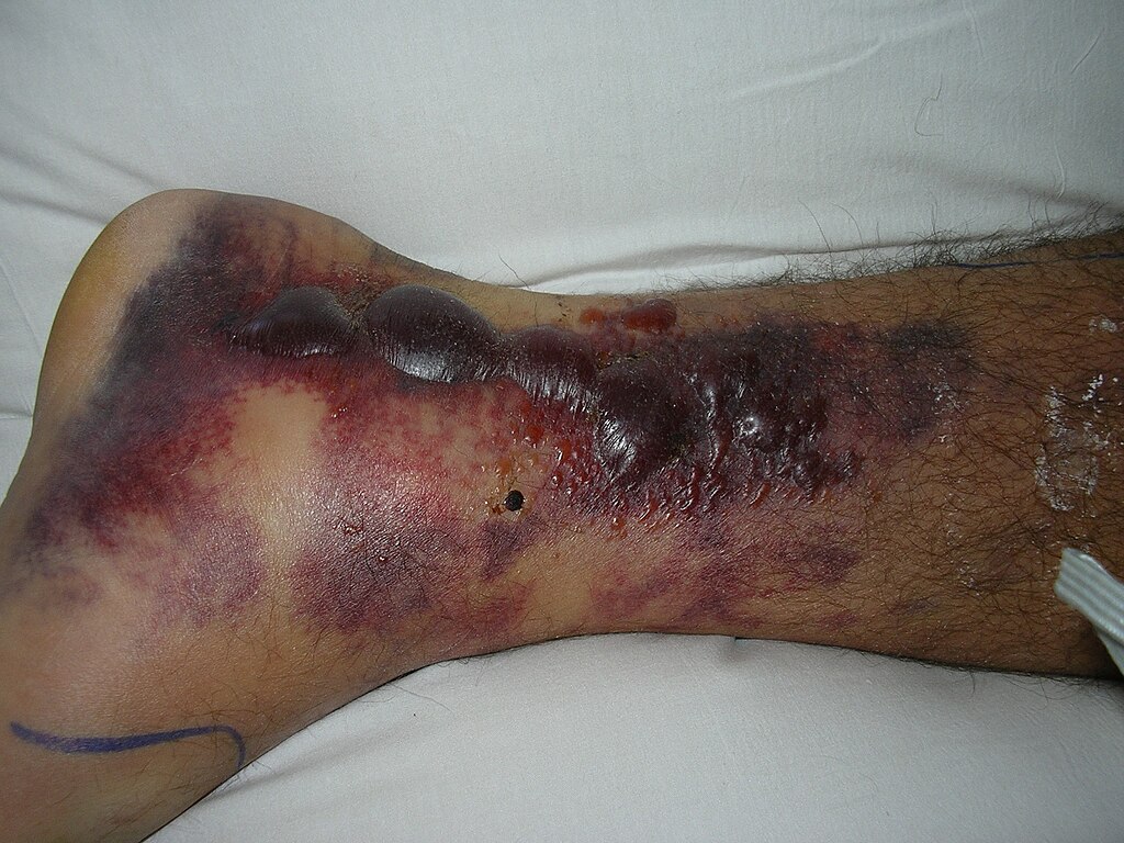
Fracture blisters due to shearing forces can occur, and are collections of exudate that occur between the dermis and underlying tissues, particularly in the leg or ankle. The exudate may be clear or contain blood. Most often they are left intact (and without surgical intervention) to preserve a sterile environment, but are estimated to delay healing time by approximately 12-16 days.
During healing of bone fractures, various complications can occur including:
Malunion is defined as the healing of the bone in an incorrect position, resulting in deformity
Delayed union is defined as healing that occurs after 3 months to a 1 year, due to infection, smoking, use of corticosteroids, or poor circulation. Often this is treated with bone graft bridging or electrical or ultrasound stimulation. Bone grafts typically involve using bone from the iliac crest, fibula or rib of the same individual. This is termed an autologous (or autogenous) bone graft.
Non-union is a term used to describe the failure of bone ends to grow together within 4-6 months, due to infection, repetitive stress, improper alignment or poor circulation. The gap may fill in with dense fibrous fibrocartilage tissue or fluid.
Exuberant callus formation is a temporary growth of bone that is quite large surrounding the fracture site may occur. This is termed an exuberant callus formation. It usually disappears overtime as the bone is remodeled.
Possible Sequelae of bone fractures
Unfortunately, bone fractures can increase the risk of developing osteoarthritis risk later in life.
Stunted growth in children can occur if the epiphyses of long bones are severely damaged in long bones.
Media Attributions
- Osteomyelitis © https://www.scientificanimations.com is licensed under a CC BY-SA (Attribution ShareAlike) license
- Deep Vein Thrombosis (DVT) © Sheikh M. Waheed; Pujitha Kudaravalli; David T. Hotwagner. is licensed under a CC BY-NC-ND (Attribution NonCommercial NoDerivatives) license
- Fracture_blisters © Cindy L. Budge is licensed under a Public Domain license
