Chapter 3 Neoplasia
Review of Cell Cycling, DNA duplication, Cell Differentiation and Errors that can lead to Cancer
More pictures coming soon!
Zoë Soon
Review of Cell Cycling, Cell Division, and DNA duplication
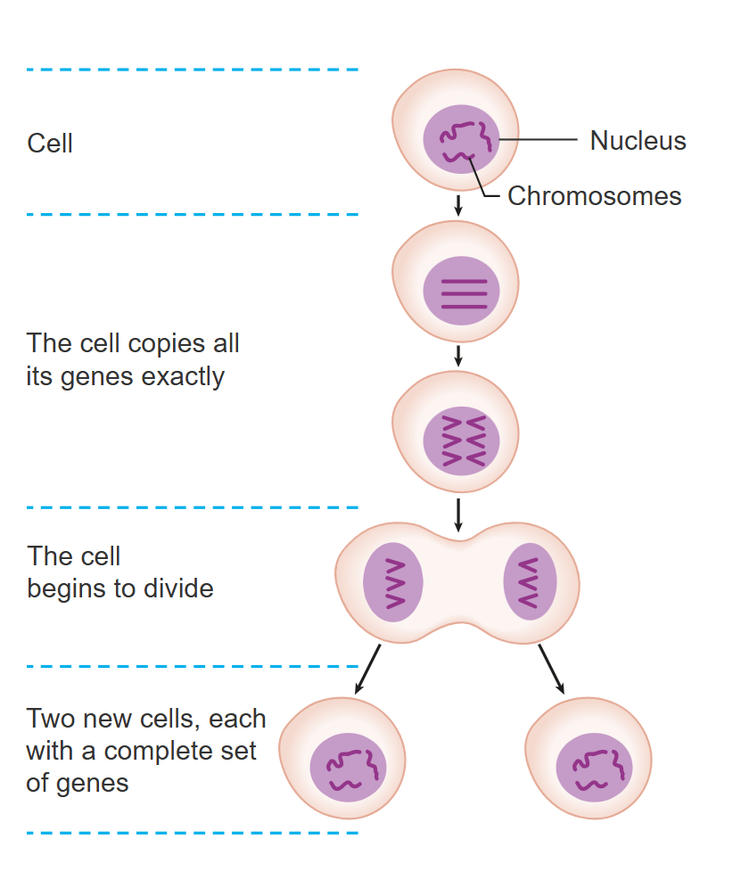
Cell cycling and cell division in humans begins during embryonic development, starting with the fertilized oocyte (zygote). Undoubtedly you don’t remember when you were this young, however, your first act as a zygote was to grow larger in size and then divide from one cell into two identical cells. This process is termed cell cycling. During cell cycling in somatic cells, the cell becomes larger, duplicates its organelles and DNA and then divides into two identical daughter cells. This process of somatic cell duplication is sometimes called cell division, or cell proliferation or simply mitosis.
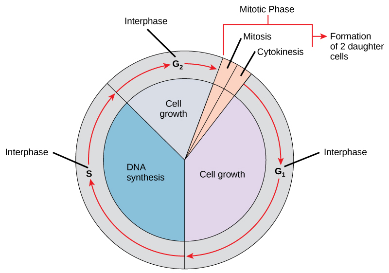
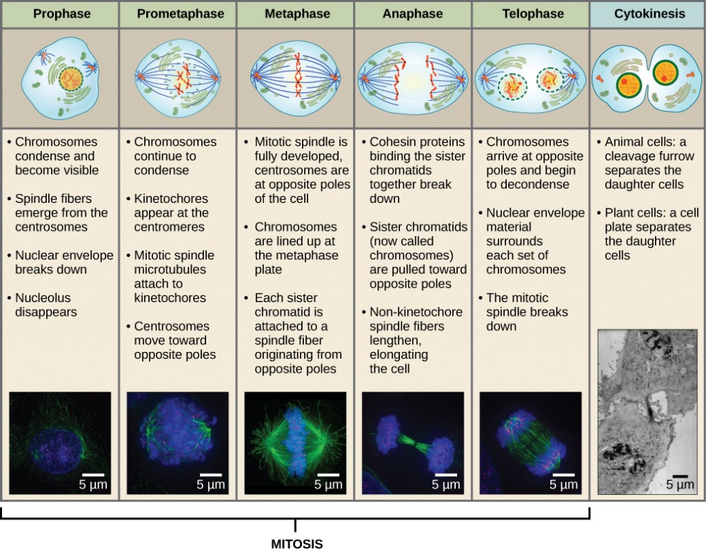
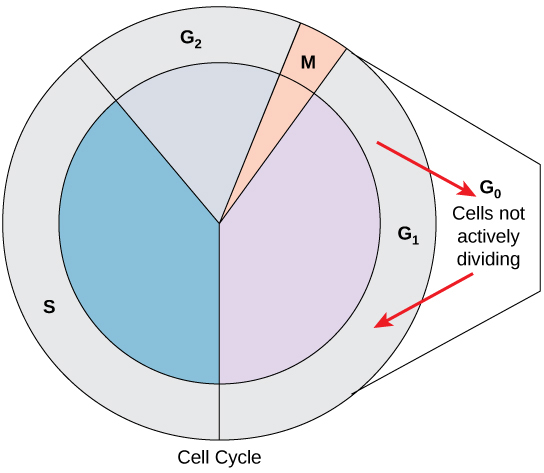
The steps of cell cycling are all equally important. The process begins in interphase and there are three distinct stages within interphase: G1, S, and G2. In G1, the cell is grows in size and duplicates its organelles. In S phase, DNA duplication occurs and in G2, the cell grows a bit more. Enzymes check DNA for errors during duplication, triggering either repair or apoptosis if mutations are found. After these 3 phases of interphase are complete, the cell enters mitosis. Within mitosis, the enlarged cell proceeds through four phases: prophase, metaphase, anaphase and telophase, finally dividing into two cells during cytokinesis, with half of its organelles and one full set of DNA (23 pairs of chromosomes) ending up in each daughter cell.
Review of Cell Differentiation
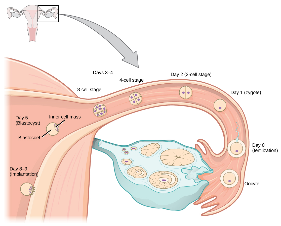
During embryonic development through the process of mitosis, a ball of cells called a blastocyst is created. At this point in time cells have begun to mature and differentiate to forming an inner cell mass which will differentiated into three unique cell lineages (endoderm, mesoderm, and ectoderm) in a process called gastrulation. Within each of these cell types, cells continue to undergo cell cycling and the embryo gets larger and larger in total size. Eventually these cells will become even further differentiated forming lineages for all 200 cell types of the human body (e.g. epithelial cells, cardiomyocytes, hepatocytes, etc.).
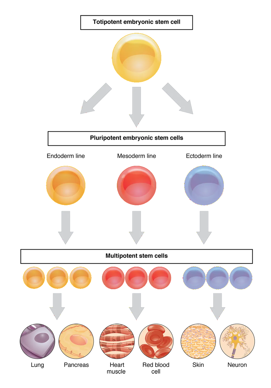
Organs will form with unique sets of these cell types becoming more functional. Within each organ and tissue, some daughter cells (termed stem cells) continue to cell cycle, producing more cells, allowing the embryo to get larger. After each round of mitosis, many daughter cells exit the cell cycle, entering the G0 phase and full mature (differentiate). Cells that become fully mature can usually no longer cell cycle and divide, and instead express specific proteins and enzymes to provide functionality to the organ or tissue that they are part of. This process of growth and maturation continues through all stages of development from embryo to fetus to newborn to child to teenager. Even as a full-size adult, many tissues contain stem cells that divide in order to allow for the replacement of mature cells that get old and die. At adulthood, most cells have exited the cell cycle and have fully differentiated to ensure that each organ and tissue is functional. Depending on the conditions, cells that do exit the cell cycle and enter G0 can enter a state of reversible inactivity (quiescent), irreversible inactivity (senescent), or differentiation (maturation). At adulthood, most cells have exited the cell cycle and have fully differentiated to ensure that each organ and tissue is functional.
What are Telomeres?
Telomeres are the end caps of chromosomes, and they shorten with each cell division, which is thought to act as a safety net to limit the number of rounds of cell cycling that is possible. Telomere shortening helps prevent excessive divisions, as each time DNA is duplicated the possibility of DNA errors and mutations increases. Limiting the number of times a cell undergoes DNA duplication and mitosis therefore reduces the risk of mutations. Once telomeres reach a certain length, apoptosis is triggered, preventing further cell division and potential cancerous growth. DNA mutations are the first step of cancer development.
DNA mutations are the first step of Cancer Development
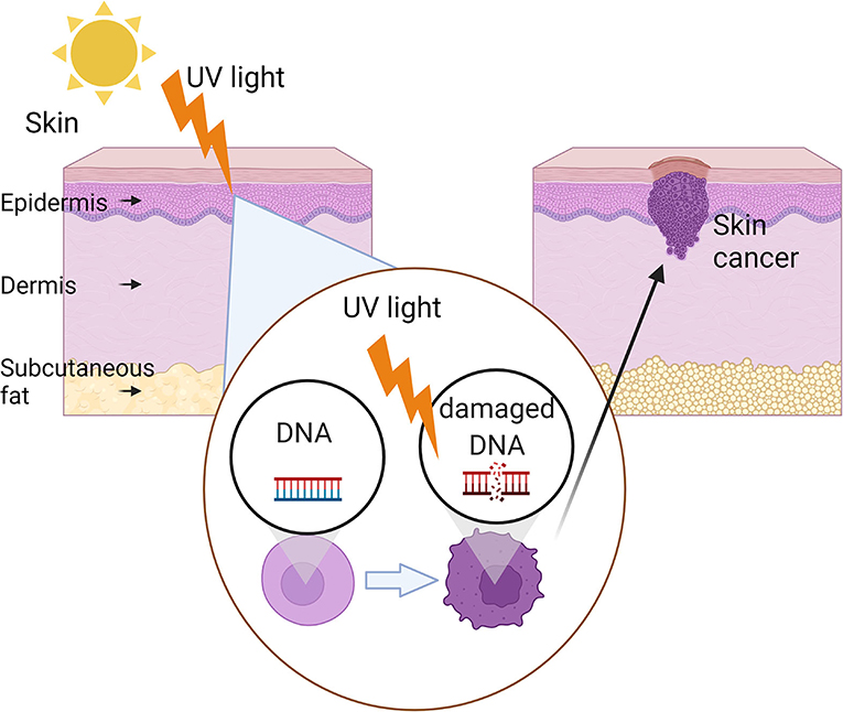
Unfortunately, during the cell cycle’s S phase, as DNA is duplicated, there is a chance for DNA errors to occur as nucleotides (adenine, thymine, guanine, cytosine) are strung together by DNA polymerases. Luckily there are several enzymes that check the DNA for errors during duplication that will trigger apoptosis if mutations are found that cannot be fixed. It is known that mutations that occur in DNA (depending on the location in the DNA) can cause cancer. It is also known that inevitably DNA errors do occur during duplication just due to the sheer number of times DNA is duplicated, not to mention the number of nucleotides within each of the 23 pairs of chromosomes (which include ~3 billion base pairs all together). It may seem obvious that the more DNA duplication events there are, the more risk there is for DNA errors to occur. Therefore, a person’s age becomes a risk factor for the development of cancer. Additionally, one can also imagine that if any of the enzymes responsible for checking DNA for errors in S phase are damaged or mutated or absent, that again person has an increased risk of accumulating mutations and therefore are susceptible to developing cancer.
The Role of Telomeres and Telomerase Re-Activation in Cancer
Telomeres, the end caps of chromosomes are maintained through childhood and adolescence through the enzymatic action of telomerase, an enzyme which continues to add telomere to the ends of chromosome. Telomerase is inactivated in adulthood, and the telomeres begin to shorten with each cell division, acting as a safety net to limit the number of cell divisions possible. At a certain length, a critical point is reached, and the cell becomes inactive (senescent) or dies. This telomere shortening helps prevent excessive divisions, reducing the risk of mutations and cancer development. Additionally, at a certain age the cell has likely become less functional or dysfunctional, potentially accumulating waste products or abnormalities and it would become detrimental to the body if it wasn’t inactivated or removed. In tissue that is regenerative, old cells can be replaced through the division of tissue-specific stem cells. In cells that die when telomeres reach a certain length, apoptosis is triggered and macrophages engulf and recycle the cellular components. Interestingly it has been found that in 90% of cancers, telomerase has been re-activated in the cancerous cells (which unfortunately helps the cancer cells to become immortal – continually adding telomere length and thereby permitting continual cell cycling).
The Roles of Telomerase Inhibitors and Activators in Future Possible Therapies for Cancer and Aging Respectively
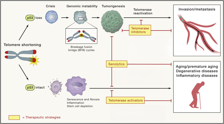
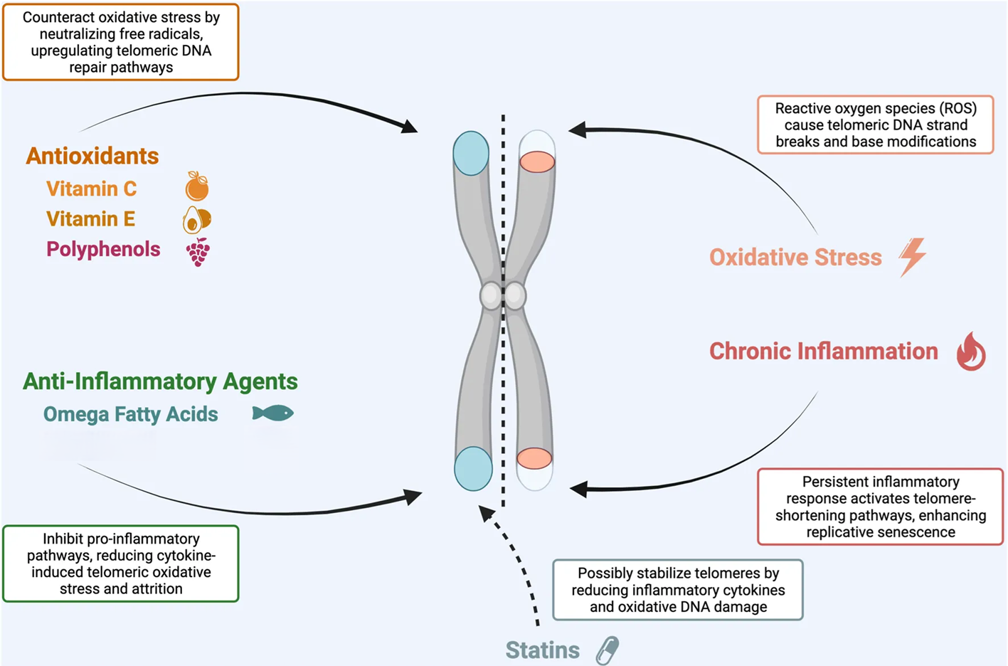
Possible future cancer therapies could involve using Telomerase Inhibitors to block the reactivation of telomerase that can occur in cancer cells. Telomerase is an enzyme that is normally inactivated in most adult cells, aside from those cells that need to divide regularly (e.g., certain adult stem cells). Telomerase lengthens telomeres which can contribute to cell immortality. In these possible future therapies, it is important to consider p53 which is called the Guardian of the Genome. p53 is a tumor suppressor gene, that codes for an enzyme that activates DNA repair and stops the cell cycle at the G1/S cell cycle checkpoint to allow time for DNA repair to occur. Additionally, p53 will initiate apoptosis if DNA damage is beyond repair. Furthermore, p53 also plays an essential role in cells becoming senescent when telomeres shorten to a specific length. Cells that have a loss of p53 expression or mutations in p53 are therefore at risk for developing DNA mutations and becoming cancerous.
As indicated in the above figure, future anti-cancer therapeutic Telomerase Inhibitors would prevent telomerase from becoming reactivated and therefore help prevent cells from maintaining long telomeres and becoming immortal cancer cells. Also, assuming cells are still protected by p53 activity, future anti-aging therapeutic Senolytics and Telomerase Activators could potentially be used to inhibit telomere shortening which may have been stimulated by processes that lead to premature aging.
Media Attributions
- Cell Division © Cancer Research UK is licensed under a CC BY-SA (Attribution ShareAlike) license
- Fertilization, Cleavage, Proliferation and Formation of Blastocyst © Charles Molnar and Jane Gair is licensed under a CC BY (Attribution) license
- Private: Cell Cycle © Charles Molnar and Jane Gair is licensed under a CC BY (Attribution) license
- Mitosis © Charles Molnar, Jane Gair is licensed under a CC BY (Attribution) license
- Stem Cells © J. Gordon Betts, Kelly A. Young, James A. Wise, Eddie Johnson, Brandon Poe, Dean H. Kruse, Oksana Korol, Jody E. Johnson, Mark Womble, Peter DeSaix is licensed under a CC BY-NC-SA (Attribution NonCommercial ShareAlike) license
- Private: Cell Cycling Regulation – Exiting of Cell Cycle to Enter Quiescent (Inactive) Stage, G0 © Charles Molnar, Jane Gair is licensed under a CC BY (Attribution) license
- UV skin cancer © Breen I and Richmond J is licensed under a CC BY (Attribution) license
- Telomeres Antioxidants Anti-inflammatories Cropped © Schellnegger, Marlies, Elisabeth Hofmann, Martina Carnieletto, and Lars-Peter Kamolz. adapted by Zoë Soon is licensed under a CC BY (Attribution) license
- Cropped Version of Original Image – Telomeres history, health, and hallmarks of aging cropped © Deepavali Chakravarti, Kyle A. LaBella, Ronald A. DePinho adapted by Zoë Soon is licensed under a CC BY (Attribution) license
