Chapter 1 Introduction to Pathophysiology; Cellular Responses to Stress, Injury, and Aging
Section 7 Cell Death – Planned and Unplanned
Zoë Soon
Cell death is a normal and essential part of life. However, not all cell death is the same. There are two fundamentally different types: planned cell death (apoptosis) and unplanned cell death (necrosis). Understanding the difference between them – and the conditions that trigger each – is central to understanding how disease affects the body.
Apoptosis: Planned Cell Death
Apoptosis (sometimes called programmed cell death) is a tightly regulated, orderly process in which aging or damaged cells are eliminated to make room for new, more functional ones. It is driven by an internal enzymatic cascade involving proteins called caspases, and it results in a clean, contained death that does not trigger inflammation.
Apoptosis in Normal Life
Apoptosis is not a sign of disease – it is a routine, continuous process that keeps tissues healthy. Consider the following examples:
- Red blood cell (RBC) turnover: RBCs live approximately 120 days. Millions undergo apoptosis every day and are simultaneously replaced by new cells produced in the bone marrow. This balance keeps the RBC population stable.
- Skin cell turnover: Skin cells replace themselves approximately every 30 days through a cycle of apoptosis and mitosis. Similarly, the epithelial cells lining the mucosa membranes of the respiratory and digestive tracts experience frequent turnover (every 30-50 days and every 3-5 days respectively).
- Embryonic development: Apoptosis plays a critical role in shaping the body before birth. For example, the webbing between fingers and toes during fetal development is removed through apoptosis, sculpting the final form of the hands and feet. Heart development and the remodeling of other organs during fetal stages also rely on apoptosis.
- Breast tissue remodeling: During pregnancy, breast tissue grows to support lactation. After breastfeeding ends, apoptosis returns breast tissue to its pre-pregnancy size.
Note that some cell types – particularly neurons – do not undergo ongoing turnover. After birth, the number of neurons a person has is largely fixed for life, which is one reason that neurological damage can be so significant.
Triggers of Apoptosis
While apoptosis is normal, it can also be triggered by a variety of stressors. Triggers are classified as either extrinsic (coming from outside the cells) or intrinsic (arising from within the cell):
| Extrinsic triggers | Bacterial LPS (lipopolysaccharide): A component of the bacterial cell membrane that activates a ‘death receptor’ on the surface of human cells initiating the caspase cascade.
Some pro-inflammatory cytokines released by White Blood Cells can also activate the ‘death receptor’ stimulating the apoptotic enzyme cascade, providing an important defence mechanism against pathogens that have infected cells. |
| Intrinsic triggers | Reactive Oxygen Species (ROS) increased levels
DNA damage Hypoxia (low oxygen / low ATP) Senescence (cellular aging) All of these activate the intrinsic pathway of the caspase cascade within the cell. |
Why Does DNA Damage Trigger Apoptosis?
When a cell detects that its DNA has been mutated, apoptosis serves as a critical safety net. A cell with a mutated genome is at risk of dividing and passing on those mutations potentially leading to either cancer or loss of normal organ function. By eliminating itself, the cell prevents this from happening. Similarly a cell experiencing chronically low ATP due to hypoxia becomes less functional and potentially more prone to mutation – making apoptosis a logical and protective response.
Telomere Shortening and Cellular Aging
Each time a cell divides, the telomeres – protective caps at the ends of chromosomes – shorten slightly. After approximately 50 rounds of cell division telomeres reach a critical minimal length. At this point, the cell recognized that it is ‘old’ and becomes susceptible to DNA damage and cancer. Telomere shortening therefore acts as a built-in trigger for apoptosis, ensuring that aged cells make way for newer, healthier replacements.
The Hayflick limit refers to the number of times a human cell can divide before it stops and enters a state of cellular senescence. The Hayflick limit varies depending on the specific cell type. Senescence is a state of cell cycle arrest, that differs from maturation and differentiation. Senescent cells remain metabolically active and while cleared by young healthy immune systems, they can remain as “zombie cells” that accumulate, secrete harmful chemicals, and contribute to tissue deterioration in chronic disease and aging.
The Apoptotic Process: What it Looks Like
Under a microscope, apoptosis follows a recognizable sequence of steps:
- Step 1 – Cell shrinkage: The cell shrinks and rounds up.
- Step 2 – Chromatin condensation: The DNA condenses and forms compact patches against the inner surface of the nuclear envelope.
- Step 3 – Nuclear disintegration: The nuclear envelope breaks down and the DNA fragments.
- Step 4 – Membrane blebbing and apoptotic body formation: The cell membrane bubbles outward forming small, membrane bound vesicles called apoptotic bodies. Each apoptotic body carries transmembrane ‘flag’ proteins that signal macrophages to engulf and recycle it.
The process is clean and efficient. Macrophages rapidly phagocytose the apoptotic bodies, recycling their components without triggering an inflammatory response – a key distinction from unplanned cell death.

Unplanned Cell Death: Necrosis
Necrosis occurs when cells are damaged so severely that they burst and die in an uncontrolled manner. Unlike apoptosis, necrotic cell death is messy – cells lyse (rupture), spilling their contents into the surrounding tissue. This triggers inflammation: white blood cells, such as neutrophils are recruited to the area and, while they help clean up the debris, they can also cause collateral damage to neighbouring cells through the release of Reactive Oxygen Species (ROS).
The Number One Cause of Unplanned Cell Death: Ischemia
Ischemia – the interruption of blood flow to a tissue – is the leading cause of unplanned cell death in humans. When blood flow is blocked or reduced (e.g., by a blocked or compressed blood vessel), three harmful conditions arise simultaneously:
- Oxygen deprivation (hypoxia): Hypoxia (hypo- = below, -oxia = oxygen) means reduced oxygen in tissues. Without oxygen, cells cannot perform aerobic cellular respiration and ATP production falls dramatically.
- Nutrient deprivation: Glucose, vitamins, and other building blocks essential for cellular function are no longer delivered.
- Waste accumulation: Metabolic waste products build up and become toxic to cells.
Together, these three factors cause far more cell injury than oxygen deprivation alone.
Which Organs are Most Sensitive to Hypoxia?
The organs most vulnerable to hypoxia are those with the highest energy demands – the brain, heart, and kidneys. None of these organs can store oxygen; they depend on a continuous blood supply. Brain cells (neurons) can survive only approximately 3-5 minutes without oxygen before they begin to die. This is why strokes and cardiac arrest must be treated immediately to minimize permanent damage.
How Ischemia Damages Cells: A Step-by-Step Breakdown
The cellular consequences of ischemia follow a predictable, escalating cascade:
- Low ATP → pump failure: Cells maintain their internal environment using two critical ion pumps that both require ATP: the sodium-potassium pump (which moves 3 Na+ out and 2 K+ in) and a calcium-sodium exchanger (which expels Ca2+). Without sufficient ATP, both pumps fail.
- Calcium accumulation: Ca2+ floods into the cell and inappropriately activates two destructive enzymes:
- Phospholipase: degrades the phospholipids of the cell membrane, cause the cell to lose membrane integrity and become leaky.
- Protease: degrades cytoskeletal proteins (the cell’s internal scaffolding), causing the cell to lose its supportive structure and shape.
- Membrane phospholipid depletion: A third enzyme – phospholipid reacylation synthase – normally replaces phospholipids in the membrane during natural turnover. This enzyme requires ATP and therefore also fails during ischemia, meaning damaged phospholipids cannot be replenished.
- Sodium and water influx: As the membrane becomes leaky, sodium and water rush into the cell. The cell swells and may ultimately burst.
- Anerobic metabolism and intracellular acidosis: in an attempt to survive, the cell has switched to anaerobic cellular respiration (glycolysis alone). The generates some ATP but also produces large amounts of lactic acid, lowering intracellular pH. Enzyme function declines as the cell becomes increasingly acidic.
Ischemia, Reperfusion, and Calcium Flooding
Restoring blood flow (reperfusion) to ischemic tissue is necessary for recovery but carries its own risks. When blood rushes back into a leaky, calcium-starved cell, the high calcium concentration of blood floods in through the damaged membrane. This triggers a surge of phospholipase and protease activity, accelerating membrane destruction and cell death.
Additionally, reperfusion brings a wave of white blood cells (WBCs) to the area. While WBCs such as neutrophils and macrophages are essential for clearing debris, they also release Reactive Oxygen Species (ROS, e.g., peroxide, superoxide, hydroxide ions) – highly reactive molecules with unpaired electrons that can damage cell membranes, proteins, and DNA. This collateral damage to neighbouring cells is known as reperfusion injury. For this reason, reperfusion is carried out at a controlled pace in clinical settings.
Reactive Oxygen Species (ROS) and Oxidative Stress
Reactive Oxygen Species (ROS) are unstable, highly reactive molecules characterized by an unpaired electron. Examples include superoxide, hydrogen peroxide, and hydroxide ions. Although ROS are produced normally by mitochondria as part of metabolic signaling (including regulation of vascular tone – the balance between vasoconstriction and vasodilation), they are kept in check by the cell’s own neutralizing antioxidants.
Problems arise when ROS production exceeds the cell’s capacity to neutralize them – a state called oxidative stress. In this scenario, ROS cause damage to cell membranes, proteins, and DNA, promoting inflammation and contributing to cell death.
| Causes of oxidative stress | Ischemia, UV radiation, ionizing radiation (e.g., gamma rays from nuclear sources), cigarette smoking, and excessive alcohol consumption. |
| Role in disease | ROS accumulation is implicated in ALS (Lou Gehrig’s disease), normal aging, stroke, heart attack, and fatty liver disease. |
| Antioxidants | Molecules that neutralize ROS. Produced naturally by cells; also found in food such as dark chocolate and green tea. Beneficial when ROS levels are elevated, but not necessarily required in excess for a healthy individual with a balanced diet. |
∗ Real World Story: The Dangers of Radiation – Fukushima and Chernobyl
In 2011, a major earthquake off the coast of Japan triggered a tsunami that severely damaged the Fukushima Daiichi nuclear reactor. The resulting release of ionizing radiation (specifically gamma rays) caused significant environmental contamination and posed serious health risks to people in the affected area. A similar disaster occurred at Chernobyl in 1986.
Ionizing radiation damages cells through three interconnected mechanisms: it causes direct DNA damage, which can lead to genetic mutations and cancer; it triggers the caspase cascade, leading to apoptosis of irradiated cells; and it stimulates the production of ROS, which cause further membrane, protein, and DNA damage, eventually leading to necrosis of large areas of tissue. Individuals exposed to high doses of ionizing radiation may develop acute radiation sickness.
Causes of Cell Damage and Death: A Summary
A variety of agents can damage or kill cells:
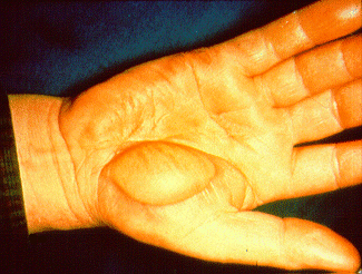
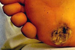
- Physical damage: Wounds, cuts, extreme heat (e.g., burns, electrical burns), and extreme cold (e.g., frostbite). Electric shocks cause burns due to heat from electrical resistance. High temperatures over 40°C induce vascular injury, disruption of cell membrane and cause blood and protein coagulation leading to cell death and downstream ischemia. During frostbite, blood vessels in exposed skin vasoconstrict to protect internal organs, depriving skin tissue of oxygen and nutrients and leading to necrosis of the affected tissue.
- Radiation: Ionizing radiation (X-rays, gamma rays) cause DNA damage, ROS production and cell death.
- Mechanical damage: Tearing or crushing of tissue
- Chemical toxins: Exogenous toxins (e.g., acids, mercury, lead) or endogenous toxic accumulations (e.g., lipids, abnormal proteins) as discussed above.
- Reperfusion injury: Restoration of blood flow to ischemic tissue causing intracellular calcium flooding, loss of cell membrane integrity, cell lysis, and ROS-mediated damage.
- Microorganisms: Bacteria, viruses, fungi (e.g., yeast), helminths, and protozoa cause cellular damage through direct infection and/or inflammatory responses
- Abnormal metabolic diseases: Rare conditions in which toxic waste products accumulate inside cells (e.g., inherited Tay Sachs Disease)
- Malnutrition: Cells deprived of essential building blocks cannot maintain normal function and may deteriorate and die.
- Fluid and electrolyte imbalance: Disruptions in the balance of water and electrolytes can be severely damaging to the heart, brain, and kidneys.
∗ A Real-World Story: Water Intoxication – ‘Don’t Wee for a Wii’
In 2007, a US radio show hosted a “Hold Your Wee for a Wii” content where contestants competed for a video game console by drinking water bottles every 15 minutes while not being allowed to use the washroom. Unfortunately, the radio station ignored calls from a nurse and other listeners about the lethal dangers of water intoxication. One contestant, a 28 year old mother of three, left the contest complaining of swollen stomach and severe head pain, dying later the same day.
What the organizers did not realize is that consuming large volumes of water in a short period of time causes a dangerous drop in blood electrolyte concentrations – a condition known as water intoxication (or hyponatremia, due to diluted blood sodium levels). The resulting electrolyte imbalance can lead to impaired heart and brain function and be fatal, by causing brain swelling or heart attacks.
This tragic case illustrate why extreme dietary behaviours – including overconsumption of water – can be just as dangerous as deprivation.
Media Attributions
- Private: necrosis_vs_apoptosis © National institute on alcohol abuse and alcoholism (NIAAA) is licensed under a Public Domain license
- Electrical_burn_on_hand © National Institute for Occupational Safety and Health (NIOSH) is licensed under a Public Domain license
- Private: Electrical_burn_exit_wound © Occupational Safety and Health Administration is licensed under a Public Domain license
