Chapter 2 Innate and Adaptive Immunity: From Cell Defense to Tissue Repair
Section 9: Hemostasis (Blood Clotting)
Zoë Soon
Hemostasis is the rapid process (taking minutes) by which a damaged blood vessel is sealed to stop bleeding. From Greek/Latin: hemo- (blood) + -stasis (motionless). Three major stages:
Stage 1: Vascular Spasm
Damaged endothelial cells (of blood vessel walls) release ADP, Tissue Factor and endothelin. Endothelin peptides are potent vasoconstrictors that trigger the smooth muscle layer of the blood vessel to contract, narrowing the vessel and minimizing blood loss.
Stage 2: Platelet Plug Formation
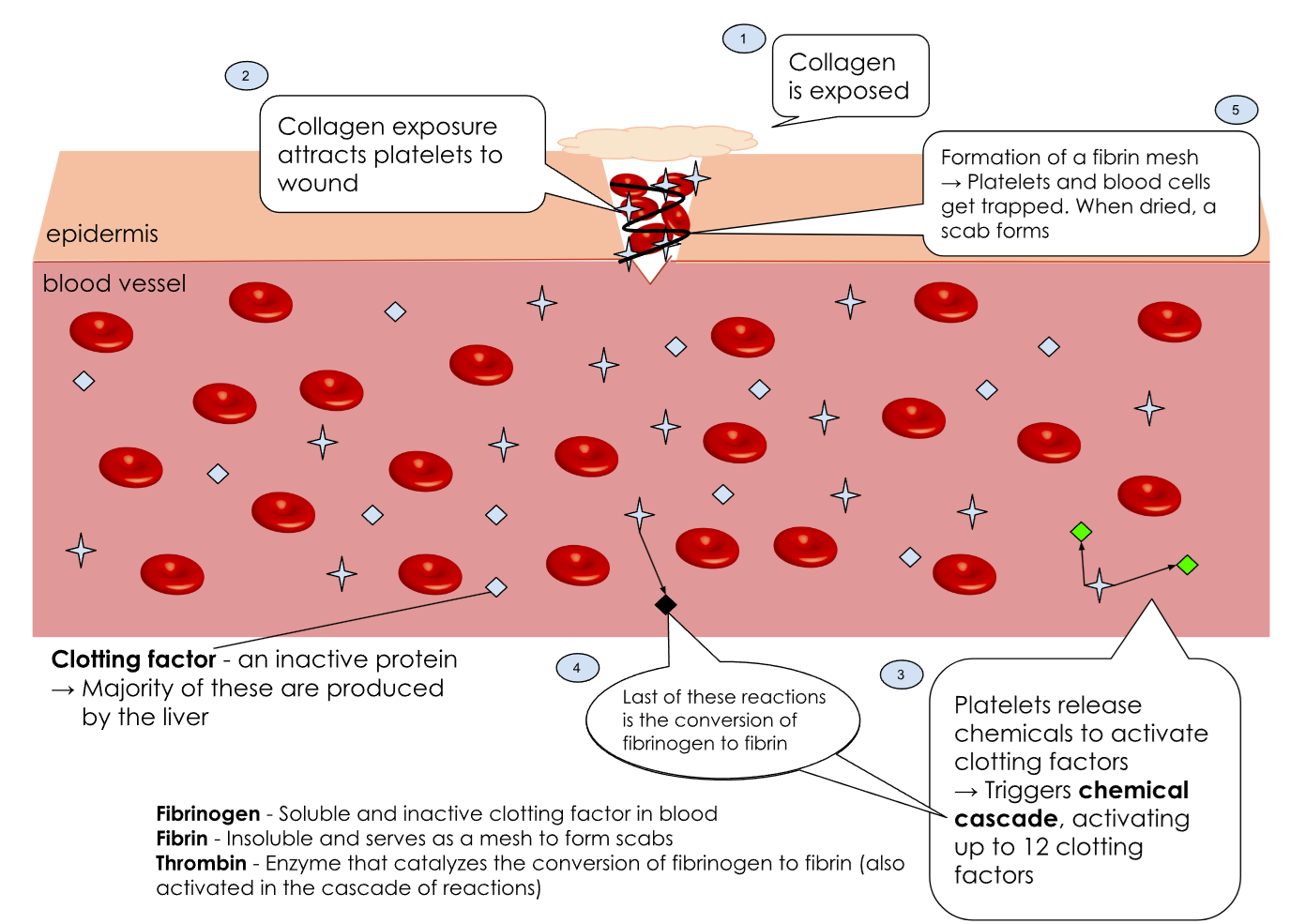
Platelets are attracted to the wound, activate, and adhere to exposed collagen of the damage blood vessel wall – forming an unstable plug within seconds to minutes. Prior to becoming activated, 30% of platelets are typically located in the spleen and 70% of platelets are circulating the blood stream. Inactive platelets travel the bloodstream and have smooth surfaces; activated platelets become spiky and sticky. Activated platelets release: ADP (attracts more platelets), thromboxane A2 (vasoconstrictor; recruits more platelets), serotonin (vasoconstrictor), clotting factors, Ca2+, prostaglandins, and PDGF (Platelet Derived Growth Factor). The recruitment and activation of more platelets is termed a positive feedback loop that ends when the wound is sealed.
Stage 3: Coagulation
Plasma protein fibrinogen (globular, water-soluble) is converted to fibrin (long, water-insoluble, rope-like proteins) that interweave through the platelet plug to stabilize it. Clot retraction then occurs as platelets contract, pulling torn vessel edges together.
Fibrin production involves two pathways running simultaneously, both activating Prothrombin Activator (Factor X) in the process:
| Extrinsic Pathway | Tissue Factor released from endothelial cells triggers a cascade (of clotting factors + calcium) activating enzyme Prothrombin Activator (Factor X) → converts inactive enzyme prothrombin → active thrombin → converts fibrinogen to fibrin.
This pathway is faster. |
| Intrinsic Pathway | Platelet Factor and calcium released from platelets trigger the activation of the intrinsic clotting factors cascade which also converges on Prothrombin Activator (Factor X) activation → converts prothrombin (inactive) → thrombin (active) → converts fibrinogen → fibrin.
This pathway is slower but also necessary. |
Fibrinolysis and Healing
Once healing begins, fibrin is dissolved through fibrinolysis. tPA (Tissue-Type Plasminogen Activator) converts plasminogen to active plasmin protease, which degrades fibrin. Macrophages remove cellular debris; platelets release PDGF to stimulate vessel regeneration; fibroblasts produce mitogens to stimulate mitosis (cell division) and tissue repair.
Platelet Regulation
Endothelial cells release prostacyclin (a prostaglandin family member) – acting as both a vasodilator and platelet inhibitor – to prevent excessive platelet aggregation.
Platelet Disorders
Thrombocytopenia: Low platelet levels (nutritional deficiency, illness, or medication).
Signs: prolonged bleeding, frequent nosebleeds, heavy menstruation, bruising, and petechiae (tiny red spots from capillary hemorrhages).
Hemophilia: Impaired blood clotting due to inherited mutations in clotting factors (most commonly Factor VIII or Factor IX, both on the X chromosome).
Thrombocytophilia (Thrombocytosis, Thrombocythemia): Abnormally high platelet counts, can lead to dangerous blood clots, increasing the risk of heart attacks and strokes.
* Did you know? Facts About Hemostasis
Surgeons may apply a topical collagen agent to attract the patient’s own platelets and stimulate natural hemostasis.
Applying direct pressure to a wound is one of the most effective first-aid measures for slowing blood loss.
Sutures (stitches) close a wound mechanically, speed recovery, and reduce scar formation.
Vitamin K (found in green vegetables, grains, and organ meats – and produced by intestinal bacteria) is required for synthesis of multiple clotting factors. Deficiency impairs hemostasis.
Media Attributions
- Blood Clot Formation © By Victdomi - Own work, CC BY-SA 3.0, https://commons.wikimedia.org/w/index.php?curid=118340457 is licensed under a CC BY-SA (Attribution ShareAlike) license
