"

Chapter 2 Innate and Adaptive Immunity: From Cell Defense to Tissue Repair

Section 6:  Lymphatic System Overview

Zoë Soon

Lymphocytes and Lymphatic System:  Overview

All three lymphocyte types – Natural Killer (NK) cells, T cells, and B cells – arise from bone marrow hematopoietic cells.  Lymphocytes have large nuclei and little cytoplasm (distinguishable under a microscope) and make up to 20-30% of circulating WBCs.  NK cells circulate the body and help to provide innate immunity through immunological surveillance, in which cells without self-antigens are lysed.  Most B and T cells reside in lymphoid tissues (tonsils, spleen, lymph nodes, thymus, bone marrow).

(A) The lymphatic system includes the primary and secondary lymphoid organs and a series of lymphatic vessels, providing a one-way drainage route from all tissues back ultimately to the blood circulation via the great veins in the neck. In the primary lymphoid organs (bone marrow and thymus) immune cell production and maturation takes place, whereas secondary lymphoid organs (lymph nodes, spleen, and mucosa associated lymphoid organs such as Peyer’s patch, tonsils and adenoids) are the sites for lymphocyte activation. The initial dermal lymphatic capillaries absorb interstitial material and fluid to make lymph which drains into lymphatic collectors. Lymph is pumped from the gut and lower half of the body to the cisterna chyli, a sac-like structure situated below the diaphragm, and then on to the thoracic duct. (B) The thoracic duct is responsible for the lymph drainage coming from most of the body with the exception of the right sides of the head and neck, the right side of the thorax and the right upper limb that drain primarily into the right lymphatic duct. Both ducts drain into the great veins of the neck. (C) The intricate dermal lymphatic capillary network drains downstream into the lymphatic collector vessels on route to the lymph nodes. (D) Oak leaf-shaped initial lymphatic capillary cells are connected via discontinuous junctions or buttons allowing the fluid to enter the system passively; the lymphatic collector endothelial cells, on the other hand, present with continuous junctions or zippers. Collectors differ from initial lymphatics by possessing intraluminal valves, smooth muscle cells (SMC) and a continuous basement membrane. Contractions of the lymphangions, the vessel segment between two valves, generate the pressure gradient ensuring the unidirectional flow of lymph. Image in (A) modified from OpenStax College under a CC BY 3.0 license. (C) modified from OpenLearn Create under a CC BY-NC-SA 4.0 license.
(A) The lymphatic system includes the primary and secondary lymphoid organs and a series of lymphatic vessels, providing a one-way drainage route from all tissues back ultimately to the blood circulation via the great veins in the neck. In the primary lymphoid organs (bone marrow and thymus) immune cell production and maturation takes place, whereas secondary lymphoid organs (lymph nodes, spleen, and mucosa associated lymphoid organs such as Peyer’s patch, tonsils and adenoids) are the sites for lymphocyte activation. The initial dermal lymphatic capillaries absorb interstitial material and fluid to make lymph which drains into lymphatic collectors. Lymph is pumped from the gut and lower half of the body to the cisterna chyli, a sac-like structure situated below the diaphragm, and then on to the thoracic duct. (B) The thoracic duct is responsible for the lymph drainage coming from most of the body with the exception of the right sides of the head and neck, the right side of the thorax and the right upper limb that drain primarily into the right lymphatic duct. Both ducts drain into the great veins of the neck. (C) The intricate dermal lymphatic capillary network drains downstream into the lymphatic collector vessels on route to the lymph nodes. (D) Oak leaf-shaped initial lymphatic capillary cells are connected via discontinuous junctions or buttons allowing the fluid to enter the system passively; the lymphatic collector endothelial cells, on the other hand, present with continuous junctions or zippers. Collectors differ from initial lymphatics by possessing intraluminal valves, smooth muscle cells (SMC) and a continuous basement membrane. Contractions of the lymphangions, the vessel segment between two valves, generate the pressure gradient ensuring the unidirectional flow of lymph.

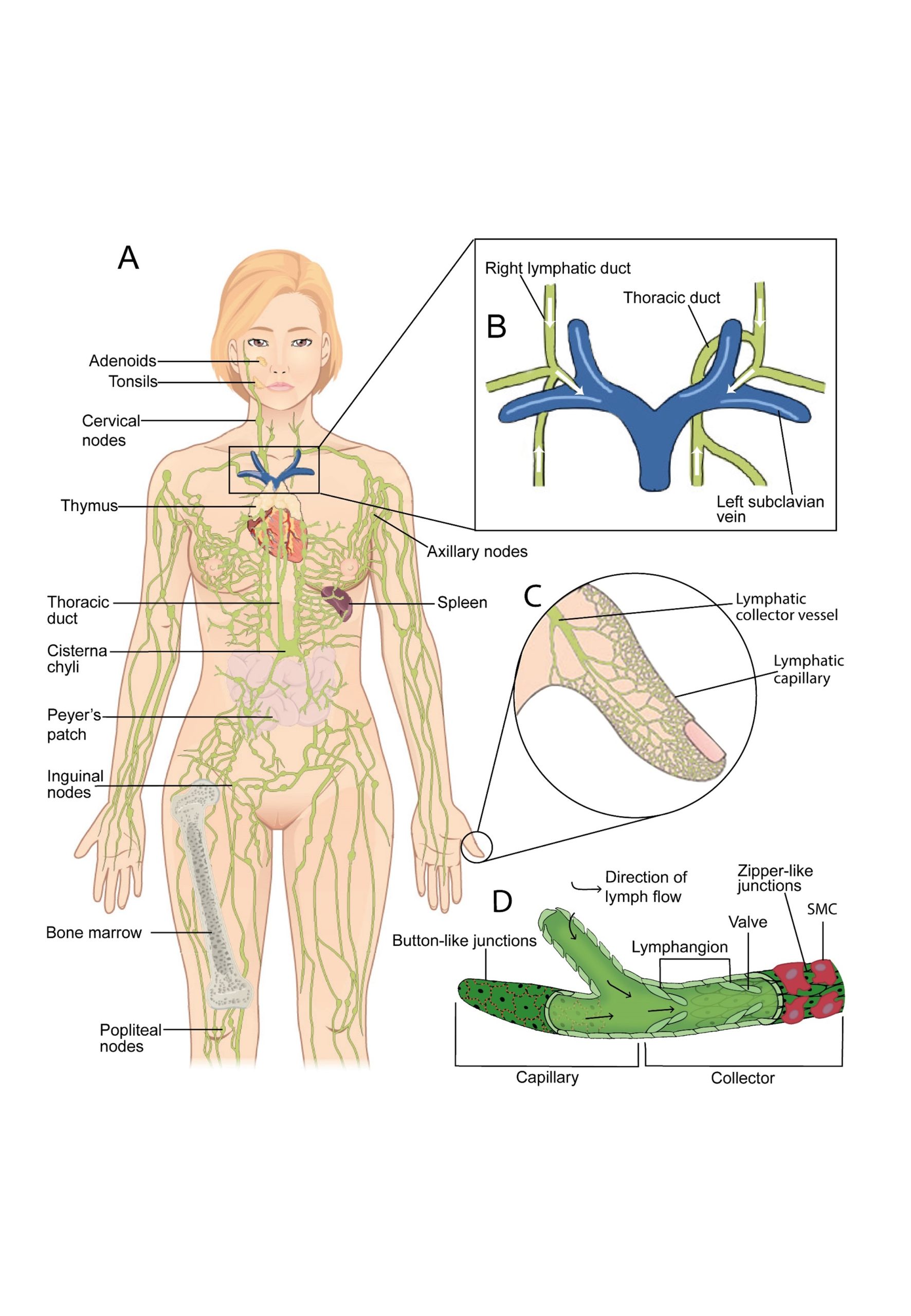
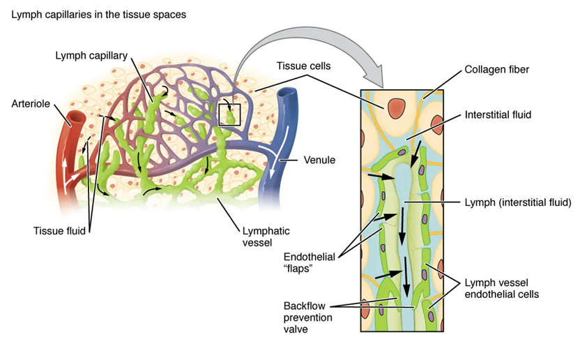
The lymphatic system continuously collects excess interstitial fluid from tissues. Beginning at blind-end lymph capillaries, lymph fluid travels through lymph vessels to lymph nodes, where B and T cells screen for pathogens. Cleansed lymph is then returned to the bloodstream through the right and left lymphatic ducts into the subclavian veins

Lymphatic capillaries are interlaced with the arterioles and venules of the cardiovascular system. Collagen fibers anchor a lymphatic capillary in the tissue (inset). Interstitial fluid slips through spaces between the overlapping endothelial cells that compose the lymphatic capillary.


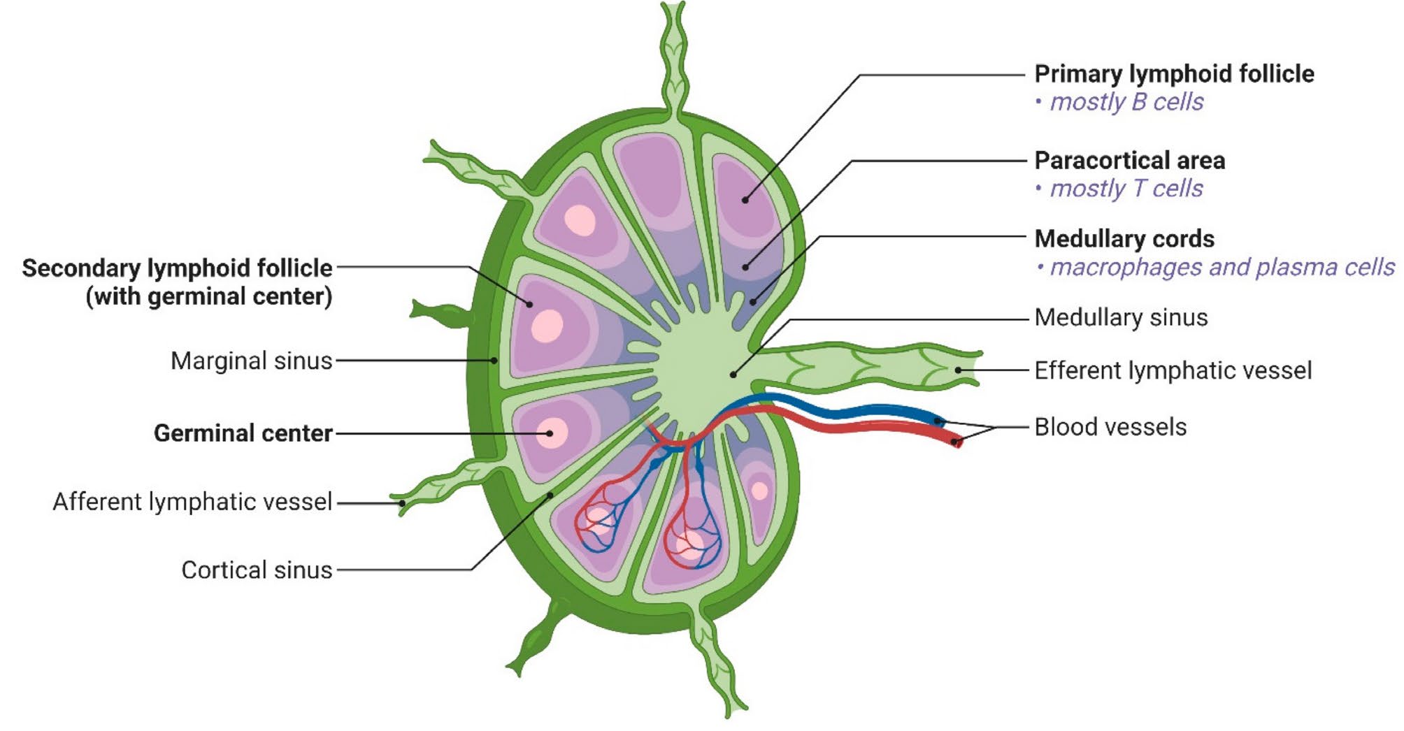
There are approximately 500-600 lymph nodes in the human body, some clustered in the groin, armpits, neck, chest and abdomen.  B cells reside in the cortex of lymph nodes; T cells are positioned in the medulla of lymph nodes.

Lymph Node
Lymph Node

GALT (Gut-Associated Lymphoid Tissue – including Peyer’s patches and the appendix) provides lymphoid screening at the digestive tract’s mucosal lining.  GALT is a division of MALT (Mucosa-Associated Lymphoid Tissue) which is characterized by populations of T cells, B cells, plasma cells, macrophages, and dendritic cells that are dispersed through the mucosal membranes of the respiratory, urogenital, and digestive tracts.  The skin and conjunctivae (of the eyes) are sometimes included when discussing this category of diffuse lymphoid tissue.

Media Attributions

License

Icon for the Creative Commons Attribution-NonCommercial-ShareAlike 4.0 International License

Section 6:  Lymphatic System Overview Copyright © by Zoë Soon is licensed under a Creative Commons Attribution-NonCommercial-ShareAlike 4.0 International License, except where otherwise noted.

Share This Book