Chapter 1 Introduction to Pathophysiology; Cellular Responses to Stress, Injury, and Aging
Section 5 Introduction to Diagnostic Testing
Zoë Soon
The Range of Diagnostic Tests
A wide variety of tests provide clues toward diagnosis, for example:
- Chemistry tests: Assess components of blood, urine, CSF (cerebrospinal fluid) and stool (e.g., lipids, pH, O2 levels, and presence of certain enzymes, normal/abnormal proteins, antibodies, waste products).
- DNA tests: Determine genetic risk factors, diagnose genetic disorders, verify transplant donor-recipient compatibility, and detect presence of specific microbial infections.
- Pressure tests: Measure blood pressure, heart contraction pressure, intracranial pressure, and ocular pressure.
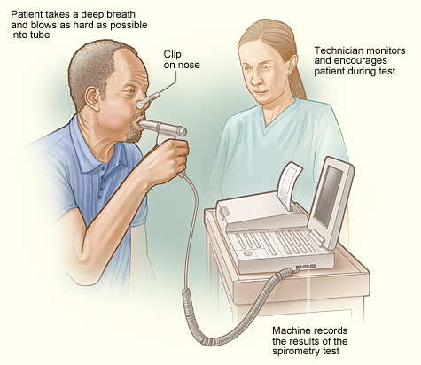
- Volume tests: Measure functional lung volumes (e.g., spirometry)
- Palpation tests: Assess heart and pulse rates.
- Microscopy tests: Analyze cellular features within biopsies, detect and identify pathogens (e.g., throat swabs, fecal analysis), and analyze for other abnormalities (e.g. crystals or protein casts in urine).
- Listening tests (auscultation): Evaluate breath, heart, and bowel sounds.
- Flow cytometry: Count WBCs and RBCs per mL of blood.
- Nerve conduction tests: Assess nerve damage through sensory and motor neuron function tests.
- Imaging:
- X-ray (radio-dense structures like bone),
- CT scans (3D x-ray), MRI (soft tissues),
- Ultrasound (heart valve movement, fetal development),
- ECG, electrocardiogram (cardiac conduction),
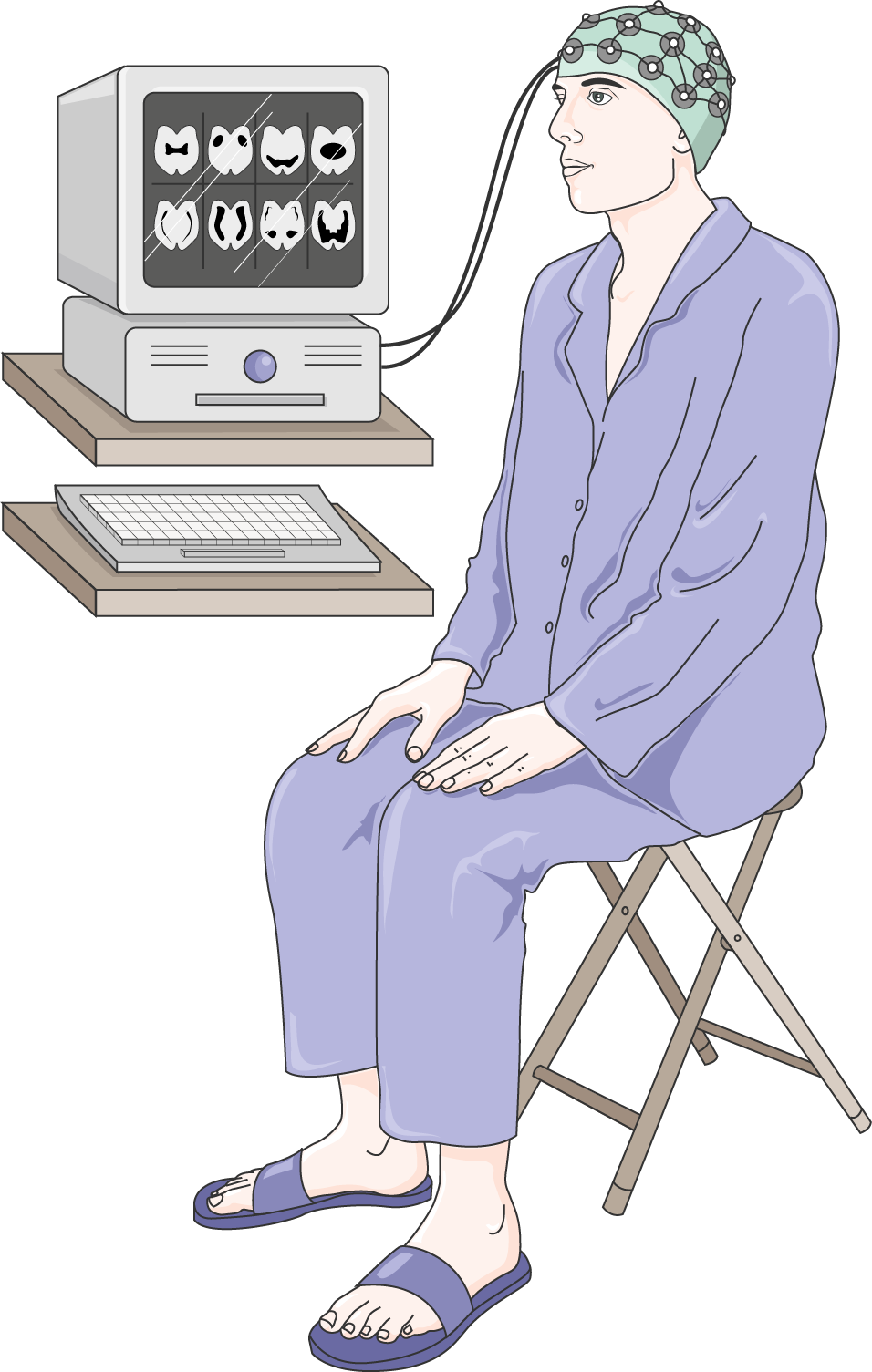
- EEG, electroencephalogram (brain electrical activity), and
- EMG electromyogram (skeletal muscle function, ability of muscle to contract when electrically stimulated).
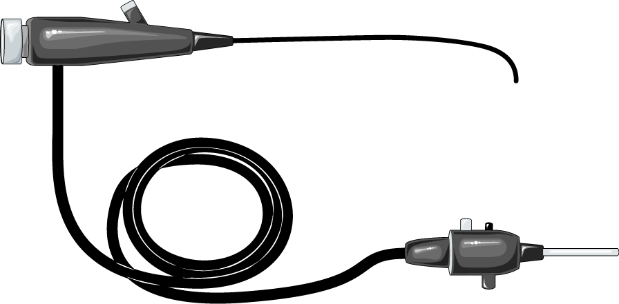
- Endoscopy: A camera on a flexible tube inserted into the body to view suspected areas of damage.
- Biopsy followed by histology: Microscopic analysis of tissue samples for changes in cellular morphology.

Normal Ranges, False Positives, and False Negatives
A bell curve is commonly used to illustrate the normal range of a test result in a given population. Results that fall outside this range may indicate disease and disruption of homeostasis, though it is important to note that abnormal test results do not always indicate disease (and normal results do not guarantee health).
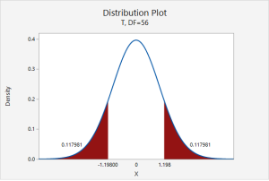
In the above figure, a typical bell curve indicates the range of test results that can be expected in a set population. In this case, resting heart rate data has been graphed for 57 college males. Even though the low and high ends of the graph have been coloured red, all of the resting heart rates in this particular population ( which are from ~60-80bpm) are in the expected normal range. Typically, if a resting heart rate is below 60bpm or above 100bpm, it would be considered abnormal, and indicative of a disruption in homeostasis. If a disease is suspected, and results fall outside of what is considered normal, usually follow-up and complementary tests are performed in order to gain enough information to make an accurate diagnosis.
| True Negative | A healthy person’s test results are normal – correctly confirming the absence of disease. |
| False Positive | A healthy person’s test results are abnormal – incorrectly suggesting disease. |
| True Positive | A sick person’s test results are abnormal – correctly identifying disease. |
| False Negative | A sick person’s test results are normal – failing to detect disease that is present. |
Most often, complimentary and follow-up tests are used together to build confidence in a diagnosis, since no single test is 100% accurate.
| Sensitivity | Can a test accurately detect True Positives? A sensitive test is positive in the presence of disease. A 99% sensitive test correctly identifies 99 out of 100 sick people. |
| Specificity | Can a test accurately detect True Negatives? A 99% specific test correctly identifies 99 out of 100 healthy people. |
The typical diagnostic strategy begins with a highly sensitive test to alert as many people with the disease as possible. A highly specific test then follows up to identify and reassure healthy individuals who received false positives from the first test.
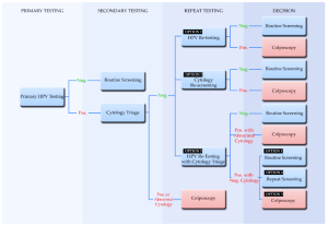
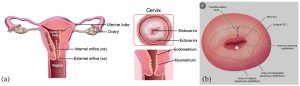
In the above figure, a new added step to the protocol for cervical testing is being developed and implemented in some countries. The newly added step is the first one, in which a Primary HPV (Human Papilloma Virus) test, a highly sensitive test is used that screens for the DNA presence of the HPV strains that are considered high risk for cancer development. The cytology test involves the Pap smear with microscopic analysis of cervical cellular morphology. Traditionally, the cytology (Pap smear) test is used as the initial test, and is also considered highly sensitive with an even greater degree of specificity than the HPV test. The third test in the above figure, the colposcopy test is considered a highly specific test.
Ethics of Diagnostic Tests
The design and use of diagnostic tests raises important ethical questions. Tests are most often developed and used with the following priorities in mind:
- A significant percentage of the population is at risk.
- Test development is feasible.
- The test will be reliable (valid and repeatable).
- The test will be sensitive and/or specific.
- The test will be inexpensive and affordable.
- The test will be easy to store and deliver.
- The test will be user-friendly and not prone to human error.
- Ideally, the test is non-invasive (which also reduces cost and time).
Examples of Non-Invasive Tests
Non-invasive tests include: blood tests, urinalysis, sputum, and stool sample analysis, x-rays (e.g., mammograms), and physical exams.
Media Attributions
- An illustration depicting an incentive spirometer. © BruceBlaus is licensed under a CC BY (Attribution) license
- Private: Equipment_-_EEG_(6199)_–_Smart-Servier © Smart Servier Medical Art is licensed under a CC BY-SA (Attribution ShareAlike) license
- Private: Parasagittal_MRI_of_human_head_in_patient_with_benign_familial_macrocephaly_prior_to_brain_injury_ANIMATED © Dwayne Reed is licensed under a CC BY-SA (Attribution ShareAlike) license
- Private: Equipment_-_Endoscopy_-_Smart-Servier © Laboratoires Servier is licensed under a CC BY-SA (Attribution ShareAlike) license
- Distribution Plot of Heart Rate is licensed under a CC BY-NC (Attribution NonCommercial) license
- Cervical Cancer Screening © Chrysostomou, A.C.; Kostrikis, L.G. is licensed under a CC BY (Attribution) license
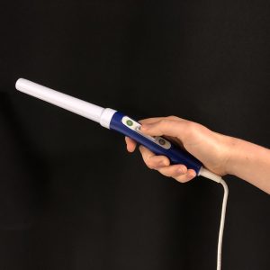
- PocketColposcope © Erica Skerrett is licensed under a CC BY-NC-SA (Attribution NonCommercial ShareAlike) license
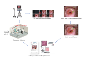
- images_large_10.1177_11769351231161477-fig1 © Dash S, Sethy PK, Behera SK. is licensed under a CC BY-NC-SA (Attribution NonCommercial ShareAlike) license
- colposcopy © Xue, P., Ng, M.T.A. & Qiao, Y. is licensed under a CC BY-SA (Attribution ShareAlike) license

_--_Smart-Servier.png)