Chapter 3 Neoplasia
Cervical Cancer
Pictures coming soon!
Zoë Soon
Cervical Cancer – What is it?
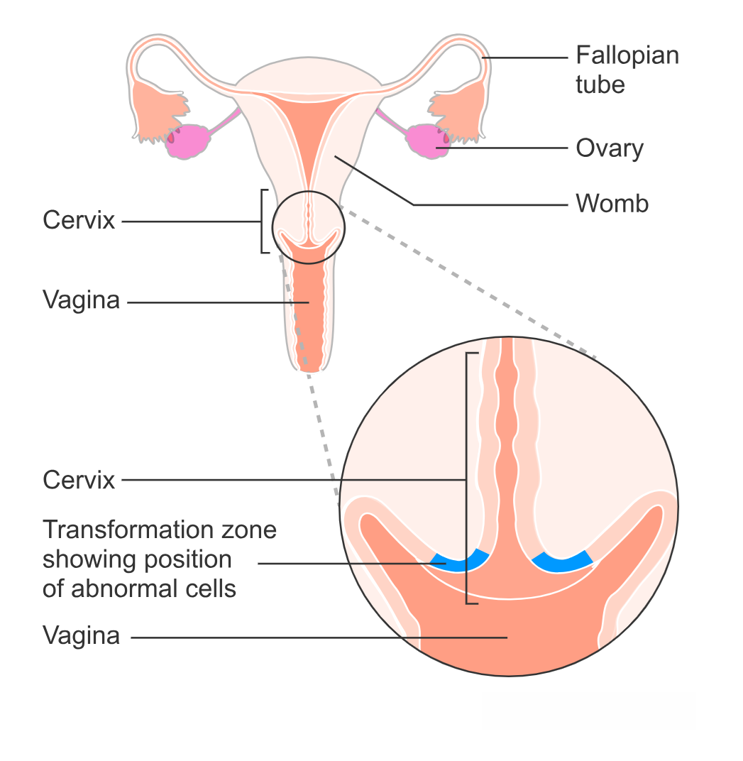
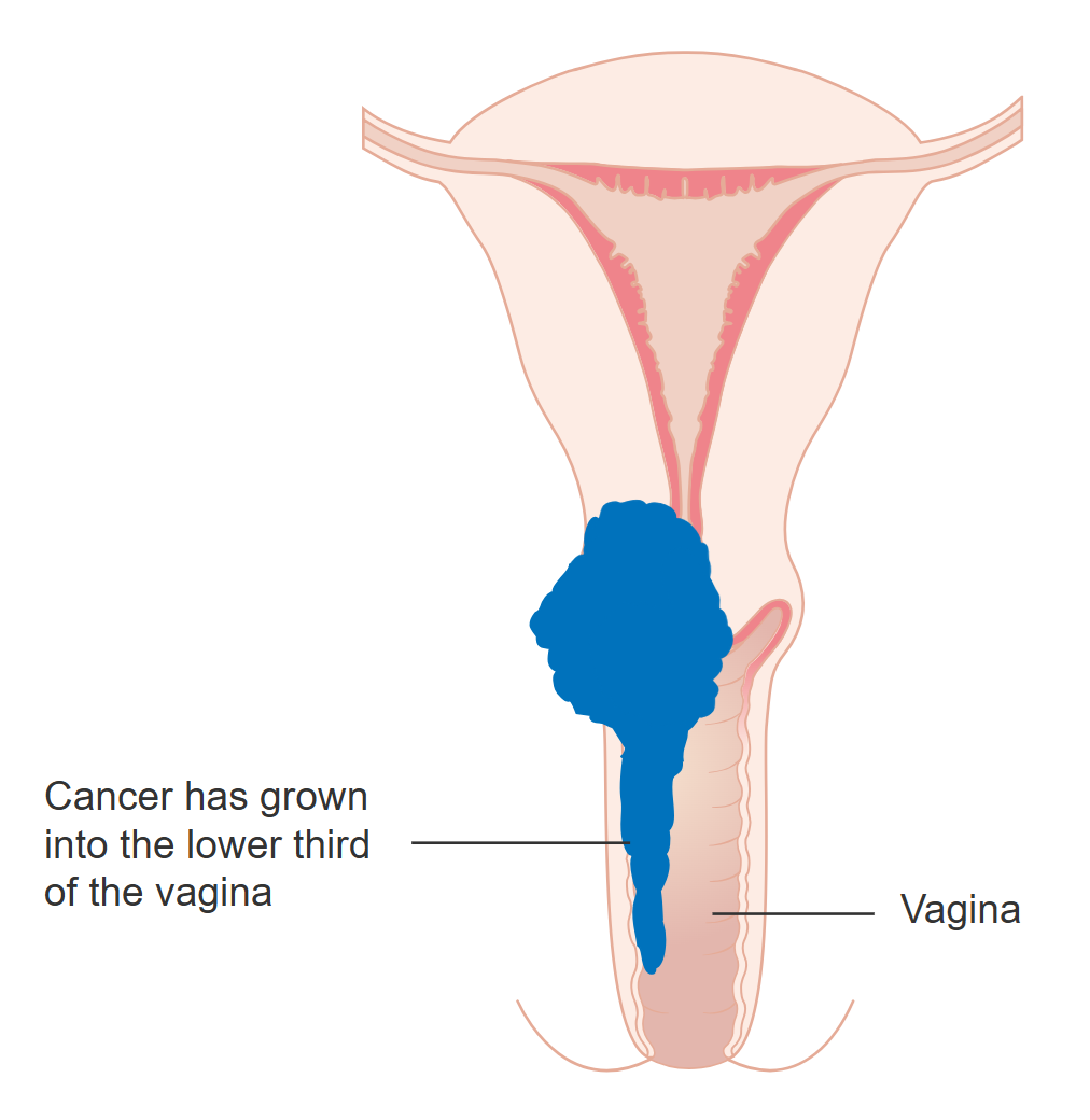
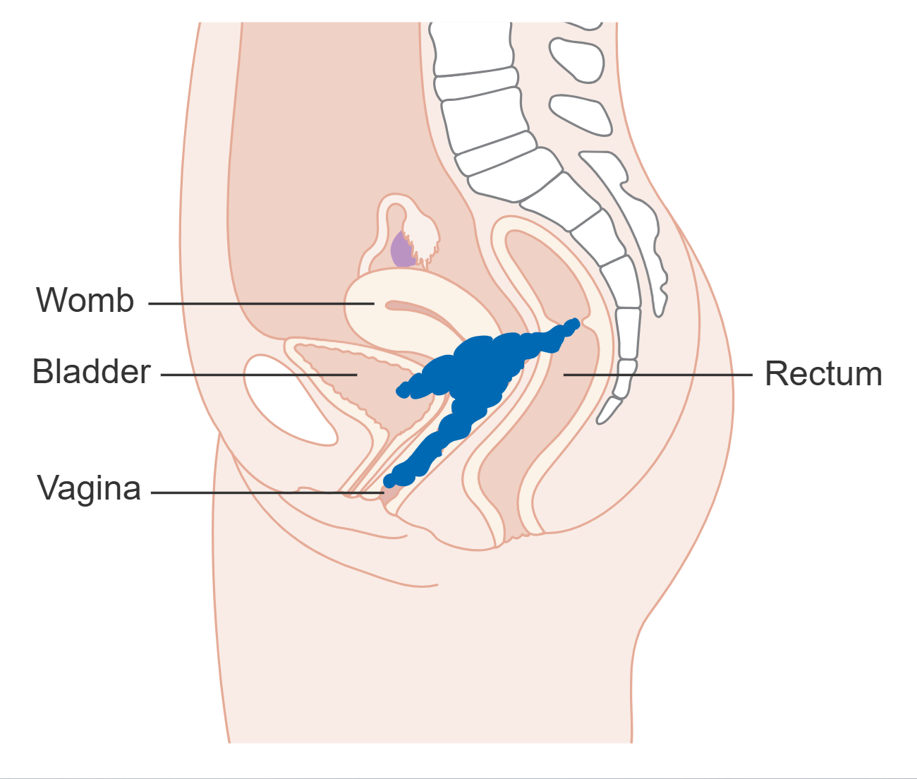
The cervix is located at the base of the uterus which opens into the superior portion of the vagina. The cervix is approximately 3-4 cm long and is composed of 3 layers of mainly connective tissue and muscle that are continuous with the endometrium, myometrium and perimetrium of the uterus. Cervical cancer typically arises when mutations occur within the surface layer of squamous epithelial cells of the cervix.
Cervical Cancer – Risk Factors and Prevention Strategies
Infection with certain strains of the sexually transmitted human papillomavirus (HPV) increase the chance of cervical cancer, as HPV is an oncovirus capable of causing cellular mutations. Age is also a risk factor, though unlike many cancers, more cases occur before the age of 50yrs. Sexual activity is a risk factor as HPV is transmitted sexually. Also, sexual activity at a young age, as damage to the cervix is thought to be a risk factor. Likewise prolonged damage or inflammation from other sexually transmitted diseases (e.g., Chlamydia trachomatis) have been found to be risk factors. As with all cancers, exposure to carcinogens (e.g., smoking) and immunosuppression (e.g., due to HIV infection) are risk factors. Prevention strategies include HPV vaccination and use of barriers (e.g., condoms). In Canada, vaccinations are recommended and available for everyone aged 9-14 years. In addition to reducing the risk of cervical cancer, HPV vaccines have reduced the risk of genital warts and other cancers (i.e., anal and oropharyngeal) which are also caused by HPV.
Cervical Cancer – Signs & Symptoms
Signs and symptoms of cervical cancer include abnormal vaginal bleeding or discharge, pain during sexual intercourse, constipating, pelvic pain, loss of appetite and unexplained weight loss.

Cervical Cancer – Diagnosis
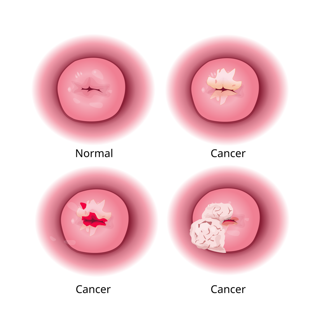
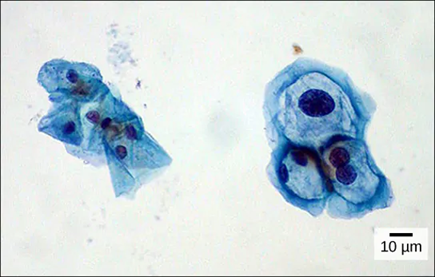
Routine Pap tests are often recommended every 1-3 years and it is likely that this will be the first clinical sign that cervical cancer may be present. During a Pap test, cervical cells are collected and sent to a lab for viewing under the microscope to check for morphological changes that indicate dysplasia or anaplasia. At the same time, typically a swab is tested for the presence of HPV using a HPV PCR test which tests for the presence of HPV genetic material (DNA or mRNA). HPV is a dsDNA virus. The presence of HPV does not indicate cervical cancer, but it can mean, depending on the strain of HPV, that a person has a higher risk of developing cervical cancer. When the Pap test reveals abnormal cells, often a more sensitive test is performed, in which colposcopy is used to closely examine the cells of the cervix. During this process, a colposcope is used and biopsies of areas of concern will often be taken for further analysis.
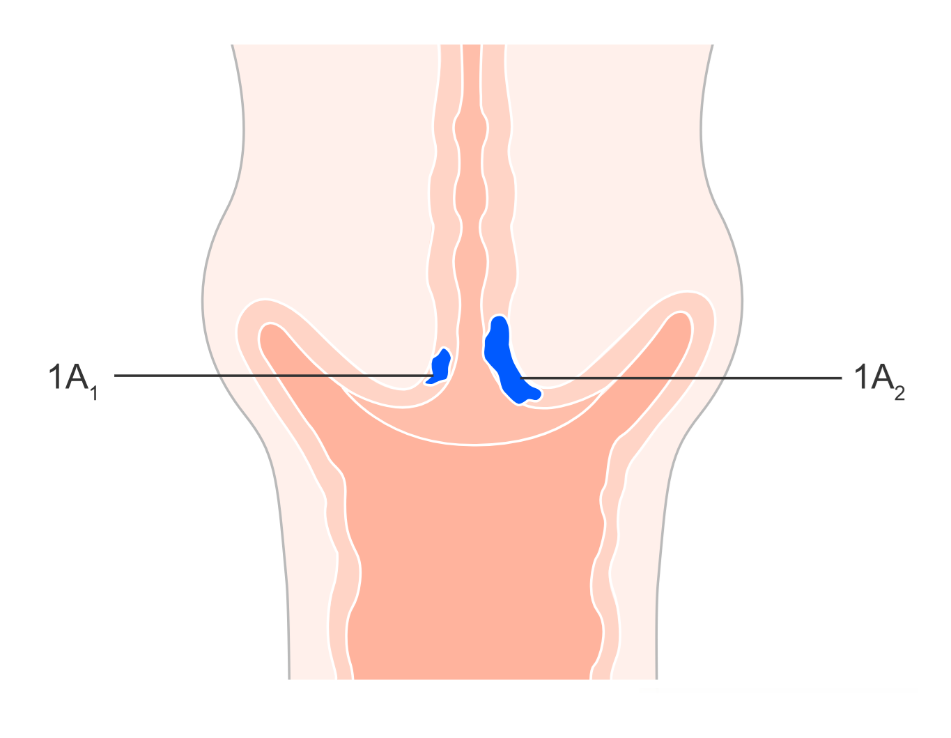
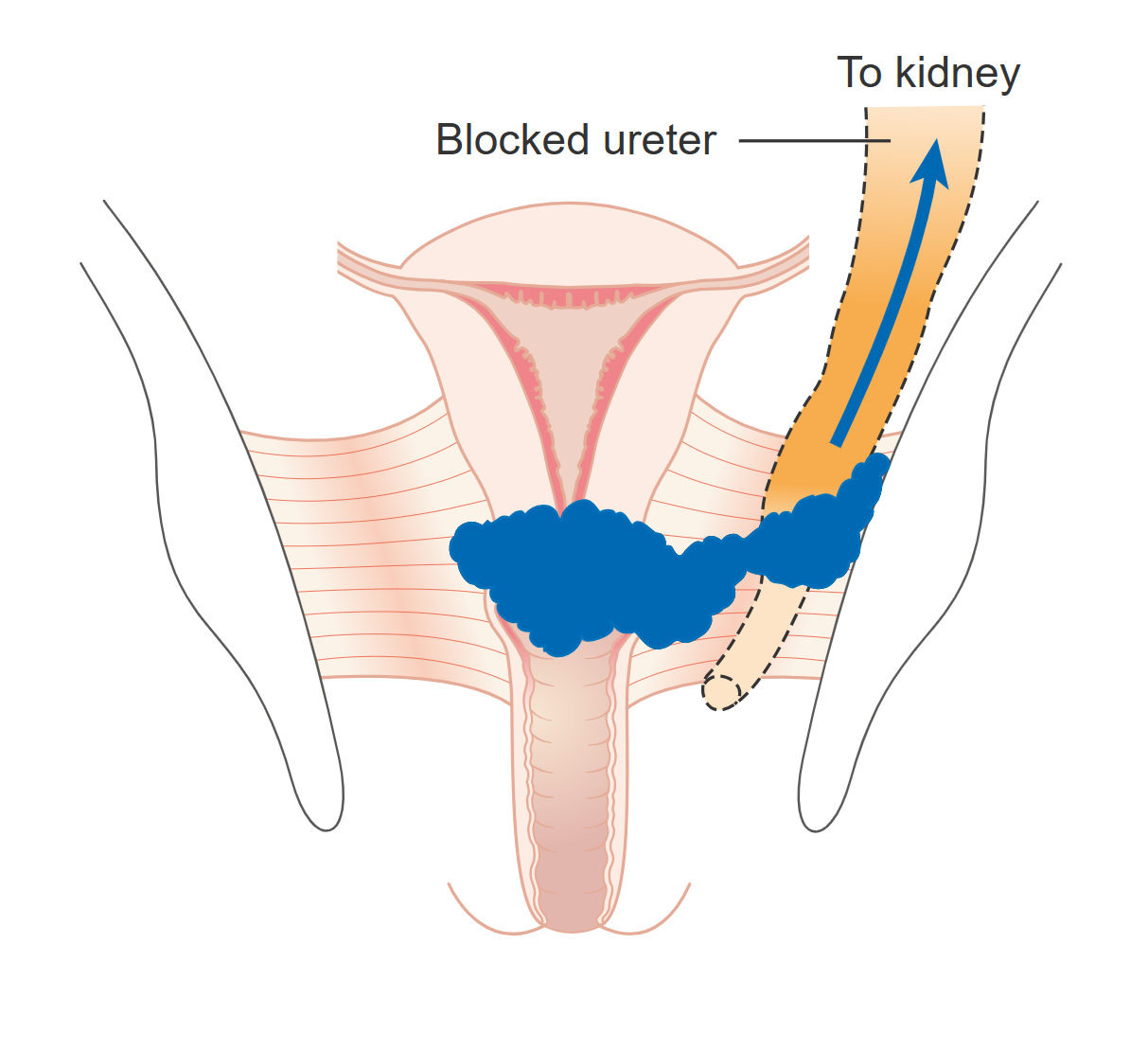
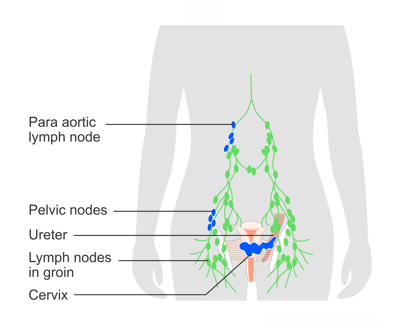
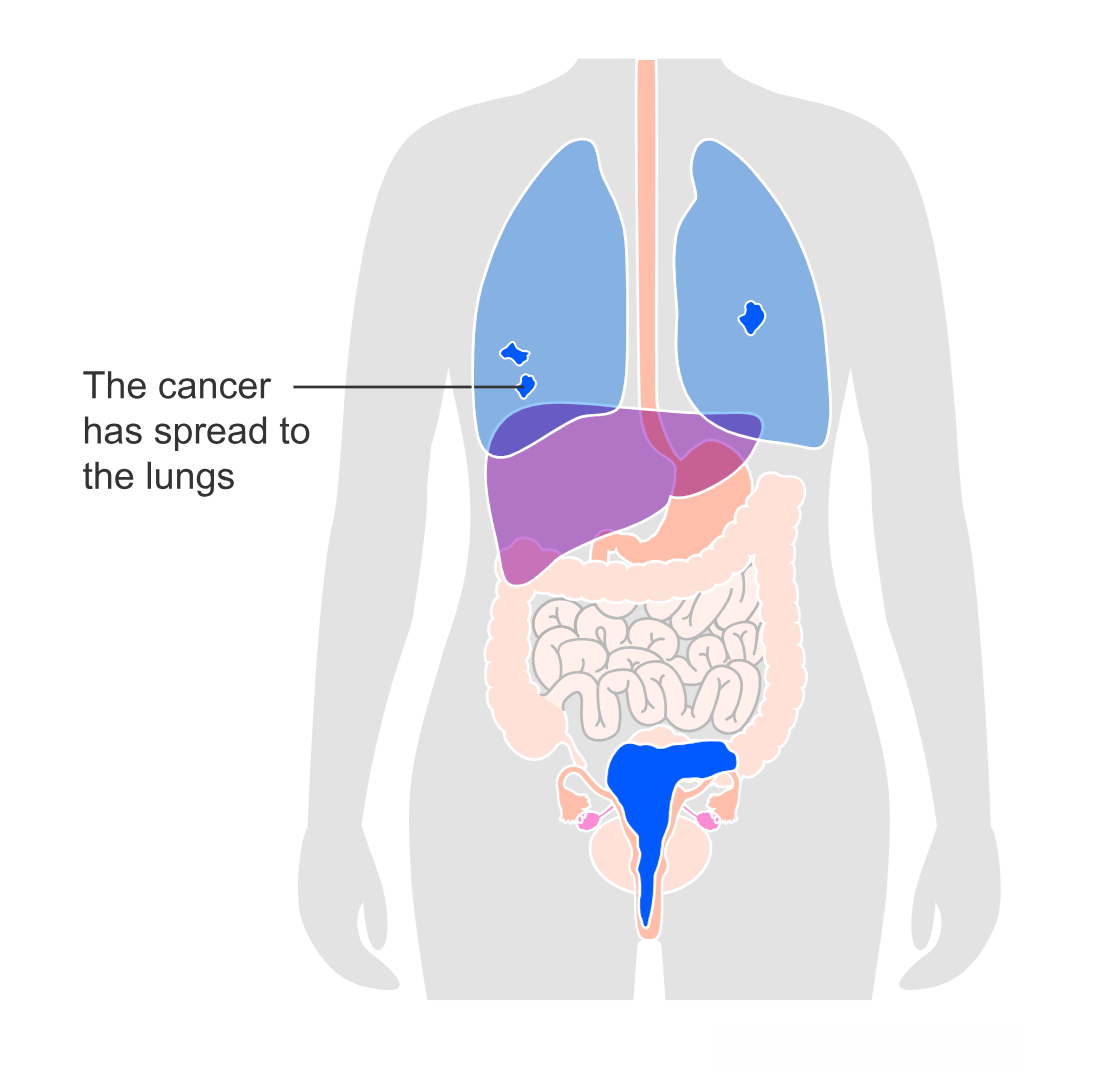
If cancer has developed within the cervix, often sentinel lymph nodes are biopsied to determine the extent to which the cancer may have spread. Further imaging (e.g., CT scan, MRI, PET scan) may also be used. Biopsy and microscopic analysis of cells is the only definitive way to diagnose cervical cancer. Fortunately, cervical cancer often grows slowly and is usually treated early with a high success rate.
Cervical Cancer – Treatment
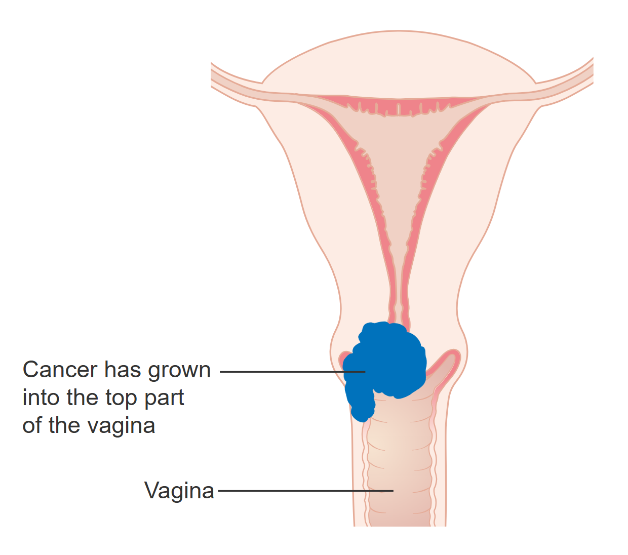
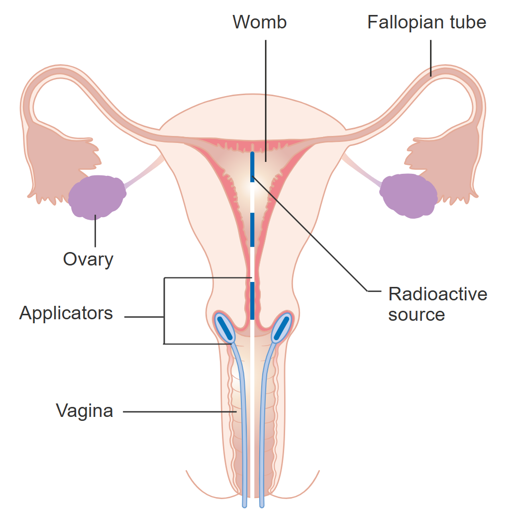
Treatments depend on the stage of cancer, but can include surgery, chemotherapy, radiation therapy, and immunotherapy.
Think about;
With regular Pap tests, cervical cancer is more likely than many other cancers to be detected as a carcinoma in situ. What is a carcinoma in situ?
Media Attributions
- Cervical Cell Dysplasia © Cancer Research UK is licensed under a CC BY-SA (Attribution ShareAlike) license
- Cervical Cancer © Hariadhi is licensed under a CC BY-SA (Attribution ShareAlike) license
- Stage 1A Cervical Cancer © Cancer Research UK is licensed under a CC BY-SA (Attribution ShareAlike) license
- Stage 1B Cervical Cancer © Cancer Research UK is licensed under a CC BY-SA (Attribution ShareAlike) license
- Stage 2A Cervical Cancer © Cancer Research UK is licensed under a CC BY-SA (Attribution ShareAlike) license
- Stage 3A Cervical Cancer © Cancer Research UK is licensed under a CC BY-SA (Attribution ShareAlike) license
- Stage 3B Cervical Cancer © Cancer Research UK is licensed under a CC BY-SA (Attribution ShareAlike) license
- Stage 3C2 Cervical Cancer © Cancer Research UK is licensed under a CC BY-SA (Attribution ShareAlike) license
- Stage 4A Cervical Cancer © Cancer Research UK is licensed under a CC BY-SA (Attribution ShareAlike) license
- Stage 4B Cervical Cancer © Cancer Research UK is licensed under a CC BY-SA (Attribution ShareAlike) license
- Cervical Cells: Normal and HPV-infected Cervical Cells: © Mary Ann Clark, Matthew Douglas, Jung Choi is licensed under a CC BY-NC-SA (Attribution NonCommercial ShareAlike) license
- Diagram showing the position of the applicators for internal radiotherapy for cervical cancer © Cancer Research UK is licensed under a CC BY-SA (Attribution ShareAlike) license
