Chapter 4 Selected Musculoskeletal Disease and Disorders, including Trauma and Rheumatic Disorders
Bone Fracture – Stages of Healing
Pictures coming soon!
Zoë Soon
Bone Fracture – Five Stages of Healing
Bones are living material and can usually heal very well.
There are five stages of fracture healing:
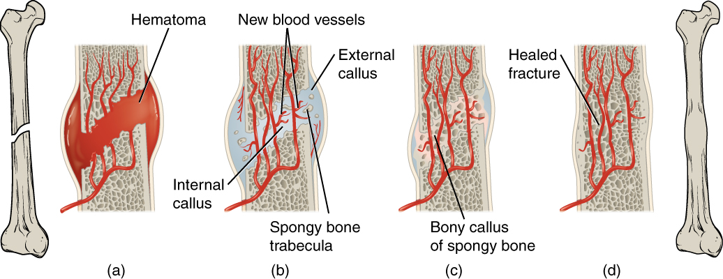
1. Hematoma formation and inflammation stage: In this stage, broken blood vessels bleed and if the fracture is closed, a hematoma (swelling of clotted blood) will form. With open fractures, external bleeding will occur. The amount of bleeding and the size of any hematomas being developed will depend on the extent of the damage to the bone and surrounding muscle and connective tissues. Vasospasm of the ends of broken blood vessels and other processes involved in hemostasis (platelet plug formation) will occur to limit bleeding. During this time, the disrupted cells and any debris will also stimulate an intense inflammatory response. The inflammatory response is characterized by vasodilation, increased capillary permeability, exudation of plasma and WBCs and infiltration of neutrophils, macrophages, basophils, and mast cells. This stage typically lasts 1-3 days.
2. Platelet and fibrous stage (organization of hematoma): In this stage, fibrin mesh and platelets have stopped the bleeding and the fibrous network has added stability to the area. The hematoma and debris are phagocytosed by WBCs (neutrophils and macro-phages). As debris is phagocytosed and as damage-signaling chemicals and cytokines are degraded and dissipated, nociceptor triggering (and pain) will diminish. The clots are retracting and granulation tissue forms characterized by neovascularization (capillary buds developing to create new blood vessels within the damaged zone). This stage lasts approximately 2 weeks.
3. Procallus formation stage (cartilage formation): This stage involves the proliferation mesenchymal stem cells which give rise to daughter chondroblasts. The chondroblasts (cartilage cells) form a procallus (fibrocartilage) which consists a gel-like extracellular matrix. The procallus (“soft” callus) bridges the gap between the broken bone ends uniting the internal and external edges of the bone. In a fractured long bone, an internal callus organized within the medullary cavity. An external procallus (enlarged collar of cartilage) forms creating a bulge around the bone encircling the fracture site. Fibroblasts contribute by secreting collagen, elastic fibers, and glyoproteins. This stage lasts approximately 2 weeks and can overlap with the preceding and subsequent stages.
*Extracellular gel of fibrocartilage contains glycosaminoglycan (GAG), proteoglycans and glycoproteins.
4. Bony callus formation stage (ossification of cartilage): Osteoprogenitor cells located in the periosteum and endosteum proliferate to produced osteoblasts. The osteoblasts invade the fibrocartilage of the procallus and produce osteoid proteins and secrete calcium (calcium hydroxyapatite) to begin ossifying the cartilage matrix of the procallus converting it into a bony callus (“hard” callus). This process relies on neo-vascularization. The newly formed bone is termed woven bone as the collagen fibers that are laid down are initially disorganized and mechanically weak. Woven bone is created when osteoblasts are producing osteoid quickly. The enlarged, mature chondrocytes that created the procallus are eventually completely replaced by bone cells. As the osteoblasts surround themselves with osteoid, they mature to become osteocytes (mature bone cells).
5. Remodeling of bone into compact and cancellous bone: The woven bone is remodeled by the slow process of creating mature lamellar (concentric/compact bone) and spongy (trabecular/cancellous bone) to replace the bone structure that was lost. The compact and spongy bone created contain organized collagen fibers and trabeculae that are able to resist more force and more forces from different directions. In long bones, compact bone is found in the diaphysis encircling a medullary cavity that is full of marrow. Compact bone is found making up the outer layer of all five other bone types (i.e., flat bones, short bones, sesamoid bones, irregular bones and sutural bones) as well. Spongy bone is found in the epiphyses of long bones and make up the interior of all 5 other bone types. As the bone is remodeled, the bulge of bony callus surrounding the bone eventually disappears, which can be observed by imaging (e.g., x-rays), which also will reveal healing of the bone (bone crossing fracture site). Remodeling requires the activities of both osteoblasts and osteocytes (to build bone) and osteoclasts (to dissolve bone). During the remodeling process, the periosteum and endosteum are also regenerated. The speed of remodeling depends on the person’s age as well as underlying factors that affect healing and health. Factors that slow healing include: old age, smoking, malnutrition, glucocorticoid use, anemia, poor blood supply, obesity, bone fracture complications (e.g., infection, extensive damage to surrounding tissues, and/or greater bone loss). In healthy adults, with simple fractures, remodeling continues for 6 months to 1 year. Children’s fracture can heal even faster, while the elderly often exhibit slower healing times. Most fractures are protected (by casts etc.) for at least 6 months until healing is sufficient to allow for weight-bearing activities.
*Side Note:
Periosteum = connective tissue that wraps around the outside of all bones. Periosteum is composed of an outer fibrous layer (consisting of a large amount of fibrous collagen protein) and an inner cellular layer (which contains fibroblast cells, osteoprogenitor cells, and osteoclasts). In general, the fibroblast cells produce extracellular collagen and maintain the outer layer and the osteoprogenitor cells (also known as osteogenic cells) and osteoclasts serve to maintain bone, repairing micro and macro fractures when necessary.
Endosteum = has the same composition as periosteum and lines the medullary cavities of long bones.
