Chapter 2 The Inflammatory Response, Fever, Healing, Cell Proliferation, Tissue Regeneration and Repair
Hemostasis (Blood Clotting)
More pictures coming soon!
Zoë Soon
Hemostasis Overview:
When a blood vessel wall is cut, hemostasis is initiated in order to create a natural band-aid that stops the bleeding and loss of blood. Hemostasis is a fast process taking minutes to occur. The word hemostasis originates from the Latin and Ancient Greek words, hemo- referring to blood, and –stasis meaning motionless. There are three major stages of hemostasis:
- Vascular Spasm: When damaged, blood vessel wall endothelial cells release a variety of chemical messengers (e.g. ADP, Tissue Factor, and endothelins). Endothelin peptides are potent vasoconstrictors, triggering the smooth muscle layer of the blood vessel to contract, narrowing the blood vessel. This serves to minimize blood loss.
- Platelet Plug Formation: In this next stage, platelets are attracted to the wounded site, becoming activated and adhering to the exposed collagen of the damaged vessel, forming an unstable plug within seconds to minutes. Prior to becoming activated, 30% of platelets are typically located in the spleen and 70% of platelets are circulating the blood stream. When inactive, the platelets have smooth unsticky surfaces. Once activated, platelets become more spikey and sticky capable to adhering to the cut edges of blood vessels and releasing from granules their own chemical messengers which include: ADP, thromboxane A2, serotonin, clotting factor proteins, Ca++, prostaglandins, platelet-activating cytokines, and Platelet Derived Growth Factor (PDGF). ADP attracts more platelets to the area. Serotonin is a vasoconstrictor. Thromboxane A2 stimulates more vasoconstriction and also recruits and activates more platelets. This recruitment and activation of more platelets is termed a positive feedback loop, which will end when the wound is sealed.
- Coagulation: In this step, the plasma protein fibrinogen (water-soluble protein) is converted to fibrin (long ropy water-insoluble) proteins which wrap around and through, stabilizing the platelet plug. At this stage, clot retraction occurs, as platelets contract pulling the torn edges of the blood vessel closer together.
During the Coagulation stage to occur, there are two pathways that simulataneously occur, the Extrinsic and Intrinsic Pathways.
- Extrinsic Pathway: In this pathway, extrinsic factors (e.g. Tissue Factor) are released from blood vessel wall endothelial cells that in a cascade of activation involving calcium and clotting factors, serve to activate Prothrombin Activator (Factor X). Prothrombin Activator converts the inactive prothrombin into the activated thrombin enzyme. Thrombin is then able to convert fibrinogen to fibrin, which is required for stabilizing the platelet plug.
- Intrinsic Pathway: This pathway’s purpose is to also activate Prothrombin Activator in order to produce fibrin for stabilizing the platelet plug. The intrinsic pathway, begins when platelets degranulate releasing intrinsic factors (e.g. Platelet Factor) that begin a cascade of activation involving calcium and clotting factors. These clotting factors again serve to activate Prothrombin Activator (Factor X). This process is slower than the extrinsic pathway though is necessary.
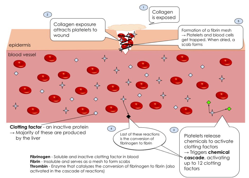
A basic diagram of the series of events that occur after a wound, starting with the attraction of platelets to the wound site, the cascade reactions, activation of clotting factors, and ending with the conversion of fibrinogen into fibrin and scab formation. (Note: Not all 13 clotting factors are explicitly pointed out; blue clotting factors are inactive, green are active, and black is the activated fibrinogen)
Fibrinolysis: Once healing has begun, the fibrin and platelet plug are removed as new blood vessel wall cells are regenerated. The digestion of fibrin is termed fibrinolysis. During fibrinolysis, tPA (Tissue-Type Plasminogen Activator) converts the inactive plasminogen to the active plasmin protease enzyme, which degrades fibrin. At the same time, macrophages are removing cellular debris.
Healing: Platelets release PDGF to stimulate vessel regeneration. In addition fibroblasts release mitogens and cell division allows for the replacement of damaged cells. Large wounds may incorporate more collagen and fewer functional cells giving rise to scar tissue.
Hemostasis and Platelet Regulation:
Endothelial cells release prostacyclin, a signalling molecule that acts as both a vasodilator and platelet-inihibitor, to ensure platelet aggregation is not excessive. Too many platelets could lead to large thrombi formations which may result in platelet-RBC-fibrin clots that break off and travel, becoming emboli, that lodge downstream in smaller blood vessels creating sites of ischemia and hypoxia.
Too few platelets can also lead to problems. Thrombocytopenia is a condition in which there are low levels of platelets, possibly due to nutrition deficiency, an underlying illness, or medicine-induced. Most common signs and symptoms involve prolonged or excessive bleeding (e.g. frequent nosebleeds, heavier menstruation, bruising, petechiae (tiny red spots caused by capillary hemorrhages).
Hemophilia is defined as an impaired ability to make blood clots, typically due to inherited mutations in one or more clotting factors involved in the intrinsic or extrinsic pathways. The most common cause of hemophilia is due to recessive genetic disorders involving mutations in either Factor VIII or Factor IX genes, both of which are on the X chromsome, meaning that more XY males are affected by hemophilia than XX females. Signs and symptoms of untreated hemophilia can involve severe and frequent bleeds internally in soft tissues and joints, even without trauma, in addition due to external cuts.
Summary
- Describe 3 stages of hemostasis: vascular spasm (role of endothelin and tunica media), platelet plug formation (extrinsic and intrinsic pathways, roles of prothrombin activator, Factor X, thrombin, clotting factors, and Ca++) and coagulation (role of fibrin)
- Discuss fibrinolysis, plasmin, prostacyclin
- Explain problems that may occur with too much platelet activity or too little platelet activity.
- Did you know that surgeons may use a topical agent containing collagen to attract a patient’s own platelets to stimulate natural hemostasis?
- Did you know that applying direct pressure to a wound slows down blood loss?
- Did you know that sutures help to close a wound will result in a quicker recovery period?
- Did you know that vitamin K, fat-soluble vitamin, found in green vegetables, grains and organ meats is required for the production of clotting factors? Vitamin K is also produced by intestinal bacteria.
- Did you know that prostacyclin, is a member of the prostaglandin family of eicosanoids (signalling lipid molecules)?
Media Attributions
- Blood Clot Formation © By Victdomi - Own work, CC BY-SA 3.0, https://commons.wikimedia.org/w/index.php?curid=118340457 is licensed under a CC BY-SA (Attribution ShareAlike) license
