Chapter 4 Selected Musculoskeletal Disease and Disorders, including Trauma and Rheumatic Disorders
Chapter 4 Musculoskeletal Systems Diseases and Disorders – Sophia
Zoë Soon
Creative Commons – Simple Pictures, Images, Video Clips, and/or Gifs that help illustrate any of the following:
*For diseases we discuss:
a) Basic Risk Factors
b) Most Common signs and symptoms
c) Basic Pathology, with basic diagnostic tools (e.g. imaging, blood tests) and basic treatment
- Musculoskeletal Trauma:
- Contusions
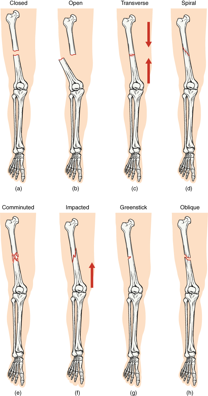
- Fractures
- Types of Fractures – oblique, simple, comminuted, open, pathologic, segmented, spiral, transverse, greenstick, impacted, Colles fracture, Pott’s fracture, compression fracture of vertebra, avulsion, stress (fatigue or insufficiency),
-
Types of Fractures - Risk factors for Fractures: occuption, lifestyle (certain sports e.g. mountain biking, snowboarding, skiing, horseback riding, impact sports), falls, workplace, automobiles, osteoporosis, bone cancer,
- 5 Stages of Fracture Healing
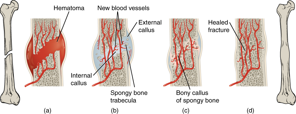
- Hematoma Formation
- Organization of Hematoma
- Procallus (Cartilage Callus) formation
- Bony Callus formation
- Remodelling (from woven/immature to lamellar/mature bone which contains both cortical/compact and cancellous/spongy/trabecular bone)
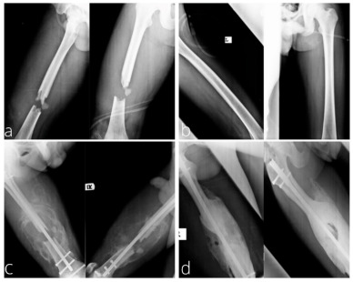
- Treatments: reduction/realignment of fracture ends, casts, pins, wires, plates, sarcoplasty, bone grafts, intramedullary nail, electrical stimulation, ultrasound stimulation
-
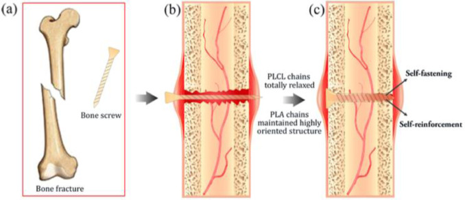
Realignment of fracture ends Intramedullary Pin
-
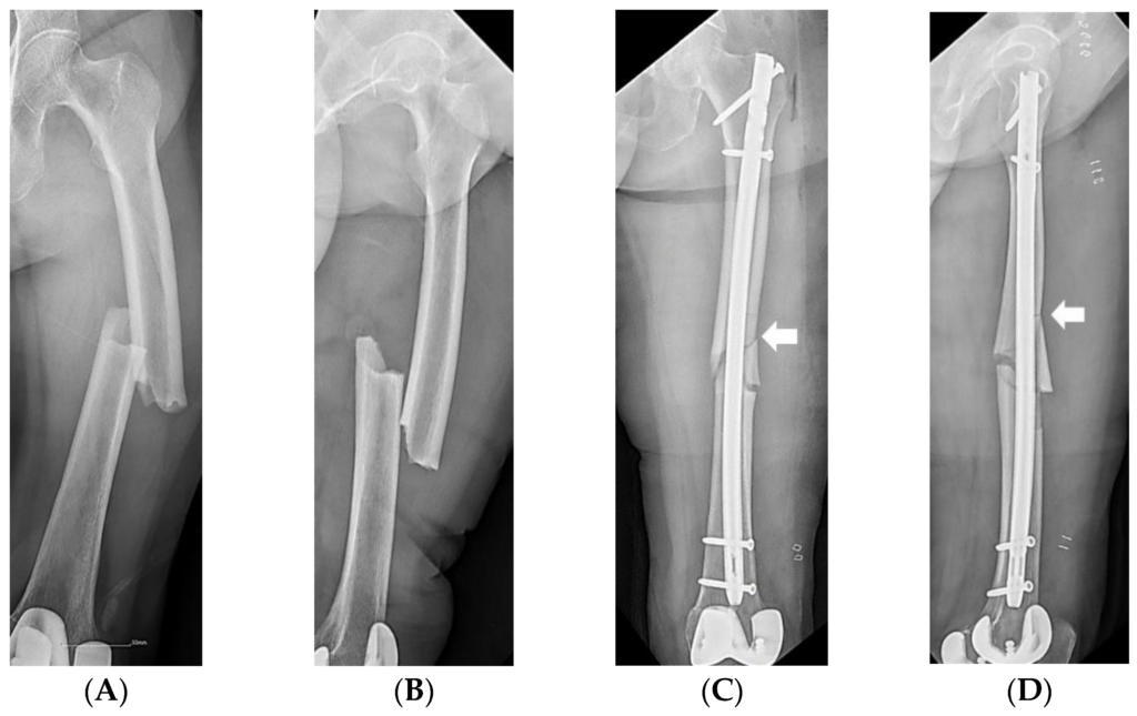
Intramedullary Pin - Possible Complications affecting Bone Fracture Healing
- Osteonecrosis
- Osteomyelitis
-
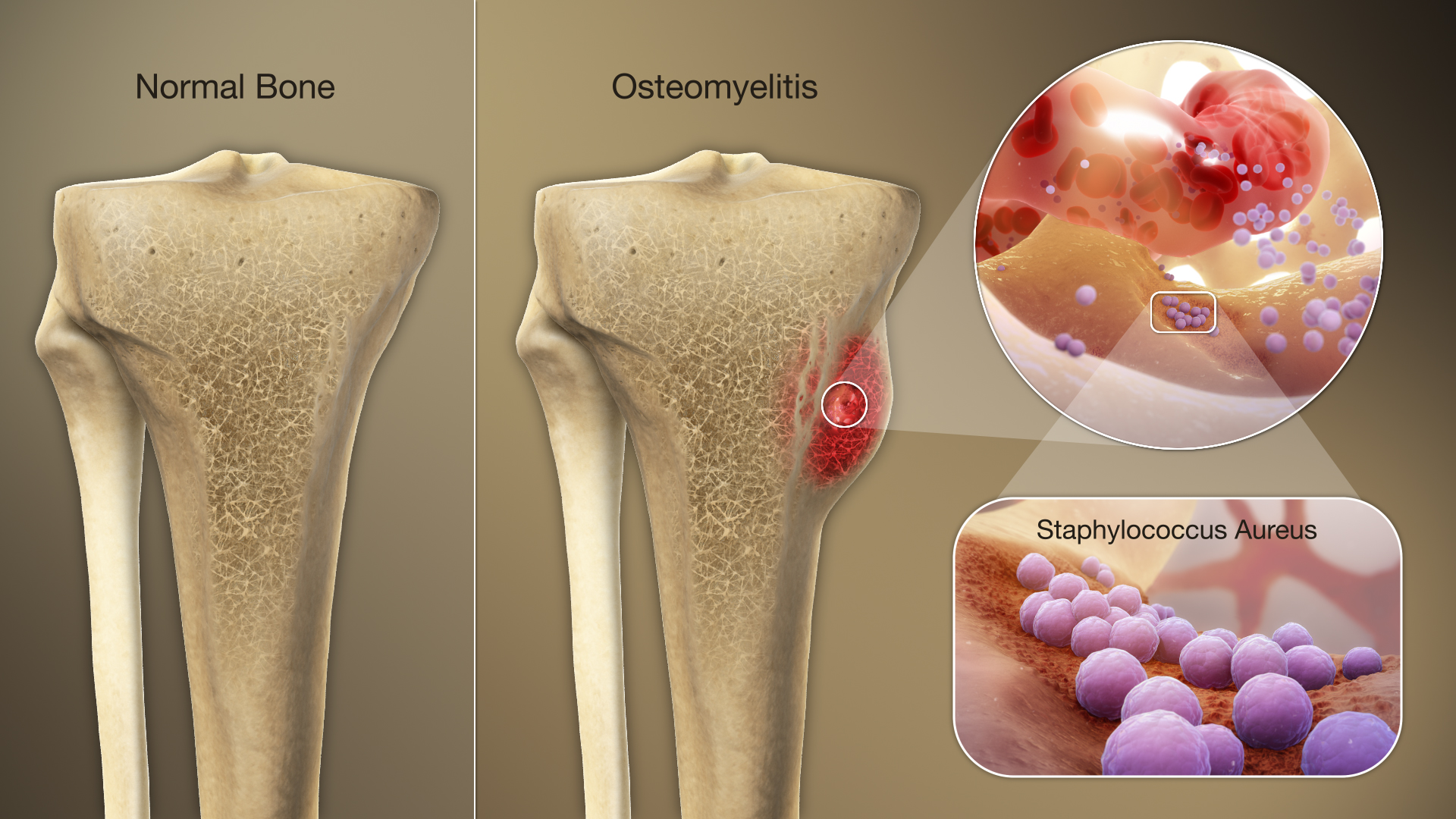
Osteomyelitis - Muscle spasms
- Ischemia,
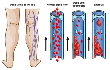
- Thrombi, emboli (fat or blood)
-
Thrombi and Emboli - Fracture blisters
-
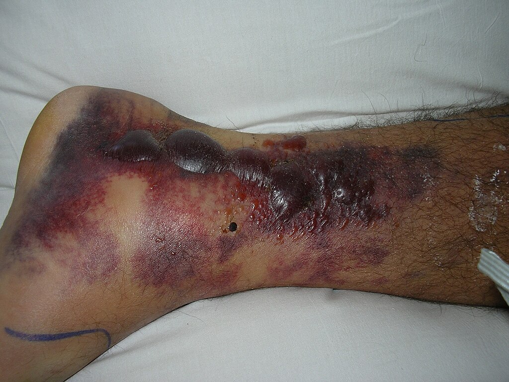
Fracture Blisters - Non-union
- Mal-union
-
Mal-union - Delayed union
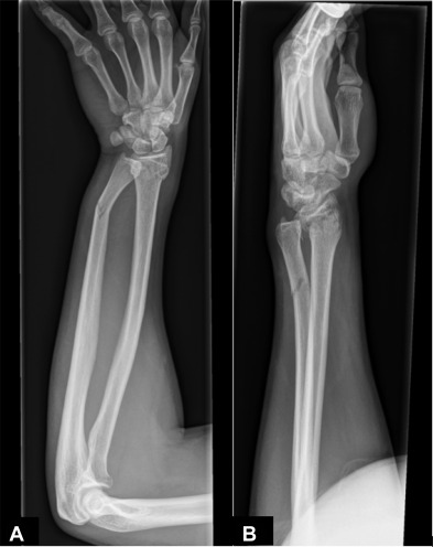
- Stunted growth (due to fracture in epiphyseal growth plate leading to premature ossification of growth plate).
-
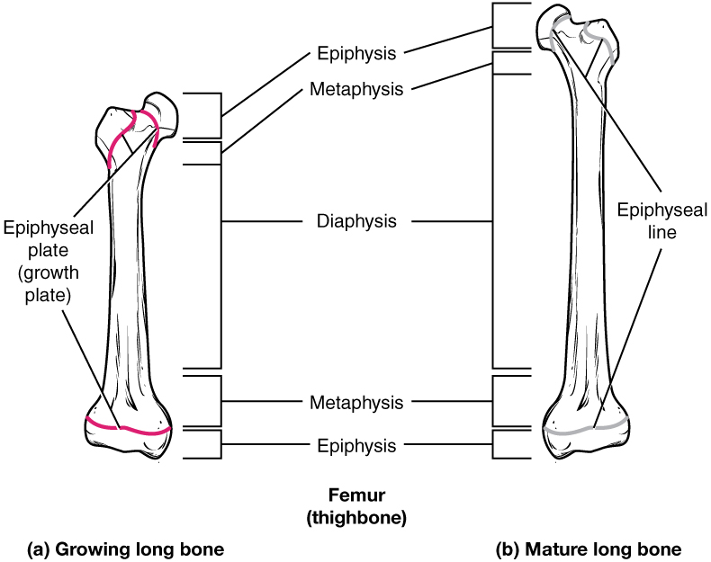
Epiphyseal plate 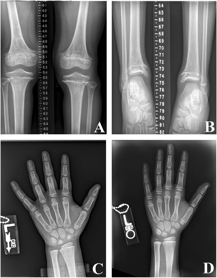
Premature Epiphyseal Closure (B) - Exuberant callus formation – typically temporary – will be remodelled to become a more typical bone shape
-
Exuberant callus formation - Osteoarthritis due to damage affecting ends of long bones – within joint space
-
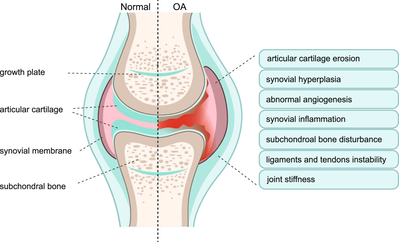
Osteoarthritis - Transchondral fractures leading to separation of articular cartilage and clicking/crepitus within join – may be limit range of motion in joint and be painful upon movement
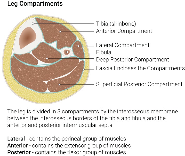
- Compartment Syndrome – damage to tissue inside muscular comparment leads to inflammation and swelling, causes increases in compartment pressure, and local tamponade (as capillaries are pinched off), ischemia and hypoxia occurs causing muscle and capillary necrosis – causing more inflammation and more edema, compartment tamponade causes increased muscle and nerve ischemia leading to nerve dysfunction and muscle infarction (death due to lack of O2), nerves and other affected tissues within the compartment can also die.
- Acute Compartment Syndrome – symptoms include 5 Ps (severe pain, paralysis, paresthesia, pallor, and pulselessness)
- Treatment – fasciotomy
-
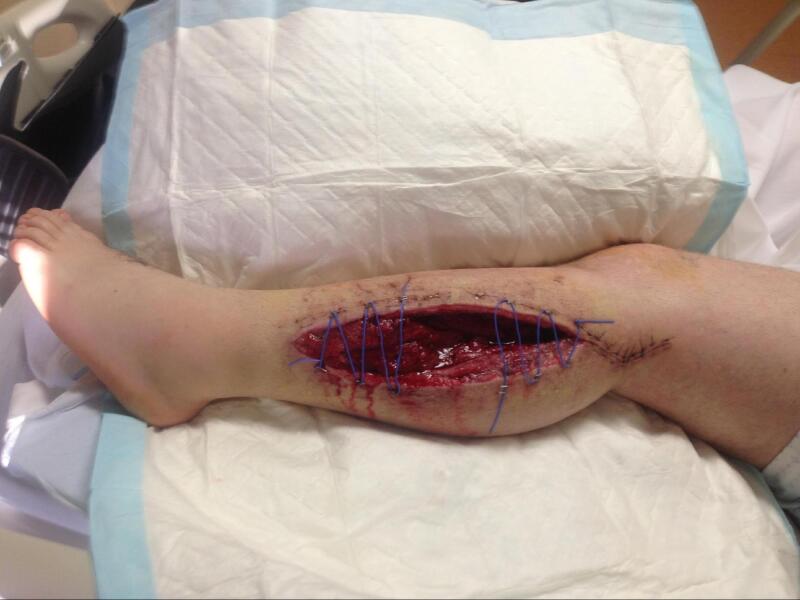
Acute Compartment Syndrome - Possible Complications: Can be fatal as:
- Death and rupture of skeletal muscle cells leads to myoglobin entering the blood stream =Myogloninemia which leads to Rhabdomyolsis, myogloinuria and renal failure. Ruptured skeletal muscle cells also release potassium causing hyperkalemia; lactic acid contributes to acidosis – electrolye and pH imbalance caus cardiac dysrhythmias (renal failure also contributes to acidosis)
- ECF shift (excessive inflammation leads to increased capillary permeability and shift of fluid from inside blood vessel to interstitial spaces, causing hypovolemia and increased compartment pressure
- Hypovolemia causes shock = not enough blood to tissues; signs of shock include: pallor, diaphoresis, hypotension, reflex tachycardia, nausea/vomiting
- Chronic Compartment Syndrome – from repeated training (e.g. gymnastics)
- Acute Compartment Syndrome – symptoms include 5 Ps (severe pain, paralysis, paresthesia, pallor, and pulselessness)
- Muscular/tendon damage – leading to scarring and shortening of tendon causing contractures (e.g. Volkmann contractures)
-
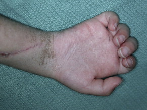
Volkmann Contracture - Neural damage – leading to temporary/permanent loss of various sensations (if sensory nerve damage) or muscle weakness (if motor neuron damage)
- Vascular damage – hemorrhaging
- Disseminated Intravascular Coagulation
- Dislocations, Subluxations
- Adhesive capsulitis
- Sprains (Grade 1, 2, 3, 4)
- Strains (1st, 2nd, 3rd degree)
- Sprain and Strain Healing
- Tendinitis
-

More details
Skeleton and bones – Tendon anatomy – Tendon Epimysium Fascicle Fiber Fibril Collagen Microfibril Perimysium Endomysium - Meniscus Tear
- Arthroscopy
- Bursitis
- Carpal Tunnel Syndrome
- Osteopenia
- Osteoporosis
- Ostemalacia
- Rickets
- Hip Fractures (femure fractures)
- Muscular Dystrophy
- Fibromyalgia
- Osteoarthritis
- Rheumatoid Arthritis
- Gouty Arthritis
Media Attributions
- Intramedullary Pin © Joo, Y.B.; Jeon, Y.S.; Lee, W.Y.; Chung, H.J. is licensed under a CC BY (Attribution) license
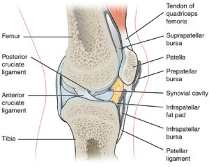
- Knee Joint – Tendon, Ligaments, Bursa © J. Gordon Betts, Kelly A. Young, James A. Wise, Eddie Johnson, Brandon Poe, Dean H. Kruse, Oksana Korol, Jody E. Johnson, Mark Womble, Peter DeSaix is licensed under a CC BY (Attribution) license
