Want to create or adapt books like this? Learn more about how Pressbooks supports open publishing practices.
Chapter 5 Selected Diseases and Disorders of the Immune System
Chapter 5 Immune System Diseases and Disorders – Portiaa
Zoë Soon
Creative Commons – Simple Pictures, Images, Video Clips, and/or Gifs that help illustrate any of the following:
Diversity Spotlight:
Dr. Susumu Tonegawa
Nobel Prize for Physiology or Medicine in 1987
Pioneered the theory of VDJ recombination, a process that allows our bodies to create the vast repertoire of antibodies needed to target the diverse array of microbial molecular patterns created by pathogens and commensal microbes. Most of his work was done at the University of Kyoto in Japan.
Diversity Spotlight:
Dr. Katalin Karikó
Nobel Prize for Physiology or Medicine in 2023
Dr. Karikó received the Nobel Prize in Medicine for her earlier work in 2005 on mRNA vaccines with her colleague Dr. Drew Weissman that led to the vaccines used to fight Covid-19.
*For diseases we discuss:
a) Basic Risk Factors
b) Most Common signs and symptoms
c) Basic Pathology, with basic diagnostic tools (e.g. imaging, blood tests) and basic treatment
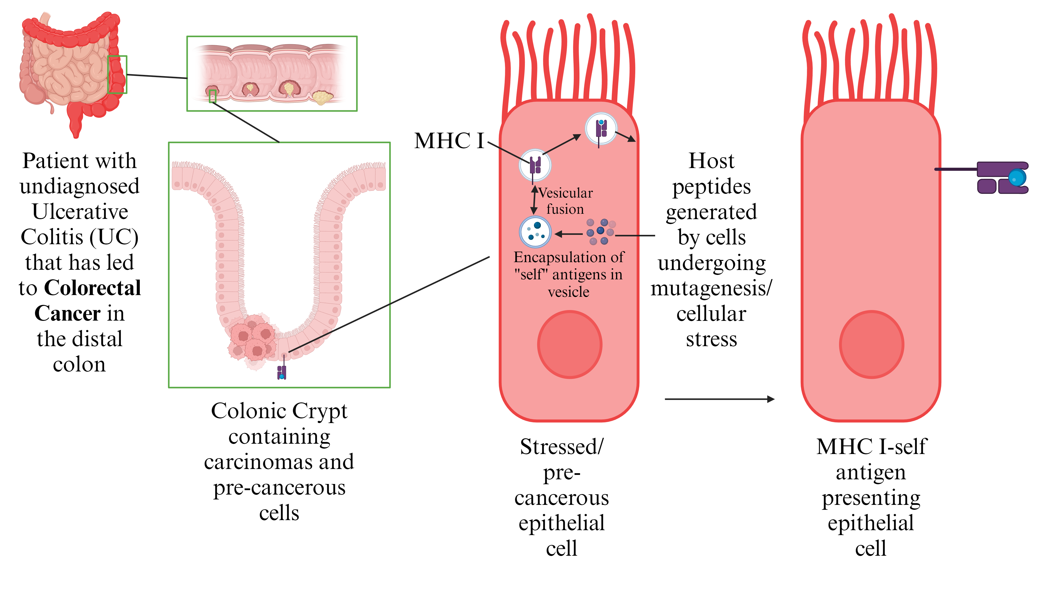
Self Antigens
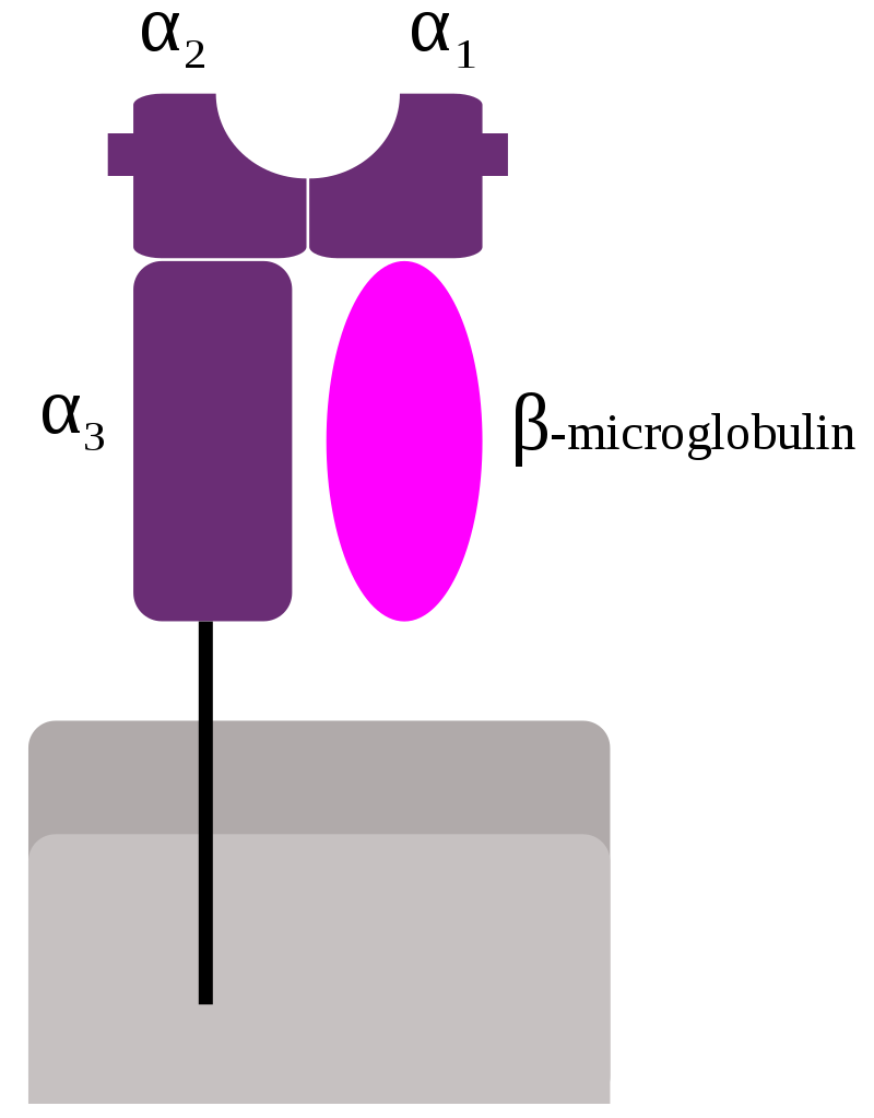
Major Histocompatibility Complex Class I
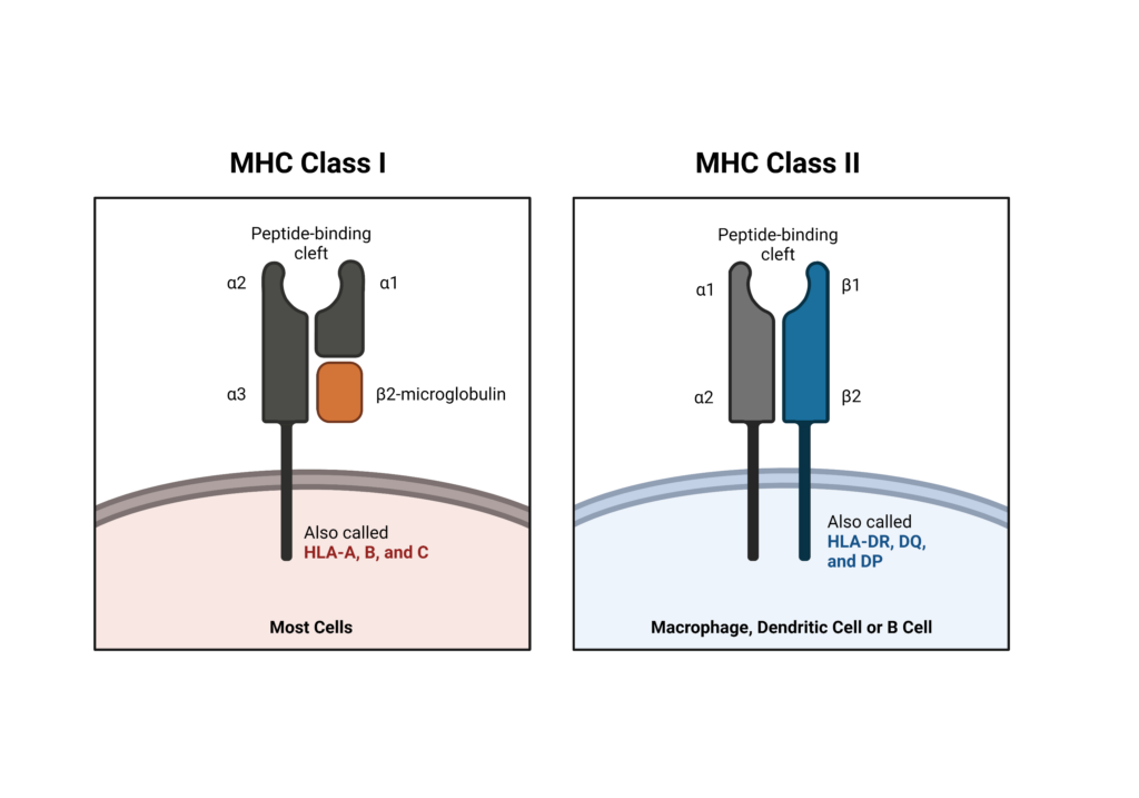
Schematic representation of Major Histocompatibility Complex (MHC) Class I molecule, consisting of three α-domains and one β2-microglobulin molecule. The peptide-binding groove is situated between domains α1 and α2.
Major Histocompatibility Complex Class II
Schematic representation of Major Histocompatibility Complex (MCH) Class II molecule, consisting of two α-domains and two β-domains. The peptide-binding groove is situated between domains α1 and β1.
Major histocompatibility complex (MHC) molecules help the immune system distinguish between self and non-self molecules by binding to and presenting peptide fragments derived from foreign cells in order to initiate attack against invading pathogens. The two structurally and functionally distinct classes of MHC proteins are class I (present foreign peptides to cytotoxic T cells) and class II (present foreign peptides to helper T cells).
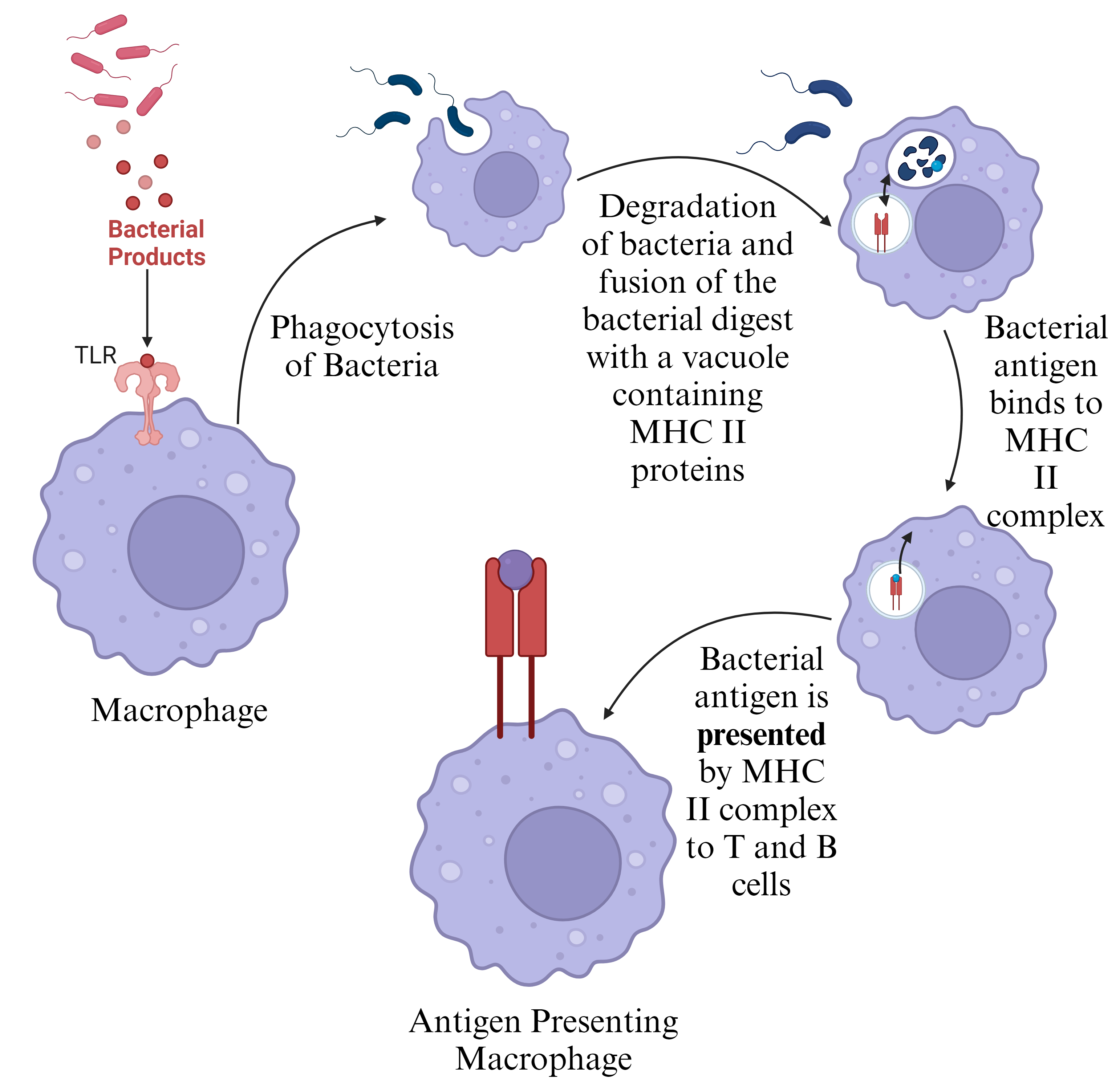
Foreign Antigens
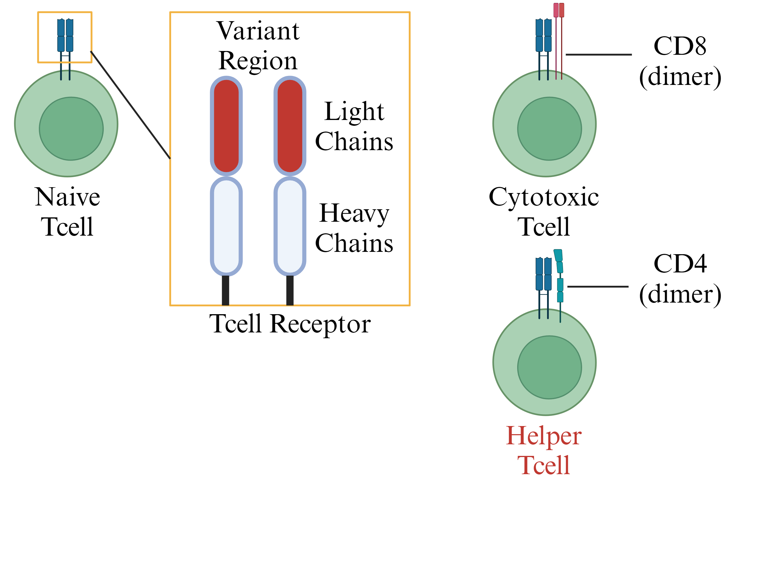
T cell Classes
T Cell Receptor (TCR) recognizes antigen (self or foreign), variant region is formed by V(D)J-recombination that is similar to B cell receptors (both are considered to be “antibodies”).
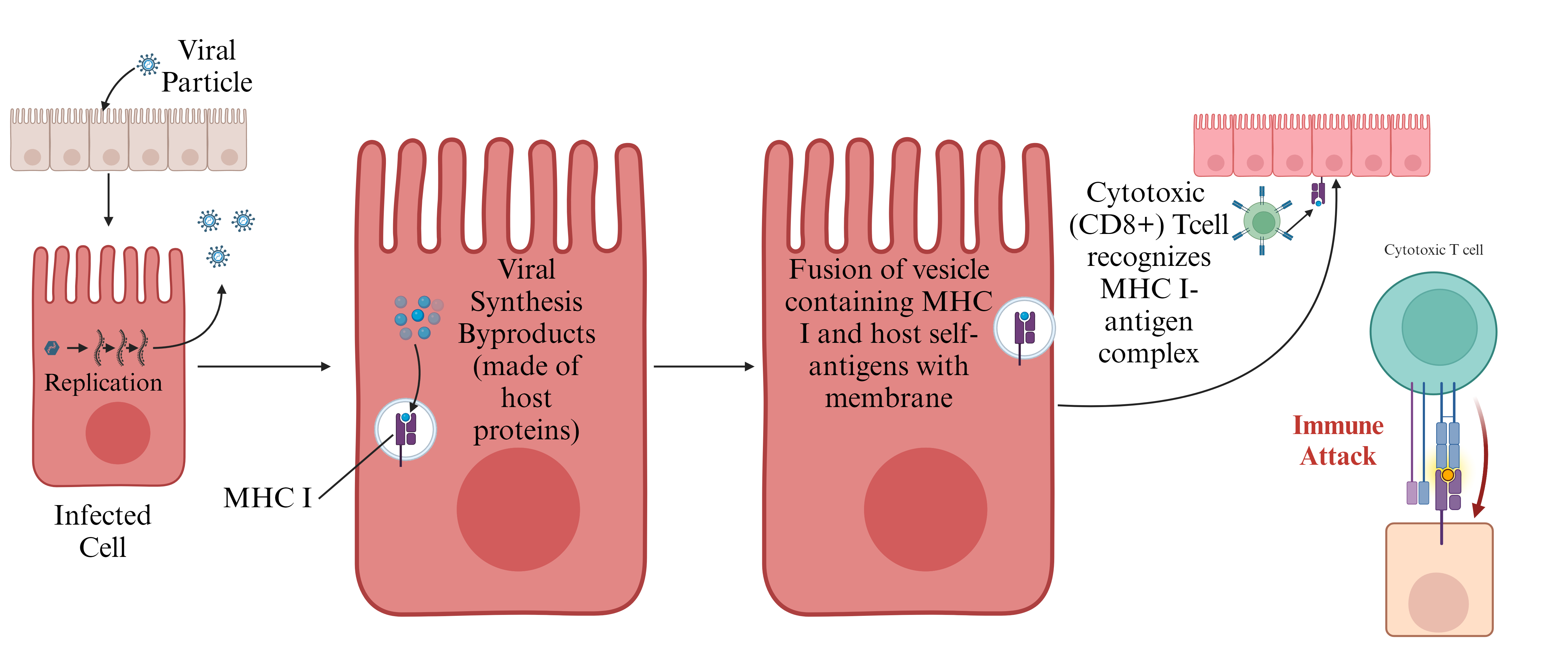
CD8+ T cells recognize MHC-1 complexes that can be expressed and presented by virtually all cell types. This helps your immune system recognized stressed, infected, or cancerous cells. These cells are degraded by CD8+ T cells and Natural Killer cells (NK cells) which both recognize MHC-1-self antigen complexes.
CD4+ T cells recognize MHC-II complexes that are mostly expressed by phagocytic cell types (macrophages, neutrophils, dendritic cells, eosinophils, epithelial cells like M cells, and to a lesser extend mast cells and basophils). Post-translation MHC-II complexes contain an invariant chain that blocks self antigens from binding to MHC-II complexes to avoid presentation of self antigens to Helper T cells as a foreign antigen (helps avoid autoimmune disease).
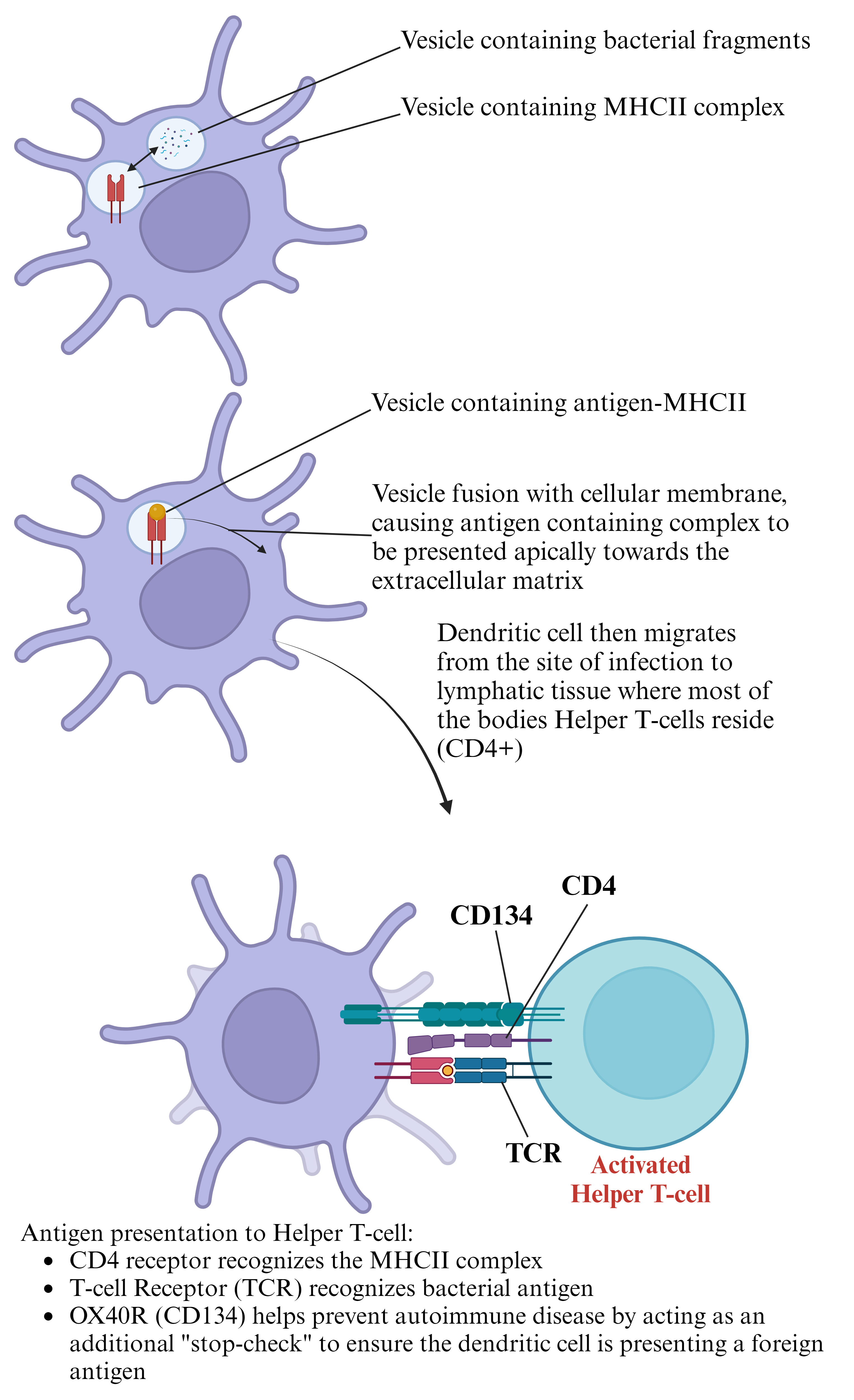
Activation of Helper T cells
Activation of Cytotoxic T cells
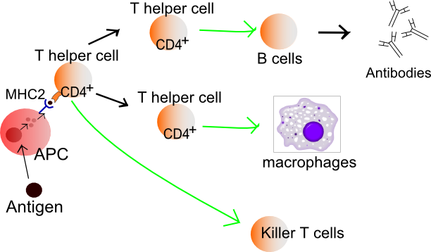
Antigen presentation stimulates T cells to activate “cytotoxic” CD8+ cells or “helper” CD4+ cells. Cytotoxic cells directly attack other cells carrying certain foreign or abnormal molecules on their surfaces. Helper T cells (Th cells) coordinate immune responses by communicating with other cells. In most cases, T cells only recognize an antigen if it is carried on the surface of a cell by one of the body’s own major histocompatibility complex (MHC) molecules.
Naive helper T cells are activated by binding to foreign peptides bound to class II MHC molecules on the surface of an antigen presenting cell (APC). Upon activation, mature helper T cells go on to activate B cells, which secrete antibodies, and macrophages, which take up and destroy foreign microbes. Furthermore, mature helper T cells help activate cytotoxic T cells (killer T cells), which kill infected target cells.
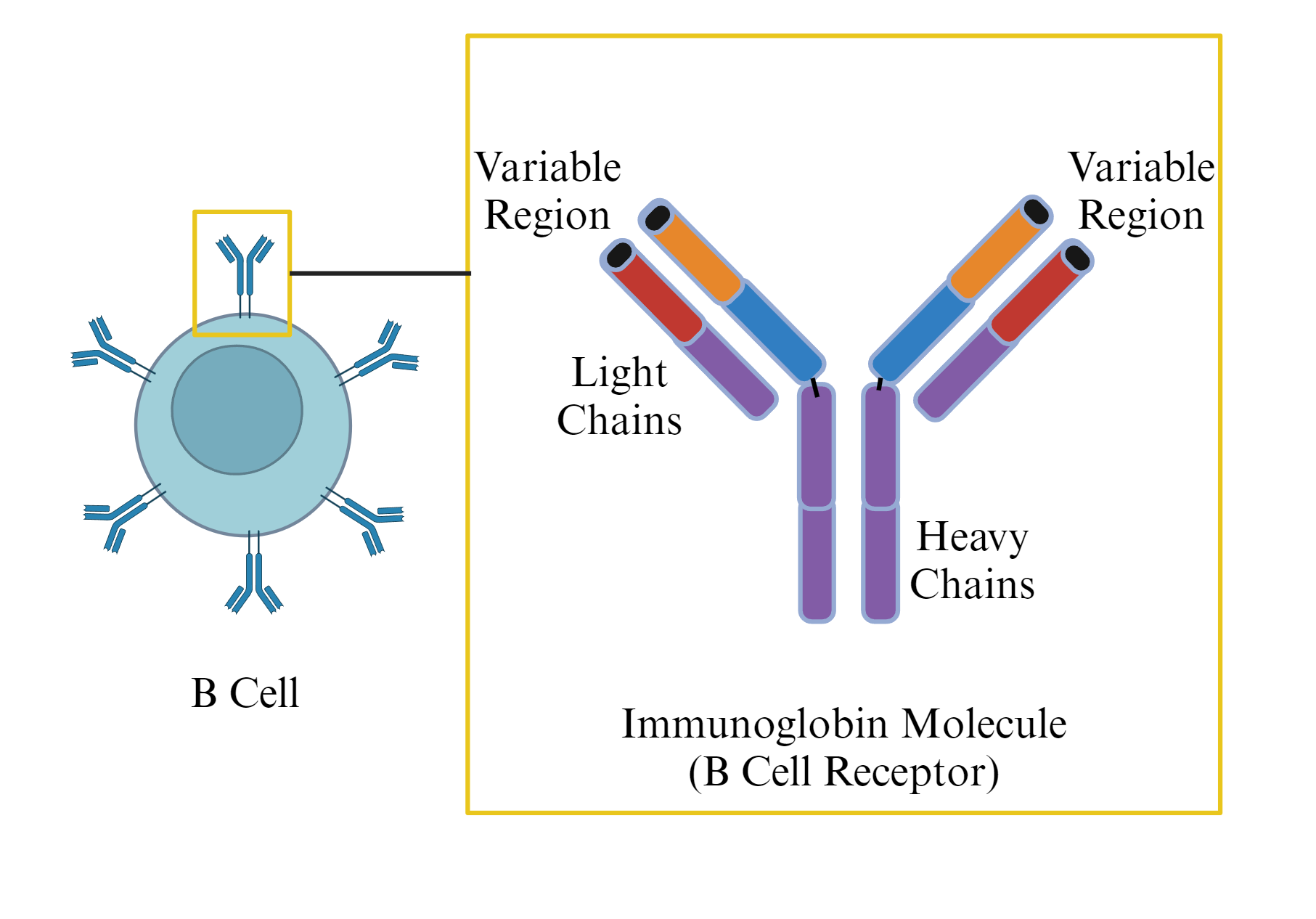
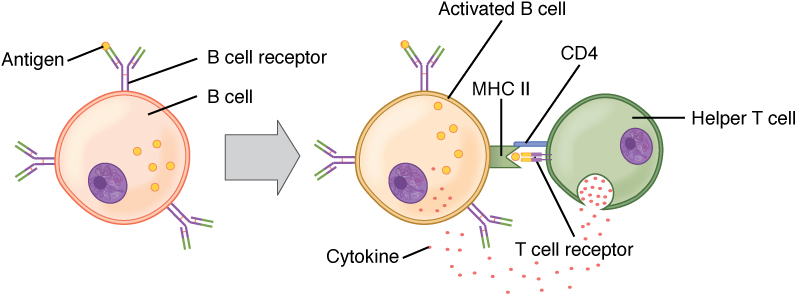
Activation of B cells B cells (B lymphocytes) are an integral part of the immune system as they are responsible for forming antibodies against invading pathogens. Antibodies can be secreted or inserted into the plasma membrane of B cells, where they form B cell receptors and control the activation of the B cell.
T and B cell binding. To elicit a response to a T cell-dependent antigen, the B and T cells must come close together. To become fully activated, the B cell must receive two signals from the native antigen and the T cell’s cytokines.
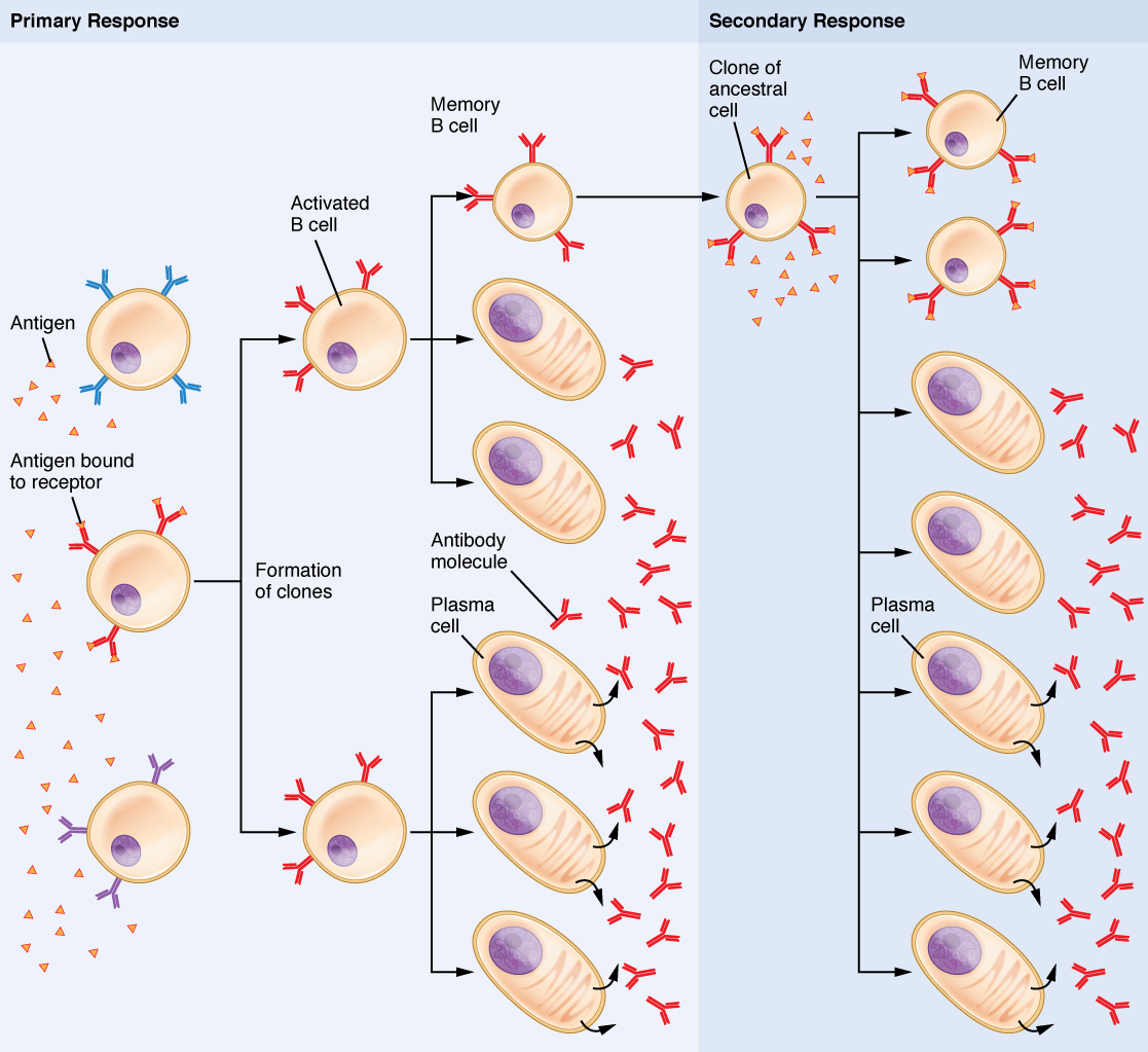
Primary and Secondary Response to Same Foreign Antigen The primary immune response occurs during the initial exposure to a foreign antigen. The secondary immune response occurs following subsequent exposure to the same antigen.
Clonal Selection of B Cells During a primary B cell immune response, both antibody-secreting plasma cells and memory B cells are produced. These memory cells lead to the differentiation of more plasma cells and memory B cells during secondary responses.
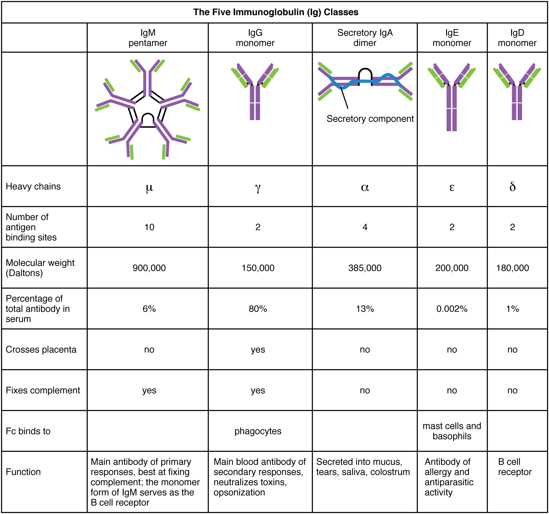
Classes of Antibodies
Antibodies (Immunoglobulins; Ig) are divided into 5 classes (IgA, IgD, IgE, IgG, and IgM) based on their physiochemical, structural, and immunological properties.
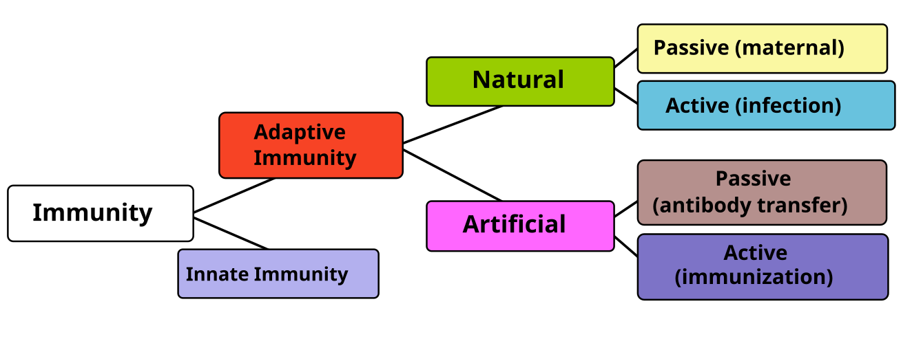
Divisions of Immunity. Natural immunity occurs through spontaneous contact with a disease-causing agent, whereas artificial immunity develops only through deliberate exposure. Both natural and artificial immunity can be further subdivided. While passive immunity (immunity obtained from outside our own immune system) provides rapid and immediate protection, active immunity (immunity built by our own immune system) takes days or weeks to develop.
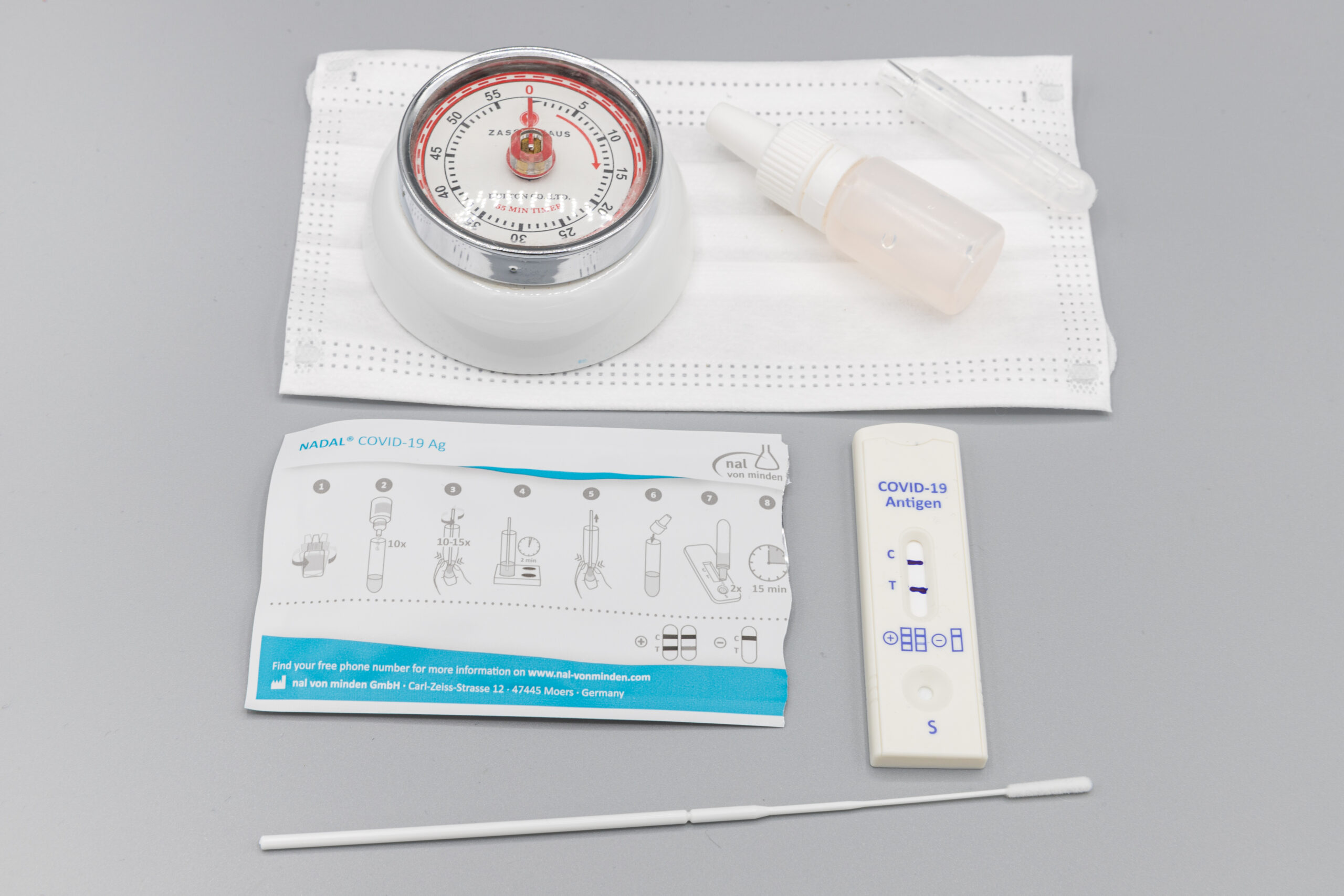
Rapid Antigen Test
COVID-19 Corona virus positive Rapid Antigen Test, which are used to test for the presence of antigens from viral infection.
Antibody Test
Administration of a COVID-19 antibody test, which checks for the presence/level of specific antibodies in the blood (in this case, antibodies against the COVID-19 virus).
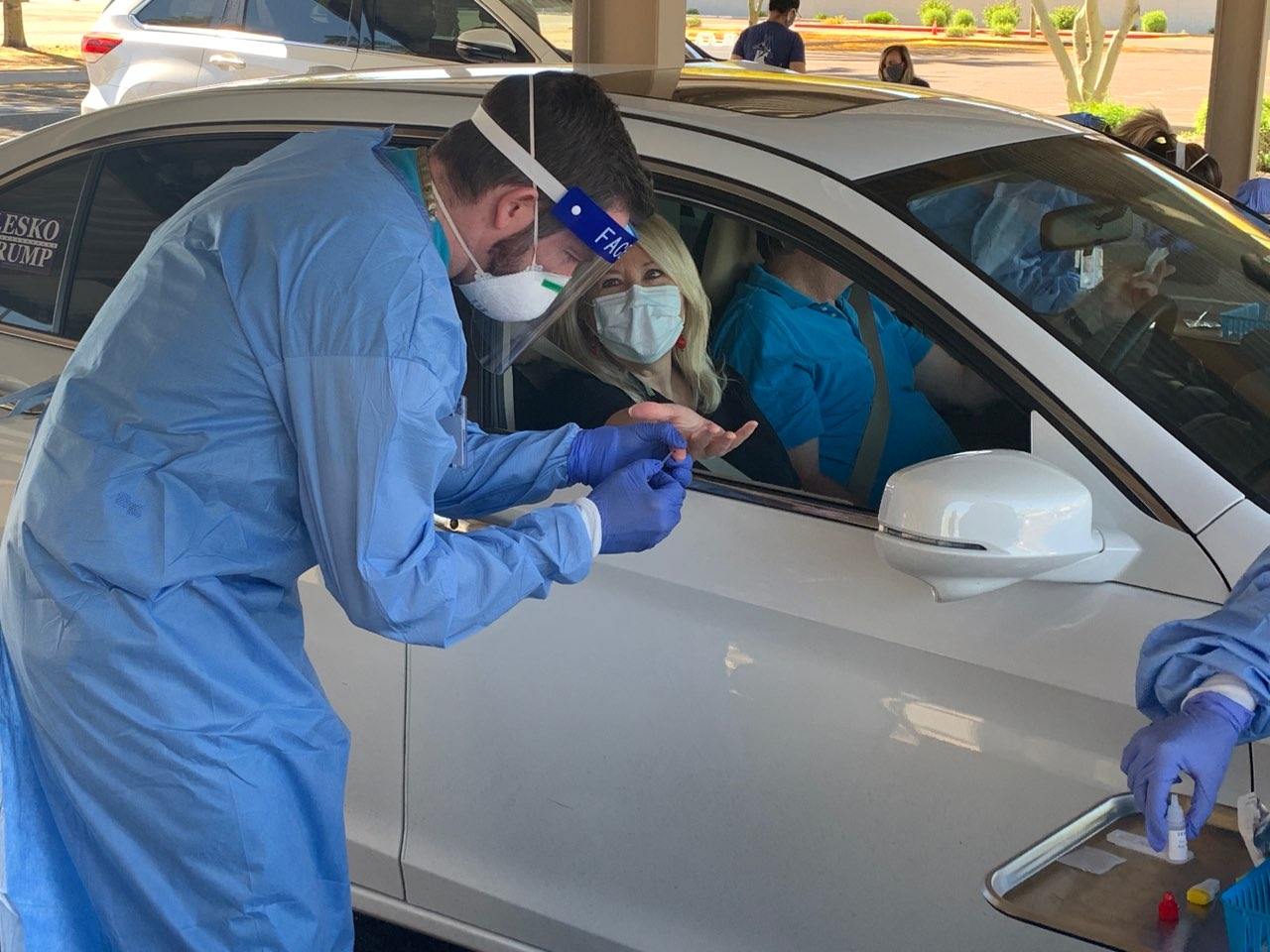
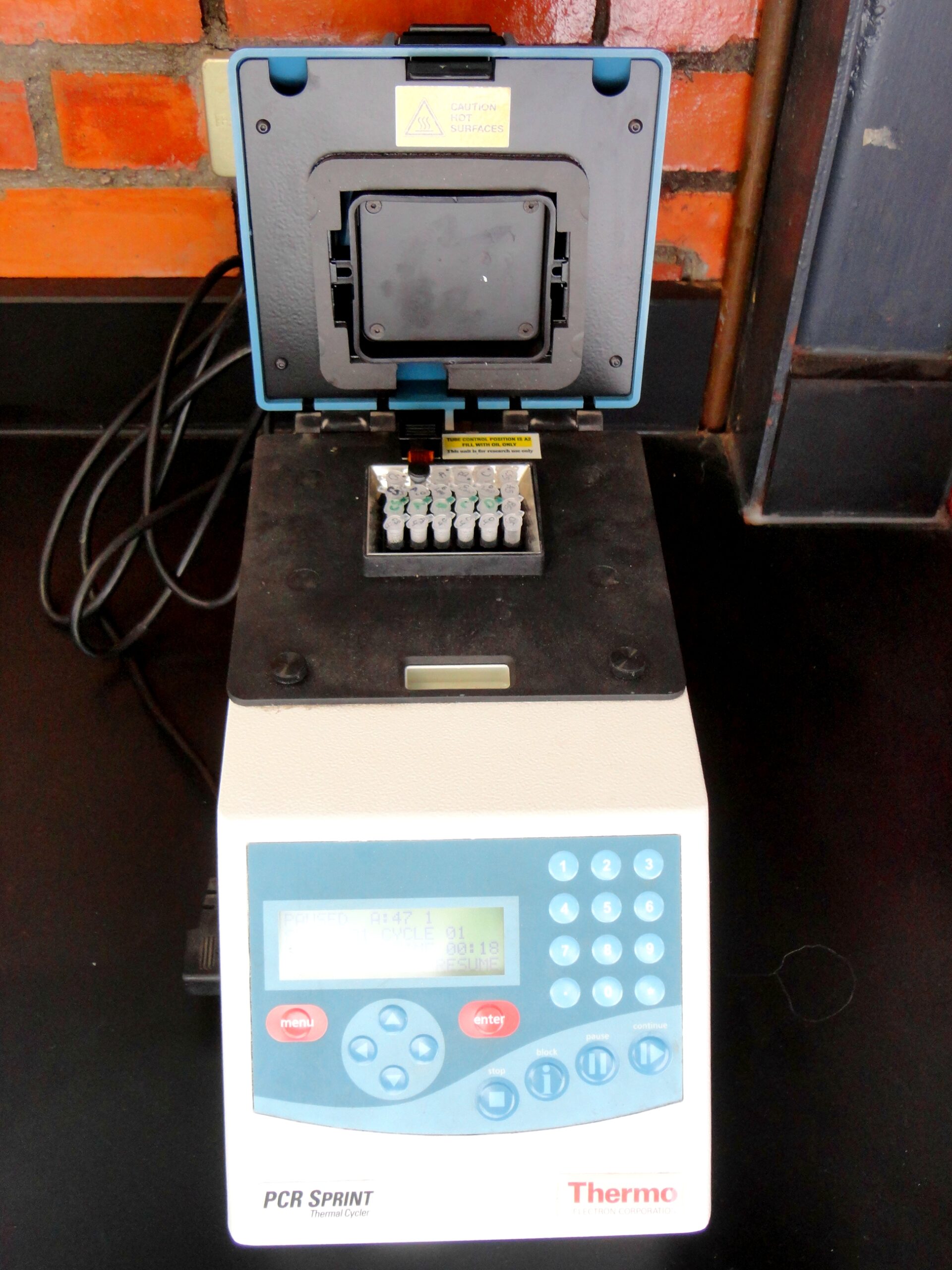
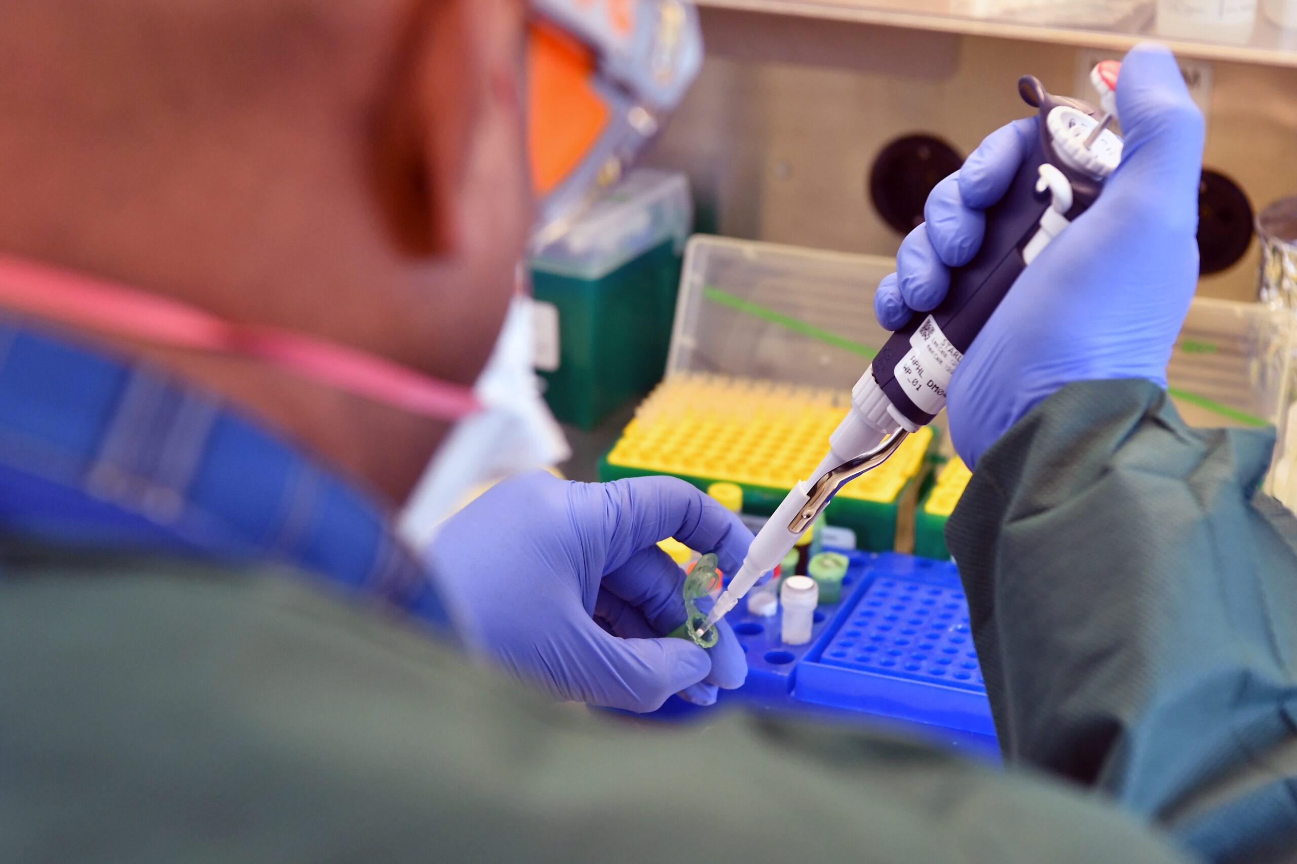
PCR Test, RT-PCR Test
Thermal cycler used to amplify segments of DNA via polymerase chain reaction (PCR).
Preparation of real-time polymerase chain reaction (RT-PCR) for rapid and sensitive diagnosis, ranging from cancer, genetic abnormalities, and infectious diseases,
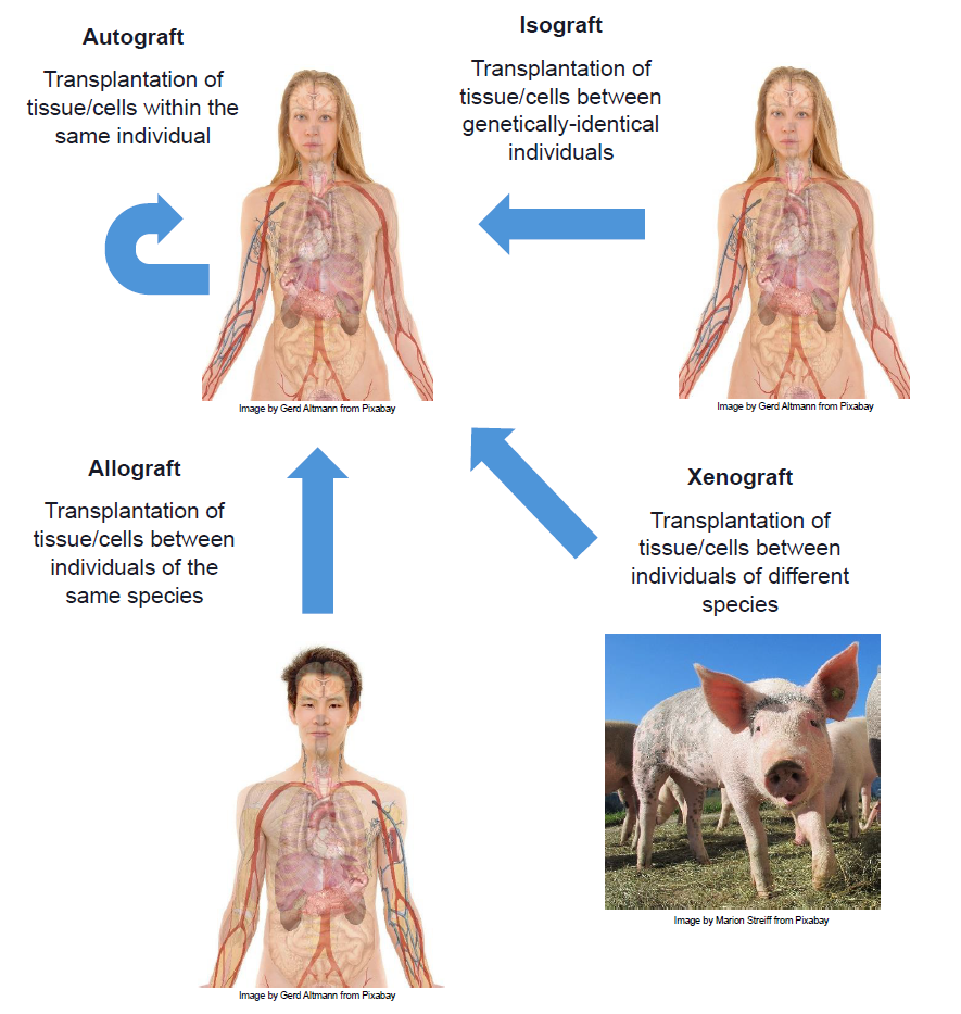
Types of transplants – allograft, isograft, autograft, xenograft
Four types of transplants: Autograft, Allograft, Isograft, and Xenograft.
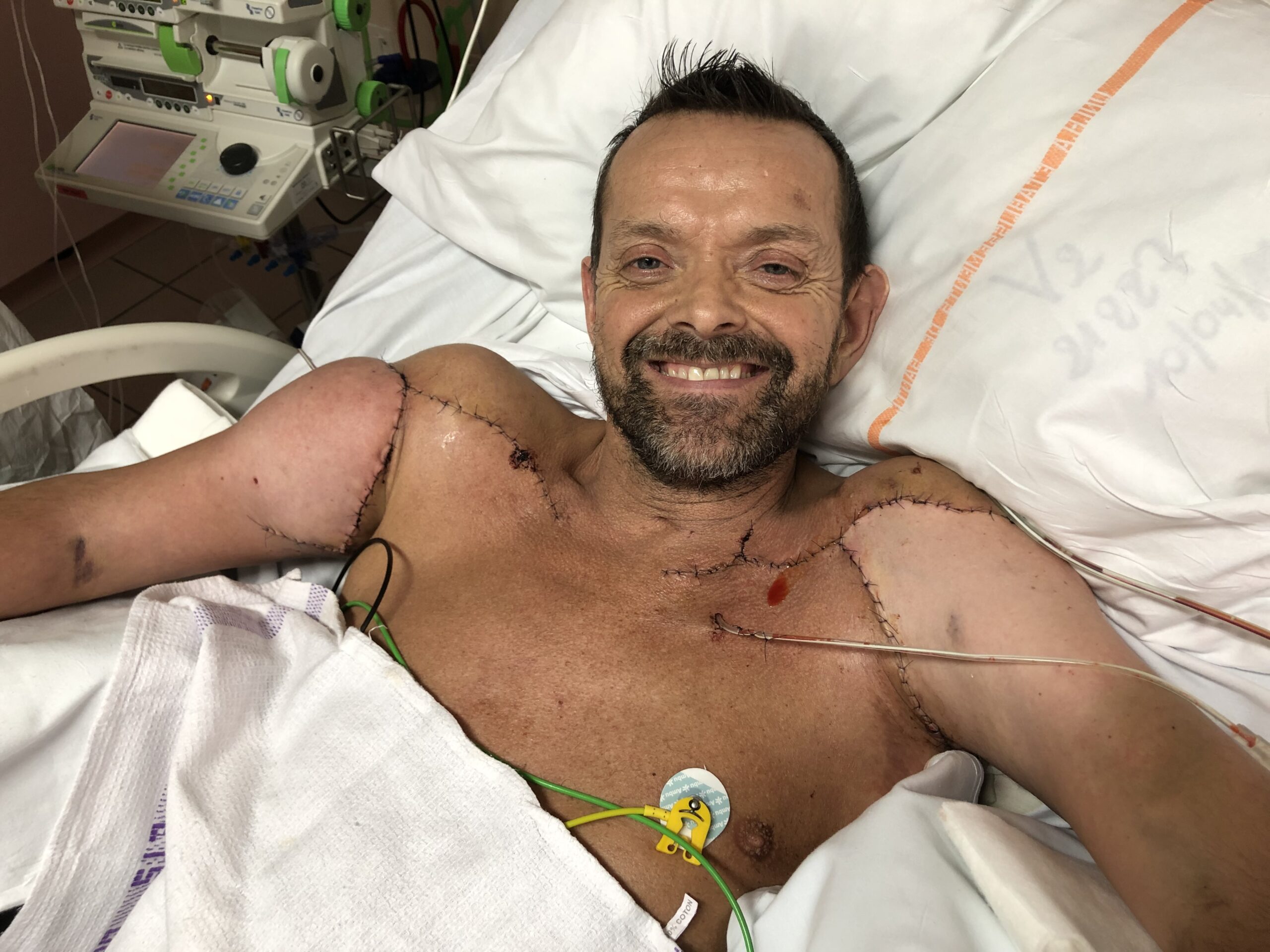
Felix Grétarsson received a double-arm and shoulder transplant at Edouard Herriot Hospital in Lyon, France in January 2021. This is an example of an allograft, a transplant of tissue from a non-genetically identical donor of the same species.
Transplant rejection – hyperacute, acute, chronic
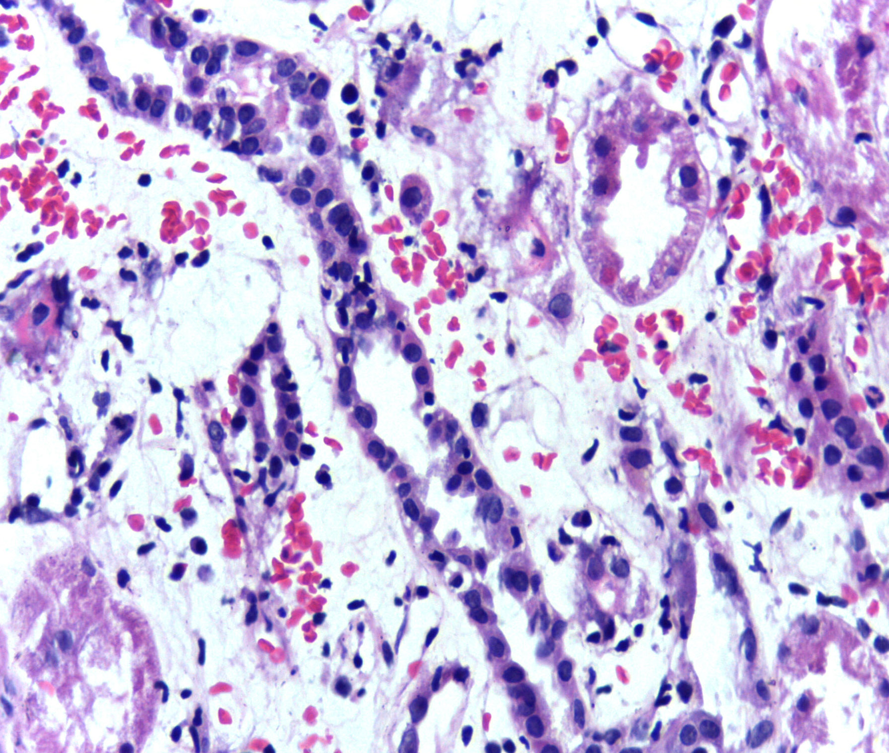
Presence of lymphocytes within tubular epithelium (tubulitis) in the kidney is one of the pathological features of acute rejection following renal transplant.
Host vs Graft
Graft vs Host
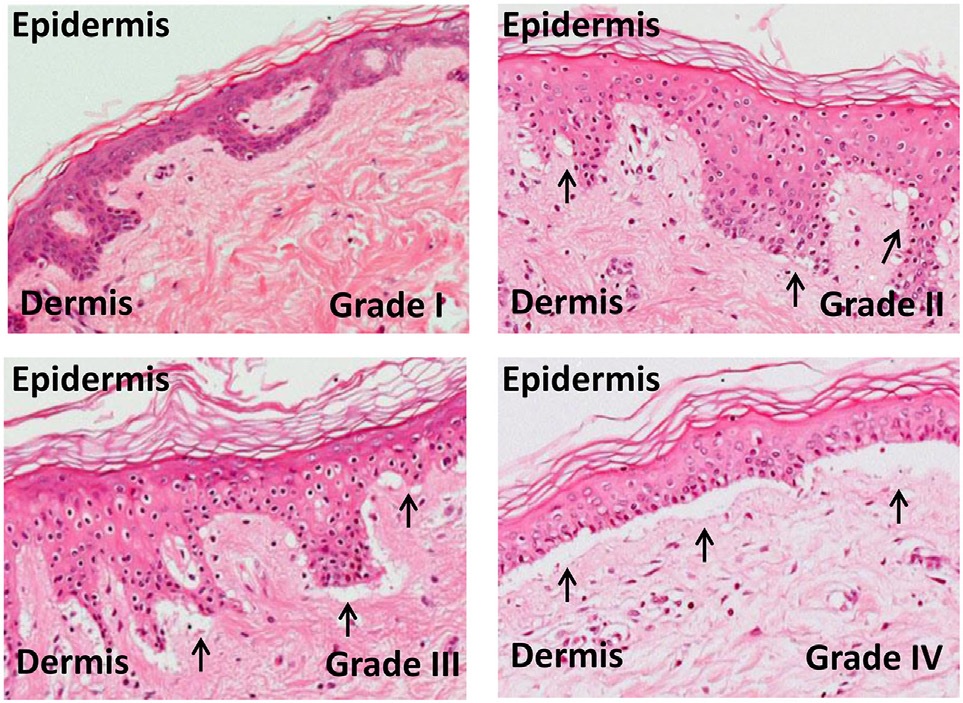
Micrographs of grades of skin graft-versus-host-disease: The outcome of the skin explant assay is histopathological damage ranging from grade I graft-versus-host reactions (GVHR), with minimal vacuolization in the epidermis, to grade II GVHR, with vacuolization and dyskeratotic bodies, to grade III GVHR, with sub epidermal cleft formation, and finally to grade IV GVHR, with separation of the dermis from the epidermis (photo by: Sakhila Ghimire, Daniela Weber, Emily Mavin, Xiao nong Wang, Anne Mary Dickinson, and Ernst Holler; license: https://creativecommons.org/licenses/by/4.0/)
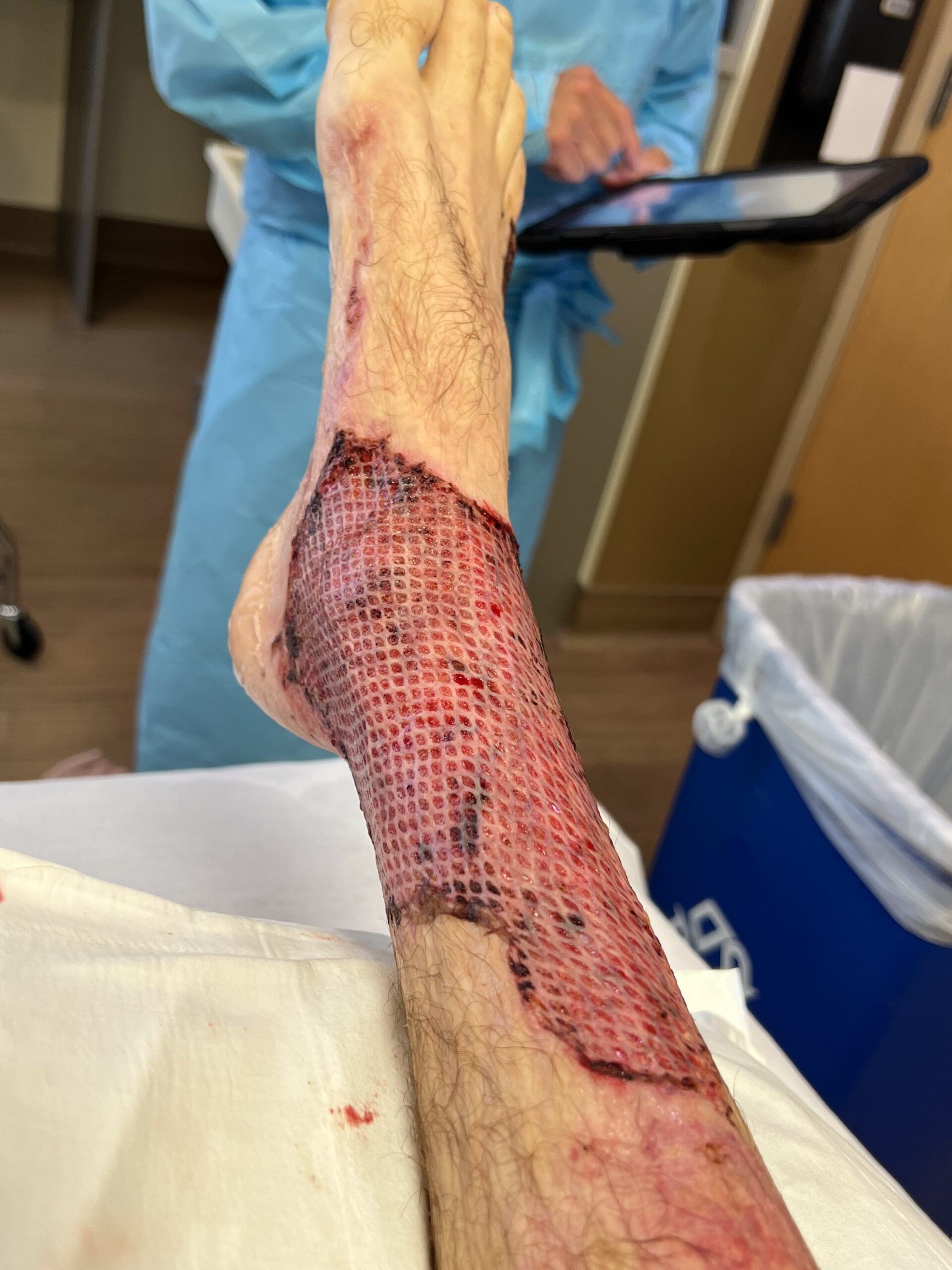
Skin graft performed on the ankle due to third degree burns (photo provided by Giftrapped, Wikimedia Commons; license: https://creativecommons.org/licenses/by-sa/4.0/)
4 Types of Hypersensitivity Reactions
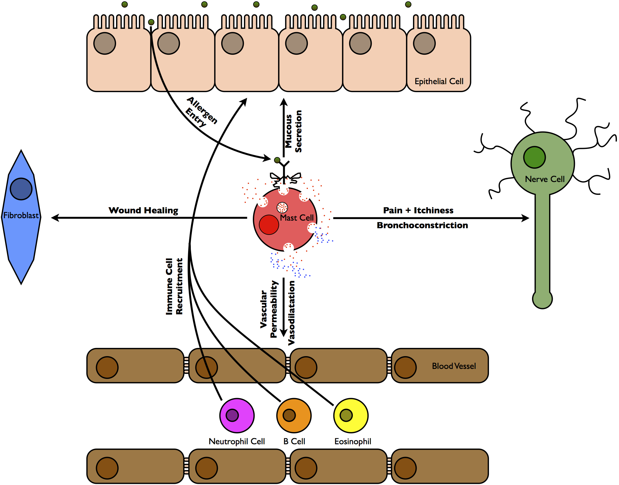
Type I – allergies
Diagram summarizing some of the tissues that are affected immediately during and following an allergic reaction (Photo by: Sari Sabban; license: https://creativecommons.org/licenses/by-sa/3.0/)
Simplified diagram showing key events that leads to allergy initiation. A. the allergen enters the body. B. an antigen-presenting cell takes up the allergen molecule and presents its epitopes, through the major histocompatibility complex (MHC) II receptor, onto its surface. The activated antigen presenting cell then migrates to the nearest lymph node C. where it activates T cells that recognize the allergen. They then give the decision for the T cell to differentiate to a type 2 helper T (Th2) cell. D. At the same time, B cells recognize the allergen and, through the activated Th2 cell, E. the B cell would be activated F. and differentiate into plasma cells, at which point they would actively synthesize antibodies of the IgE isotype. G. The IgE antibody, that now recognizes epitopes of the allergen molecule, circulates around the body through the lymphatic and cardiovascular systems and finally binds to a receptor (FcεRI) on mast and basophil cells. H. When the allergen re-enters the body at a later time it binds to the IgE, which is on the cell surface, resulting in an aggregation of the receptor causing the cells to release pre-formed mediators. One of these mediators is histamine, which causes the 5 symptoms of allergic inflammation: heat, pain, swelling, redness and itchiness. Another mediator is IL-4, which affects more B cells to differentiate into plasma cells and produce more IgE and thus the vicious cycle continues.
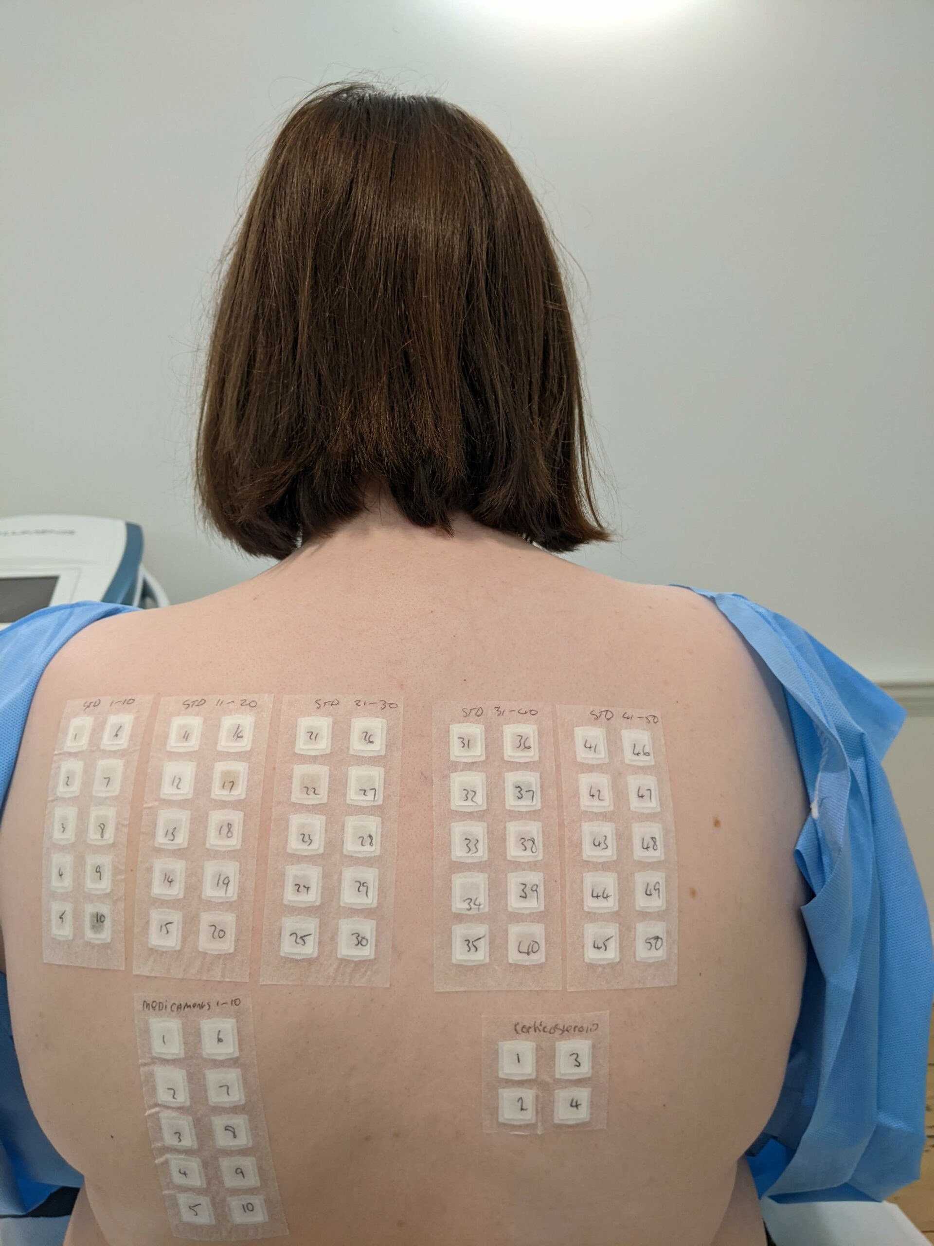
Skin patch allergy test on the back of a patient (Photo by: Smirkybec, Wikimedia Commons; license: https://creativecommons.org/licenses/by/4.0/)
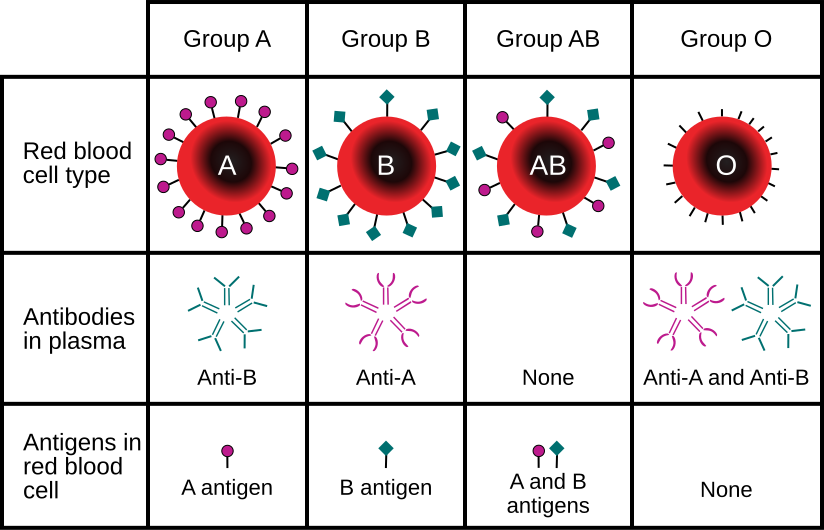
Type II – ABO blood incompatibility, Hemolytic Disease of the Newborn
The four different blood groups in the ABO system (A, B, AB, and O) and the IgM antibodies present in each.
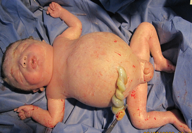
Newborn infant with severe hemolytic disease. Hemolytic disease of the newborn occurs when there is an incompatibility between the blood types of the newborn and the mother, resulting in the newborn’s red blood cells to be attacked and broken down by the mother’s antibodies.
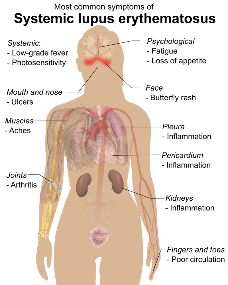
Type III – Systemic Lupus Erythamatosus, Glomerulonephritis
Main symptoms of systemic lupus erythematosus (SLE), the most common form of lupus. SLE is an autoimmune disease resulting in widespread inflammation and organ damage due to the immune system attacking the body’s own tissues.
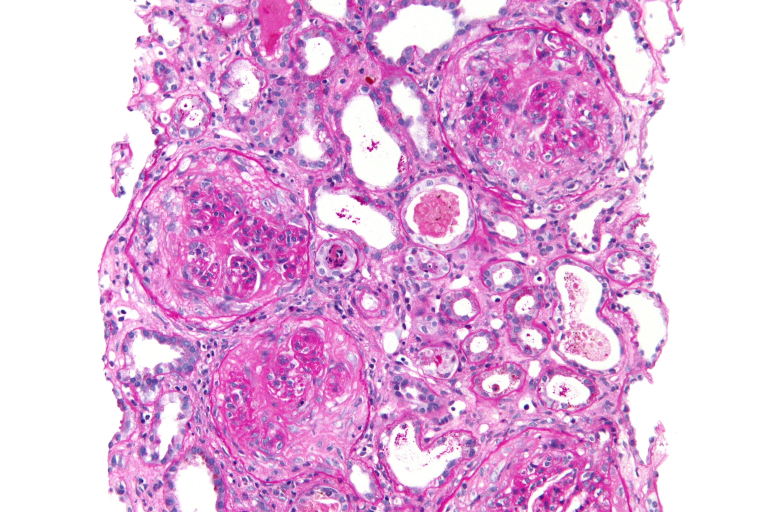
High magnification micrograph showing crescentic glomerulonephritis, also known as a rapidly progressive glomerulonephritis (inflammation and damage to the glomerulus, the filtering part of the kidneys). Glomerulonephritis can also develop progressively over time. In both instances, the kidney’s filtering function into the urine is damaged; therefore, toxins, metabolic waste, and excess fluid build up in the body, leading to fatigue and widespread inflammation. Other diseases and acute infections can lead to glomerulonephritis.
Type IV – Contact Dermatitis; Delayed Transplant Rejection, Mantoux TB skin test
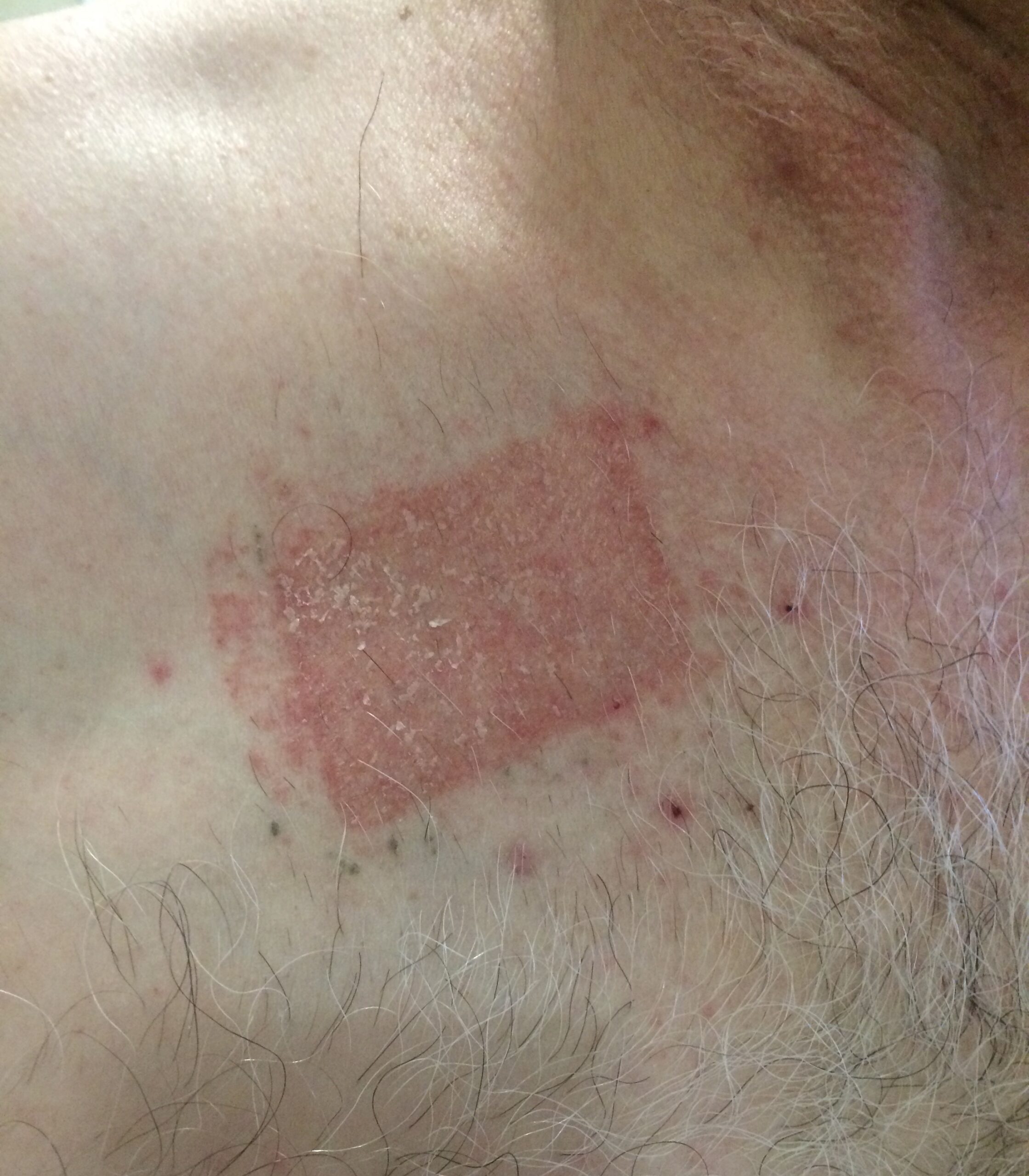
Patient with severe contact dermatitis at the application site of a 15mcg/hr buprenorphine transdermal (Norspan) patch. Contact dermatitis, caused by direct skin contact with a substance the patient is allergic to, is a non-contagious, itchy, swollen rash.
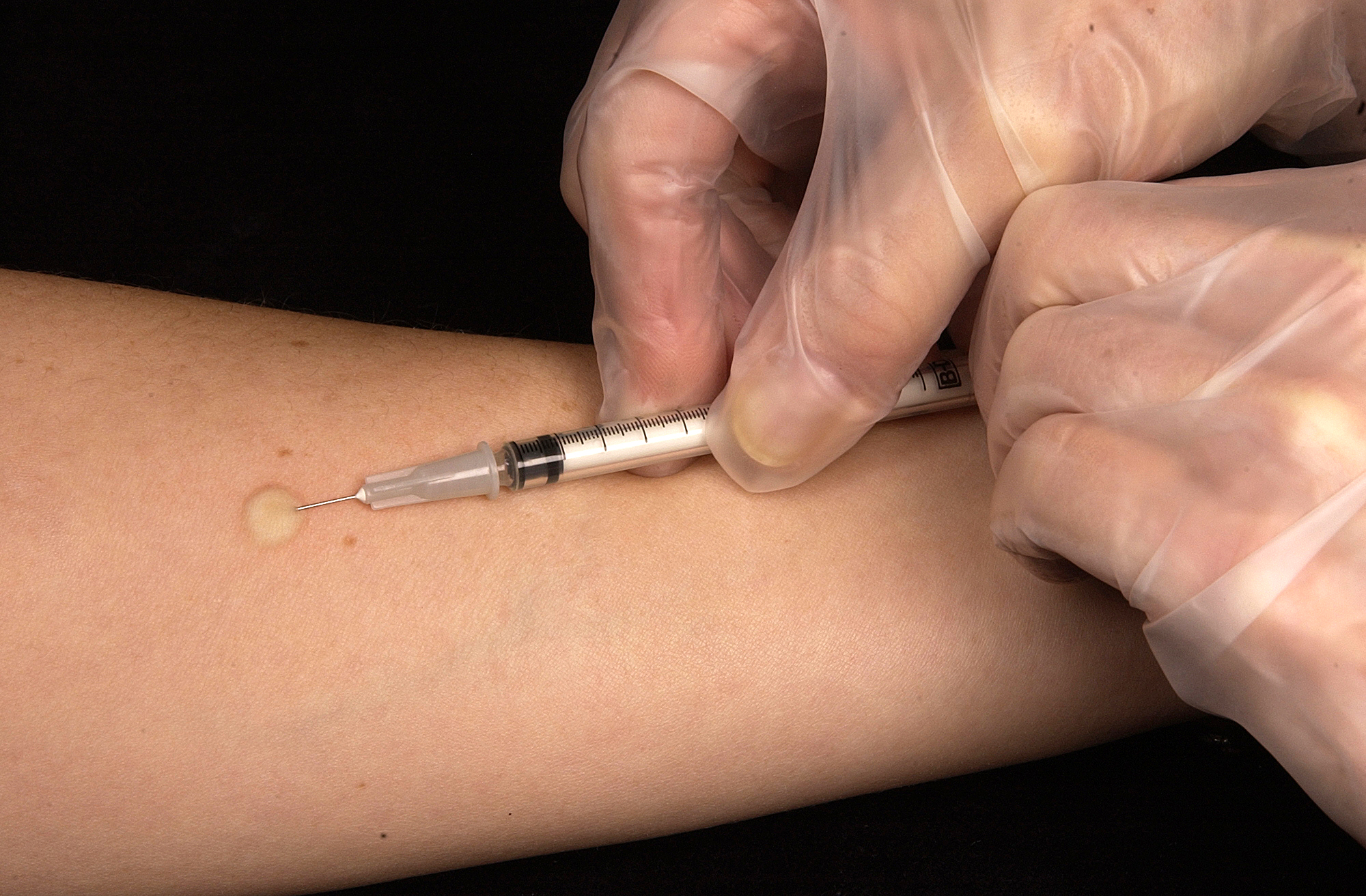
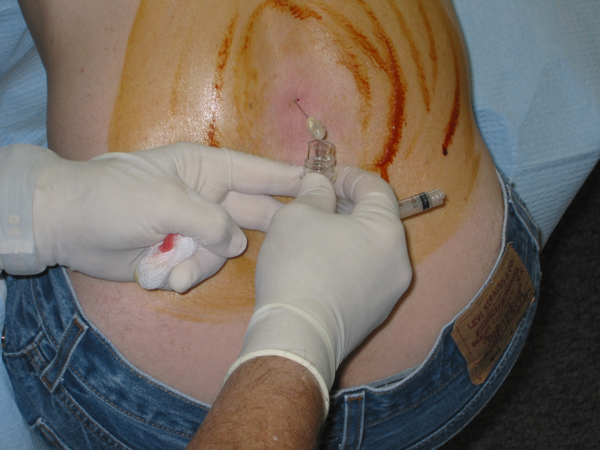
The Mantoux tuberculin skin test is used to evaluate people for exposure to Mycobacterium tuberculosis, the bacterial causal agent of tuberculosis (TB). A small amount of fluid (tuberculin) containing TB proteins (antigens) is injected under the top layer of skin on the inner forearm, Within 48-72 hours, the injection site reaction must be evaluated by a healthcare professional. A positive or negative results is determined based on the size of size of the hard, raised area, as well as swelling.
Common Autoimmune Diseases
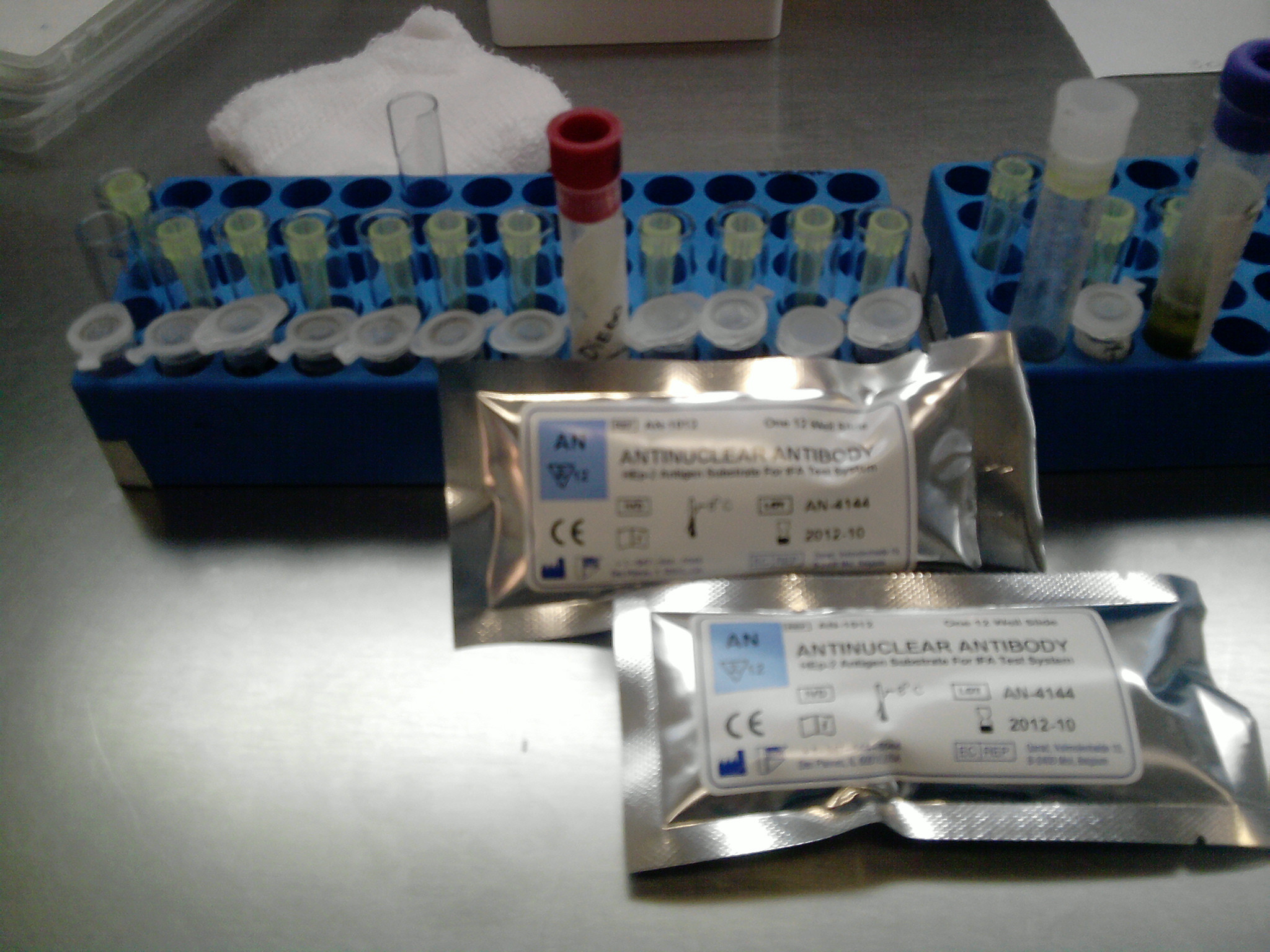
Kit for carrying out a test to detect antinuclear antibodies in the blood. Rather than regular antibodies that fight foreign pathogens, antinuclear antibodies attack your own healthy cells. The presence of antinuclear antibodies indicates an autoimmune reaction, therefore testing for antinuclear antibodies is a standard evaluation for possible autoimmune disease.
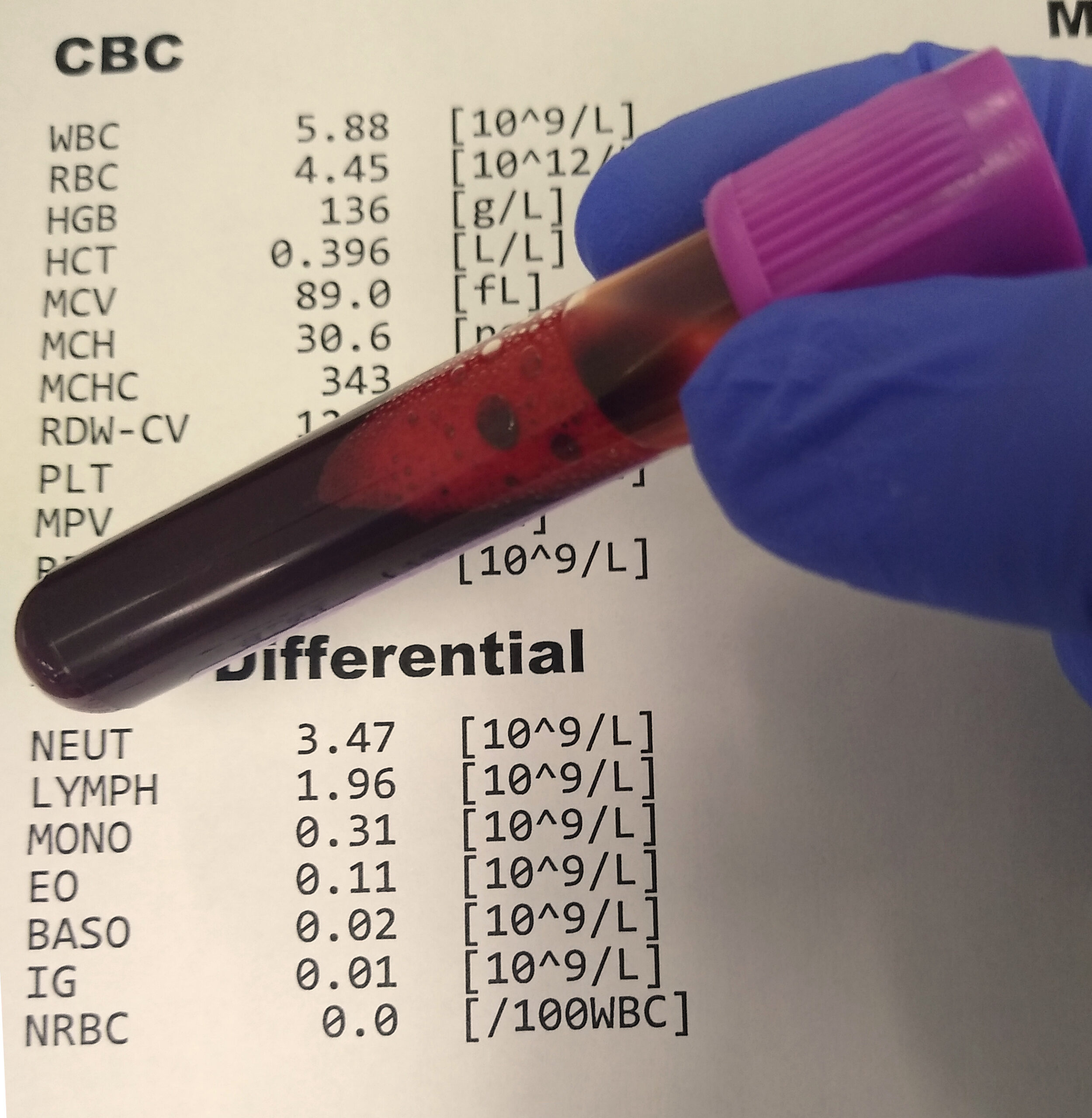
Complete blood count (CBC) and differential tests. A CBC test measures the total numbers of white and red blood cells, while a differential test measures the number of each type of white blood cell. These tests can help detect a variety of disorders, including autoimmune diseases.
A lumbar puncture being performed. A lumbar puncture (spinal tap) is performed on the lower back (lumbar region), where a needle is inserted between two vertebrae in order to collect cerebrospinal fluid (CSF). CSF is then analyzed to assess for abnormalities (for example, an elevated white blood cell count) that can help diagnose a variety of different conditions, including several autoimmune disorders, infections, and/or cancers.
Collage showing the procedure of urinalysis. Left: A urine test strip is immersed into the sample. Top right: A urine sample is about to be examined under a phase-contrast microscope using a Neubauer counting chamber. The urine is under the cover slide, in the upper segment formed by the H-shaped grooves. Bottom right: Phase-contrast microscopic image showing many white blood cells in the urine (pyuria). Urinalysis is a common test when diagnosing autoimmune disorders in order to assess renal injury, proteinuria, hematuria, or inflammatory markers.
A C-reactive protein (CRP) test is used to measure inflammation in the body and can be used to find or monitor inflammation due to infection, injury, or chronic diseases, including autoimmune disorders.
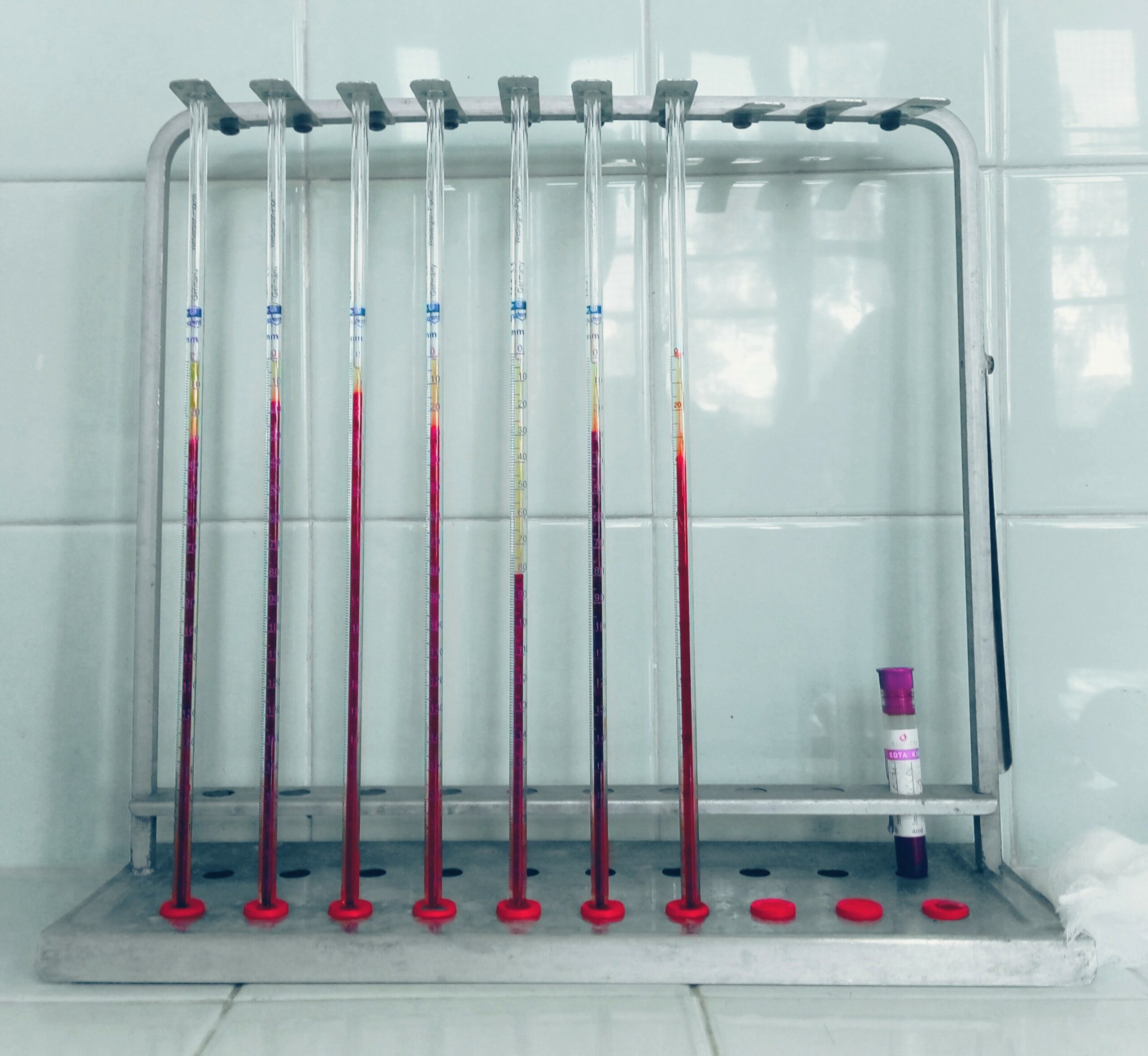
An erythrocyte sedimentation rate (ESR) test determines the rate at which red blood cells in anticoagulated whole blood descend in a standardized tube over a period of one hour. The presence of an inflammatory condition would result in an elevated ESR, which may be indicative of an autoimmune disorder.
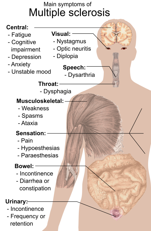
Multiple Sclerosis
Common symptoms of multiple sclerosis (MS). MS is a chronic autoimmune disease affecting the central nervous system. Symptoms vary greatly between patients, from mild to severe.
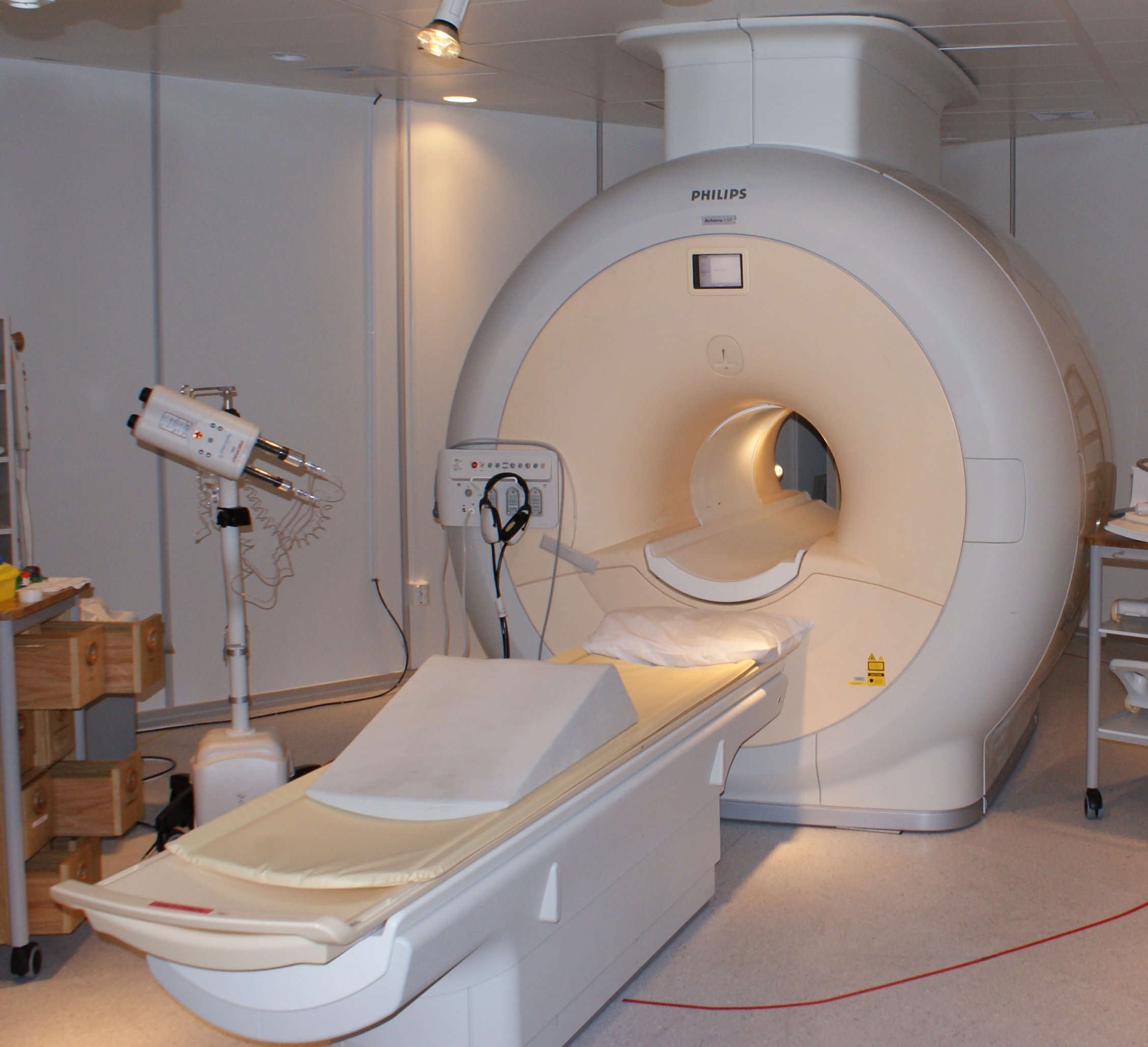
Magnetic resonance imaging (MRI) is a medical imaging technique that uses a magnetic field and high frequency radio waves to produce detailed cross-sectional images of parts around the body. MRIs are commonly used to confirm the diagnosis of multiple sclerosis (among other conditions) by revealing brain lesions.
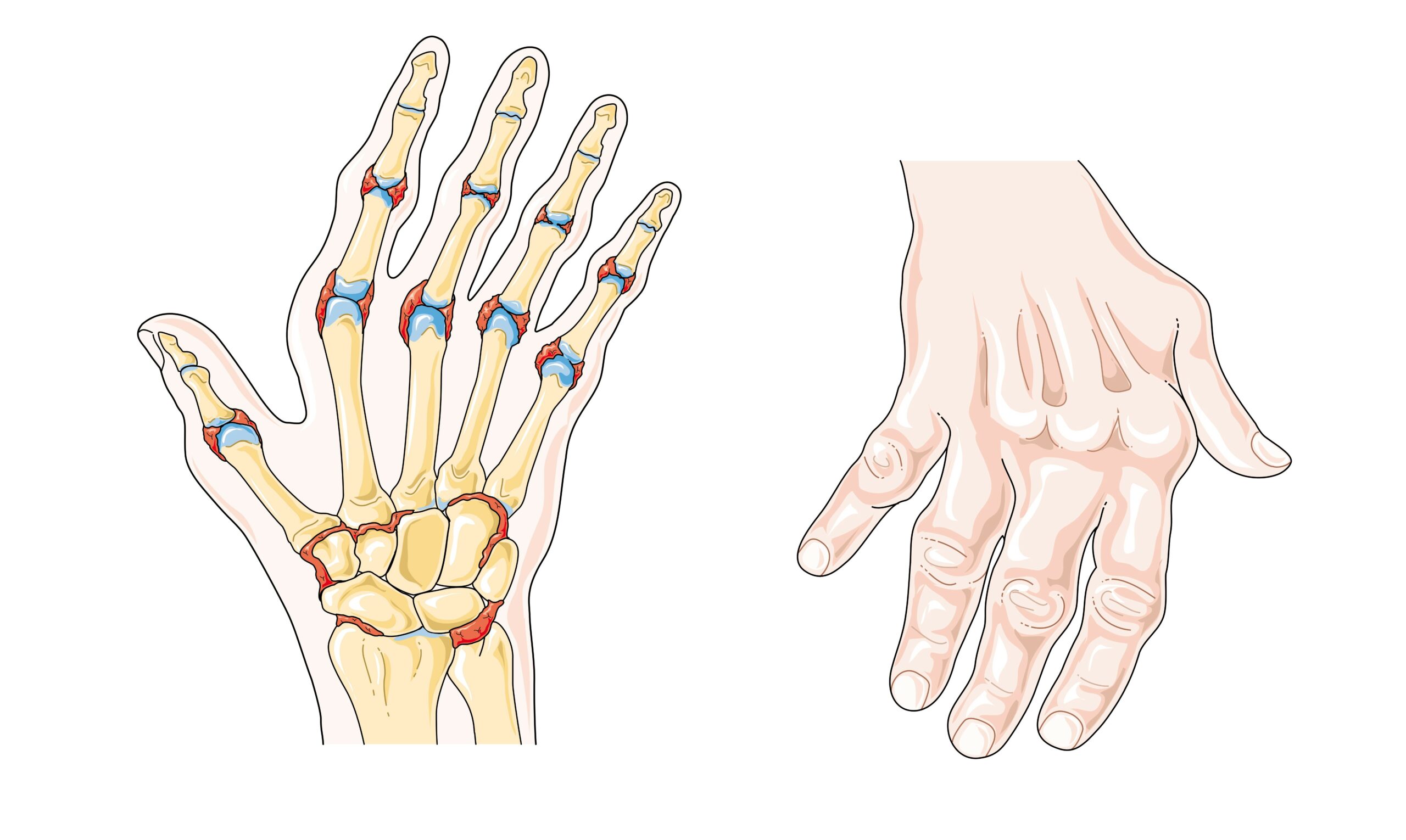
Rheumatic Arthritis
Rheumatoid arthritis is an autoimmune disease that causes the immune system to attack healthy cells of the body, primarily affecting joints resulting in painful inflammation.
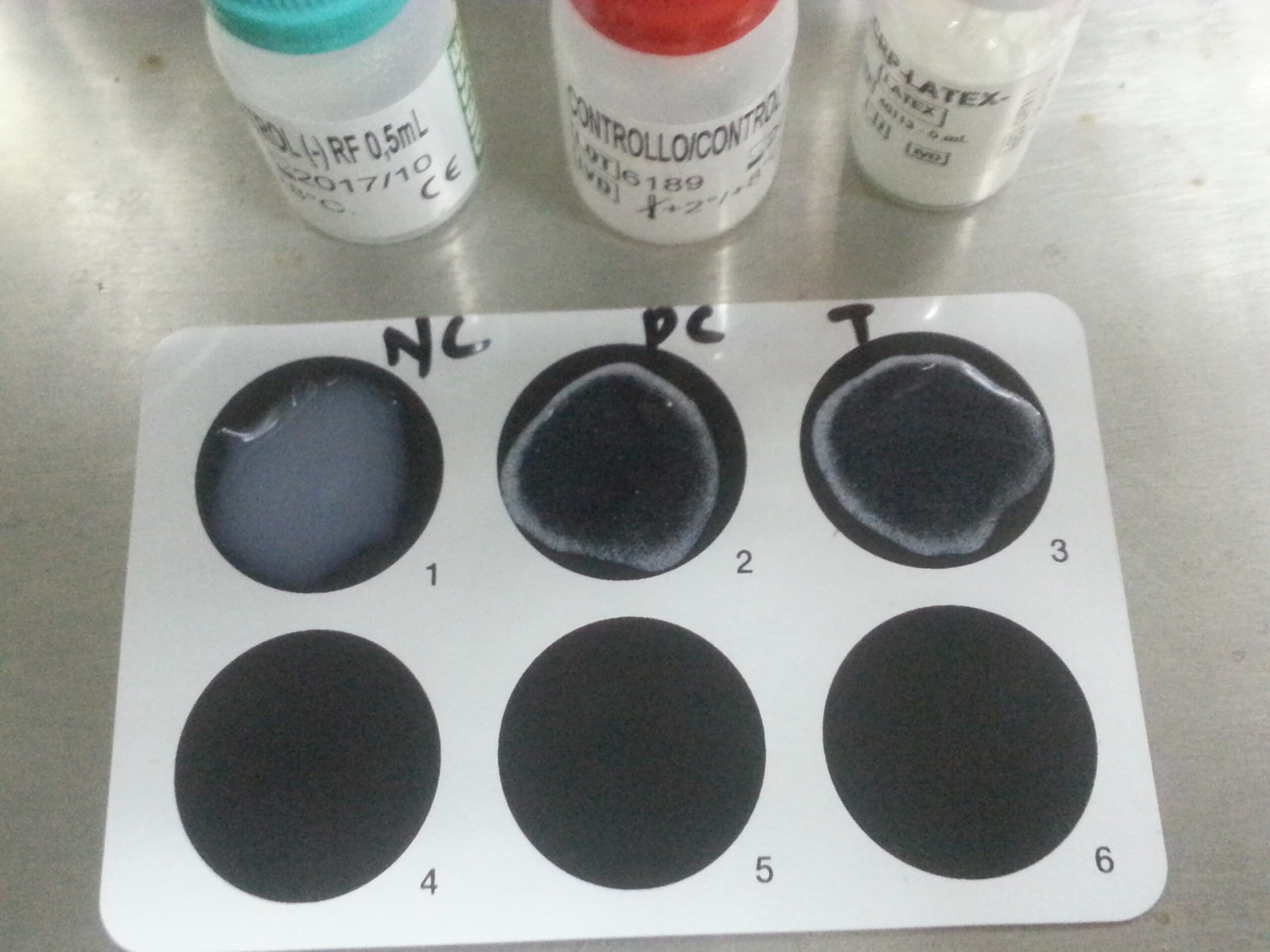
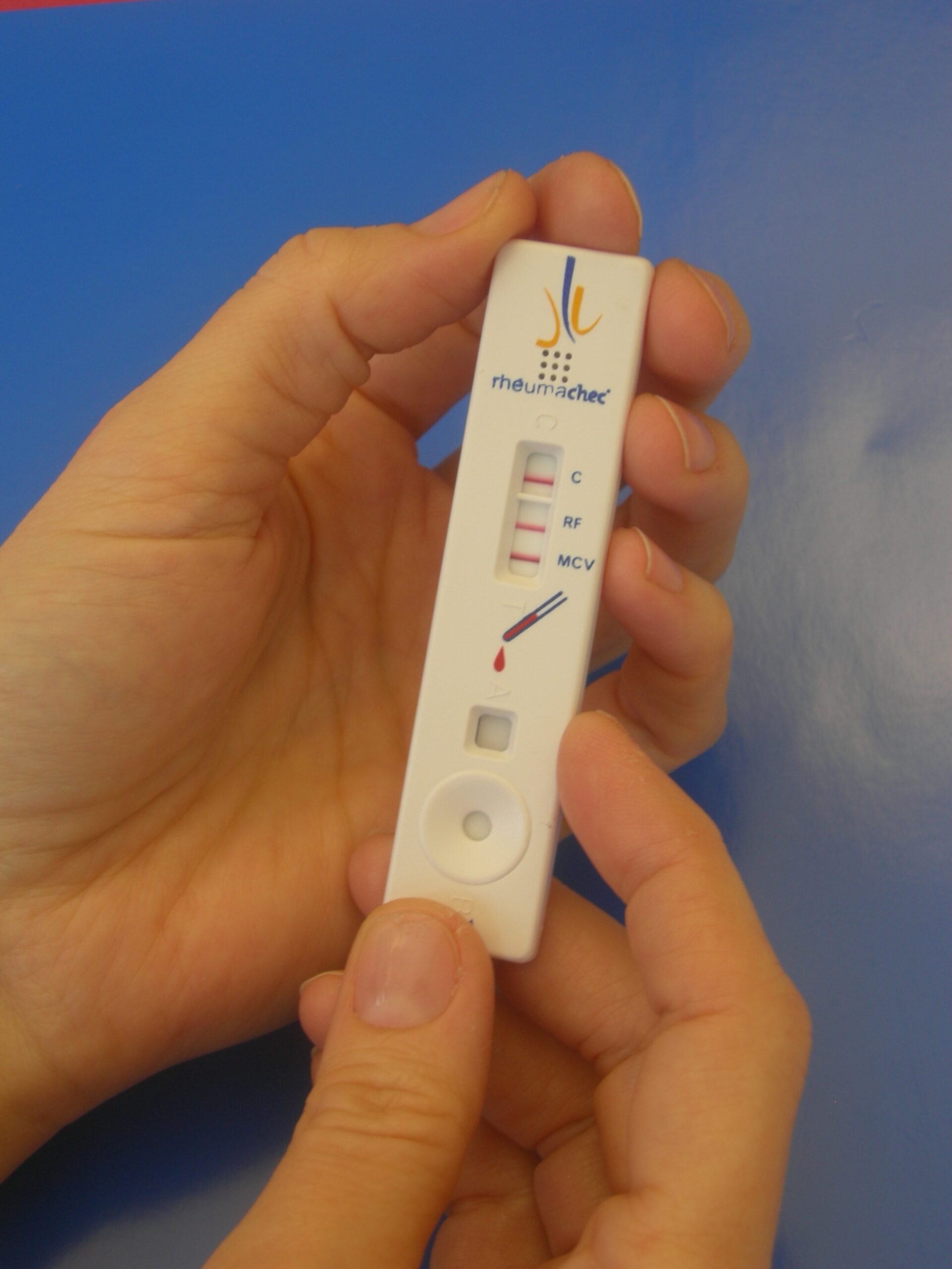
A rheumachec ® rapid test is a membrane-based flow assay for rapid detection of rheumatoid factors (RF) and autoantibodies against mutated citrullinated vimentin (MCV) in whole blood. These are two serological markers used in early detection of rheumatoid arthritis.
X-rays may be used to help track the progression of rheumatoid arthritis in the joints over time.
SLE
Systemic lupus erythematosus (SLE) is an autoimmune disease where the immune system attacks its own healthy body tissues, causing widespread inflammation in affected organs.
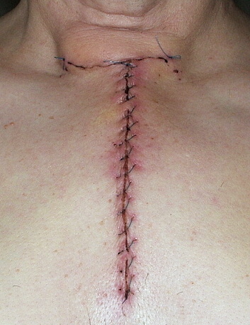
Myasthenia Gravis
Muscle weakness in the voluntary muscles of the eyes and other muscles of the body is common in patients suffering from myasthenia gravis (MG), an autoimmune disease in which the immune system attacks the communication between nerves and muscle.
A thyectomy (surgical removal of the thymus gland) is sometimes recommended in patients with myasthenia gravis to improve disease symptoms and reduce steroid and immunosuppressant use.
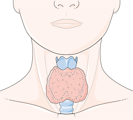
Hashimoto Thyroiditis
Hashimoto thyroiditis is an autoimmune disease in which the immune system attacks healthy cells of the thyroid, which results in a reduction in thyroid hormone production. Enlarged thyroid glands (goiters), weight gain, lethargy, and muscle weakness are common symptoms.
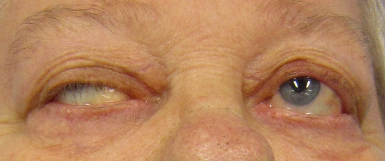
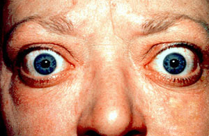
Grave’s Disease
Grave’s Disease is an autoimmune disease that can result in a hyperactive thyroid (hyperthyroidism). Some patients can develop thyroid eye disease, where their eye muscles and tissues swell, leading to proptosis and eyelid retraction.
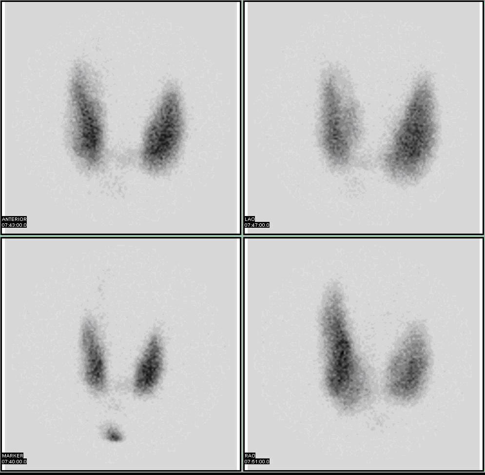
A radioactive iodine uptake (RAIU) test estimates thyroid function by determining the amount of radioactive iodine (taken by mouth) that the thyroid gland takes up. A patient ingests radioactive iodine in liquid or capsule form, and after a certain amount of time (6 and 24 hours later) the radioactivity is measured. Elevated iodine uptake can help diagnose for hyperthyroidism or Grave’s disease.
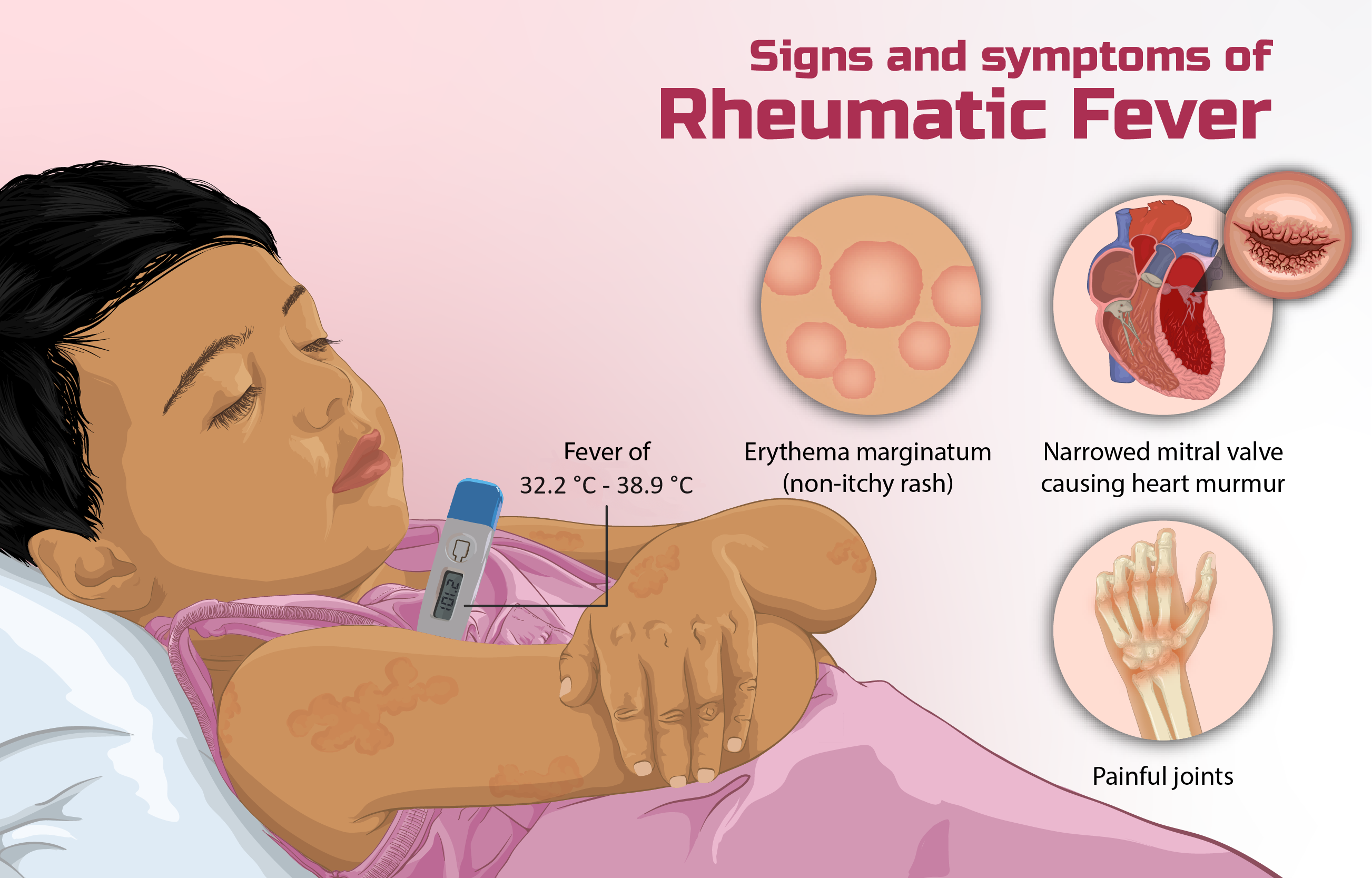
Rheumatic Fever
Rheumatic fever is characterized by inflammation, primarily of the heart, joints, skin, and brain. It is an immune response following a previous infection.
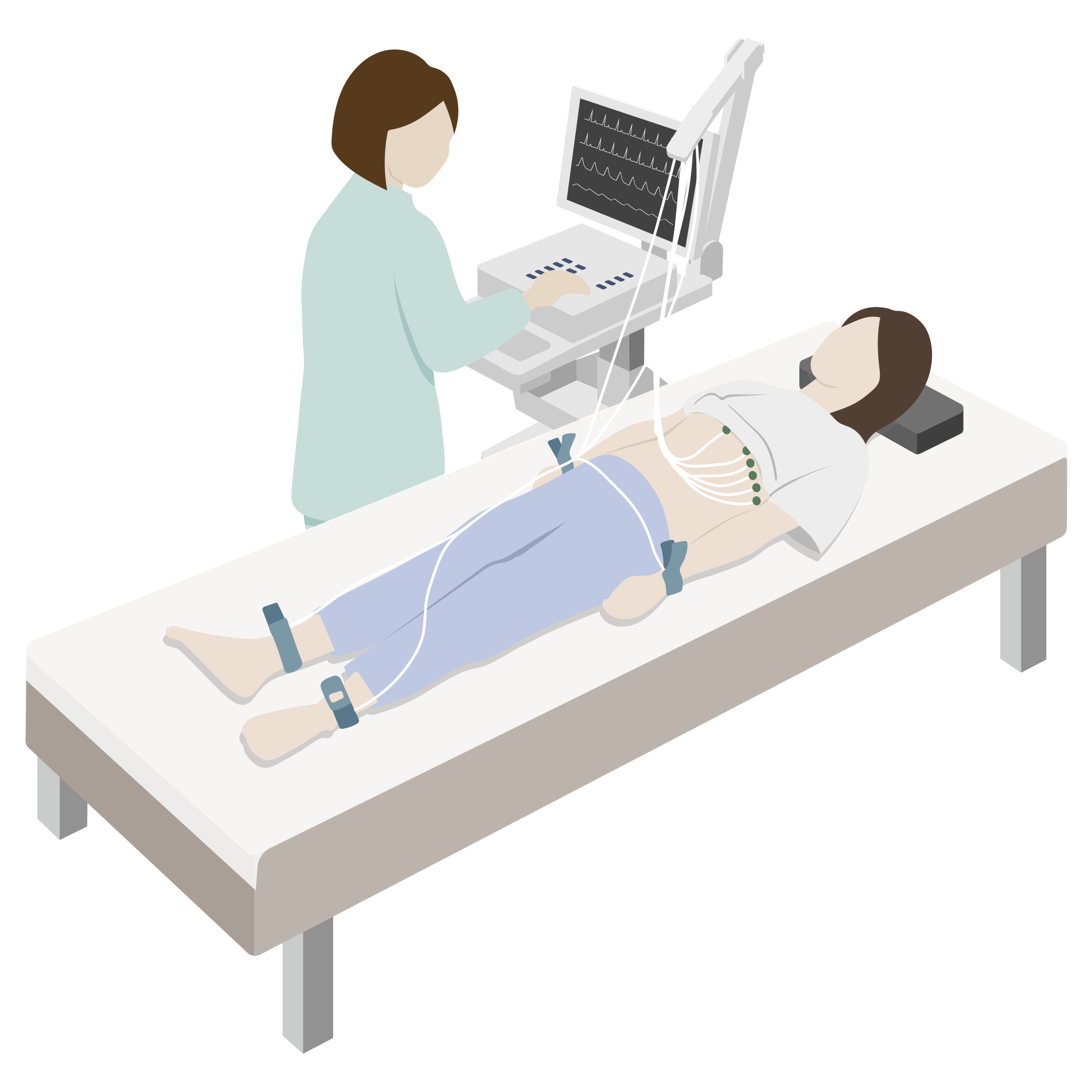
An electrocardiogram is a test that assesses how the heart is beating. It can be used to diagnose irregular heartbeats, one of the hallmark symptoms of rheumatic fever.
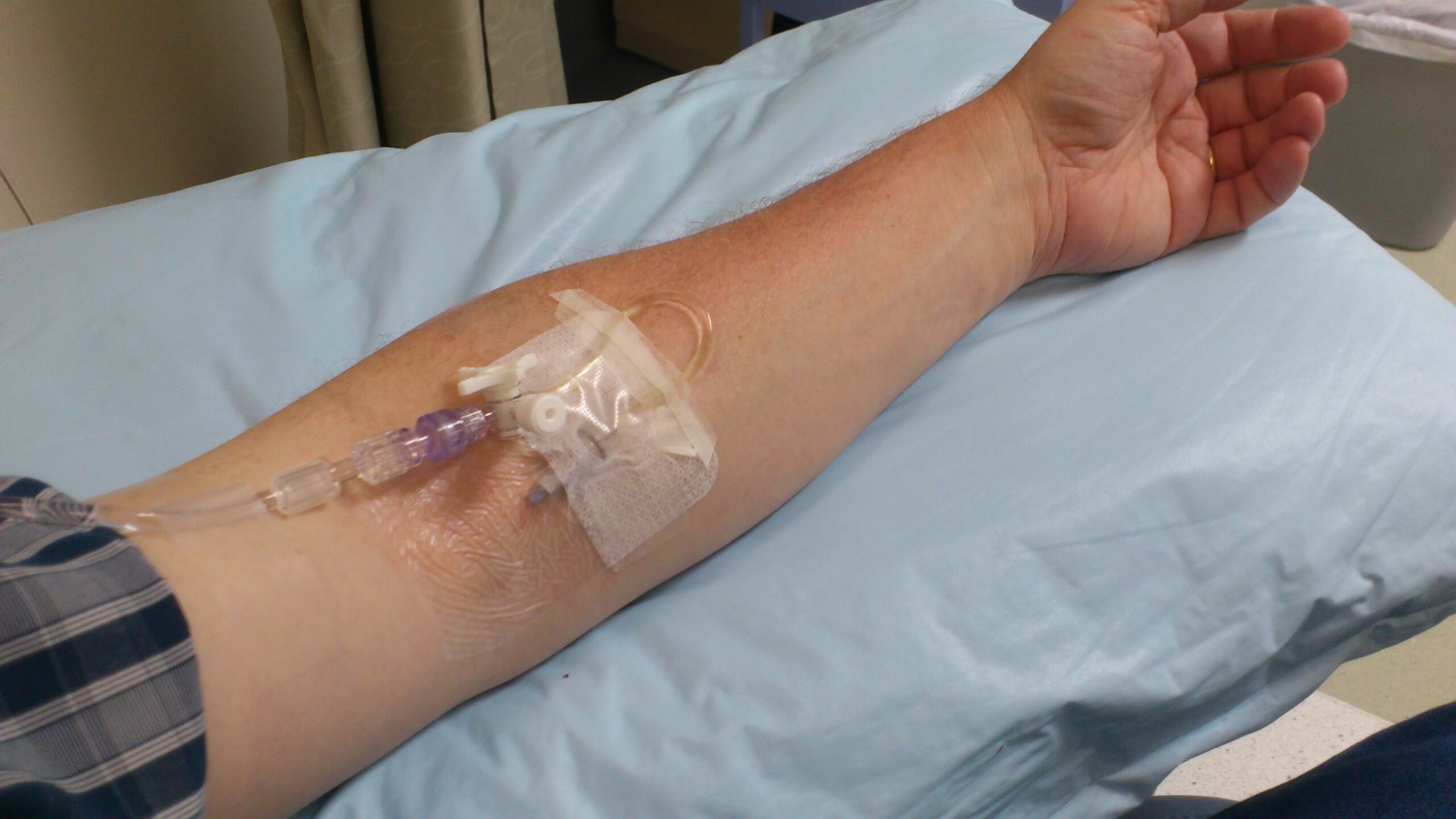
An Intragam infusion (intravenous immunoglobulin replacement) as treatment for Common Variable Immunodeficiency (CVID). CVID is a genetic condition that weakens the immune system due to low antibody levels. As such, patients with CVID typically experience frequent infections and therefore require lifelong treatment.
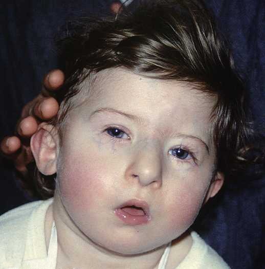
A patient with DiGeorge syndrome, showing characteristic facial appearance, with tubular nose and carp-shaped mouth. DiGeorge syndrome is a genetic condition present from birth caused by a missing portion of chromosome 22. In addition to causing distinct facial features, the condition also affects other organ systems of the body, primarily the heart and immune system.
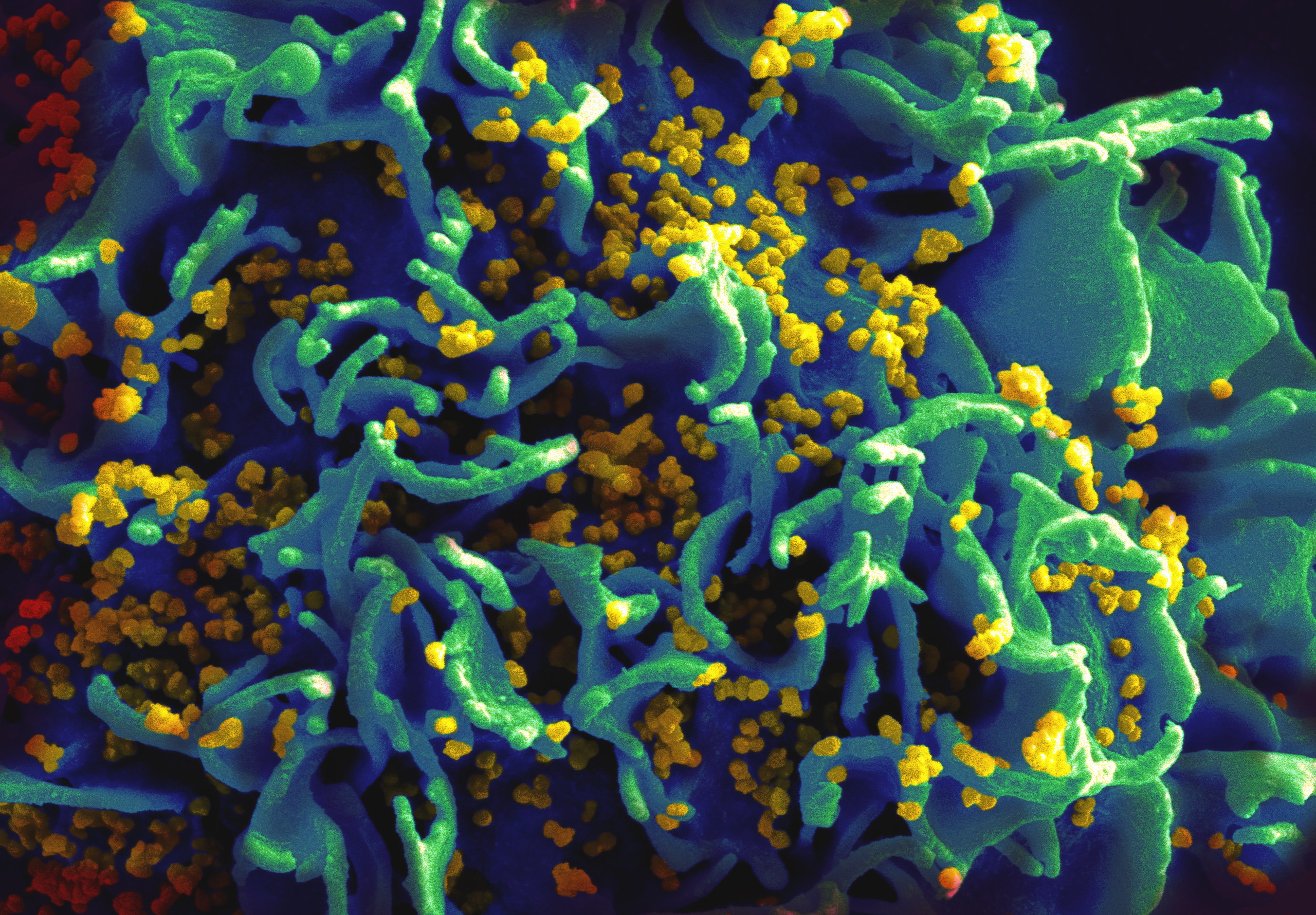
Scanning electron micrograph of human immunodeficiency virus (HIV) particles (yellow) infecting a human T cell. HIV infection of CD4+ T cells results in destruction of those cells, severely impacting the body’s immune response to intracellular pathogens.
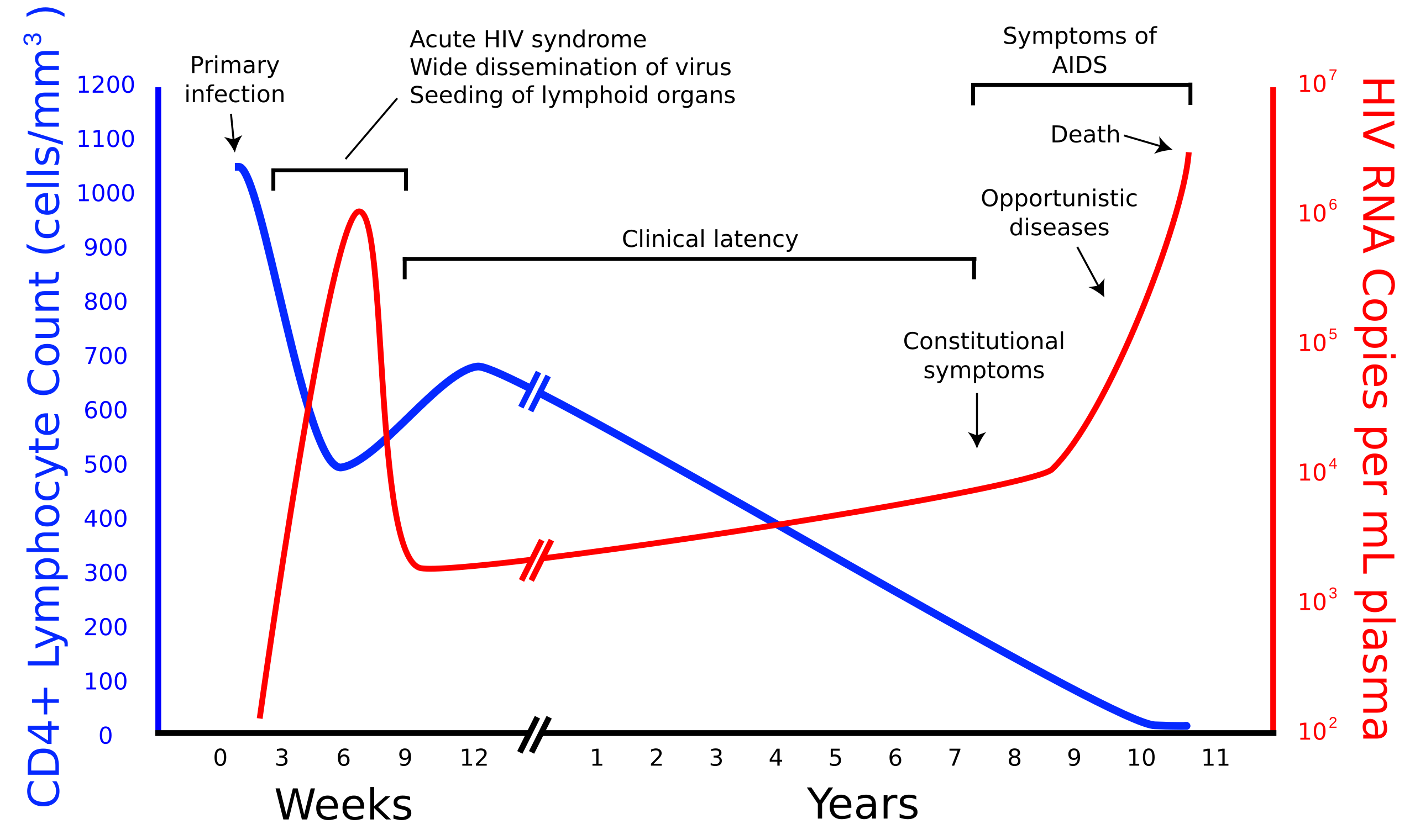
Human immunodeficiency virus (HIV) viral load in relation to CD4+ lymphocyte count and the associated onset of symptoms of infection and subsequent development of Acquired Immunodeficiency Syndrome (AIDS).
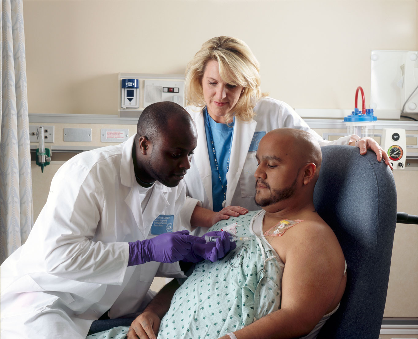
Patient receives chemotherapy drug treatment through a port that is placed in his chest. In addition to their use in cancer treatment, chemotherapy drugs can be administered in low doses to help suppress an overactive immune system.
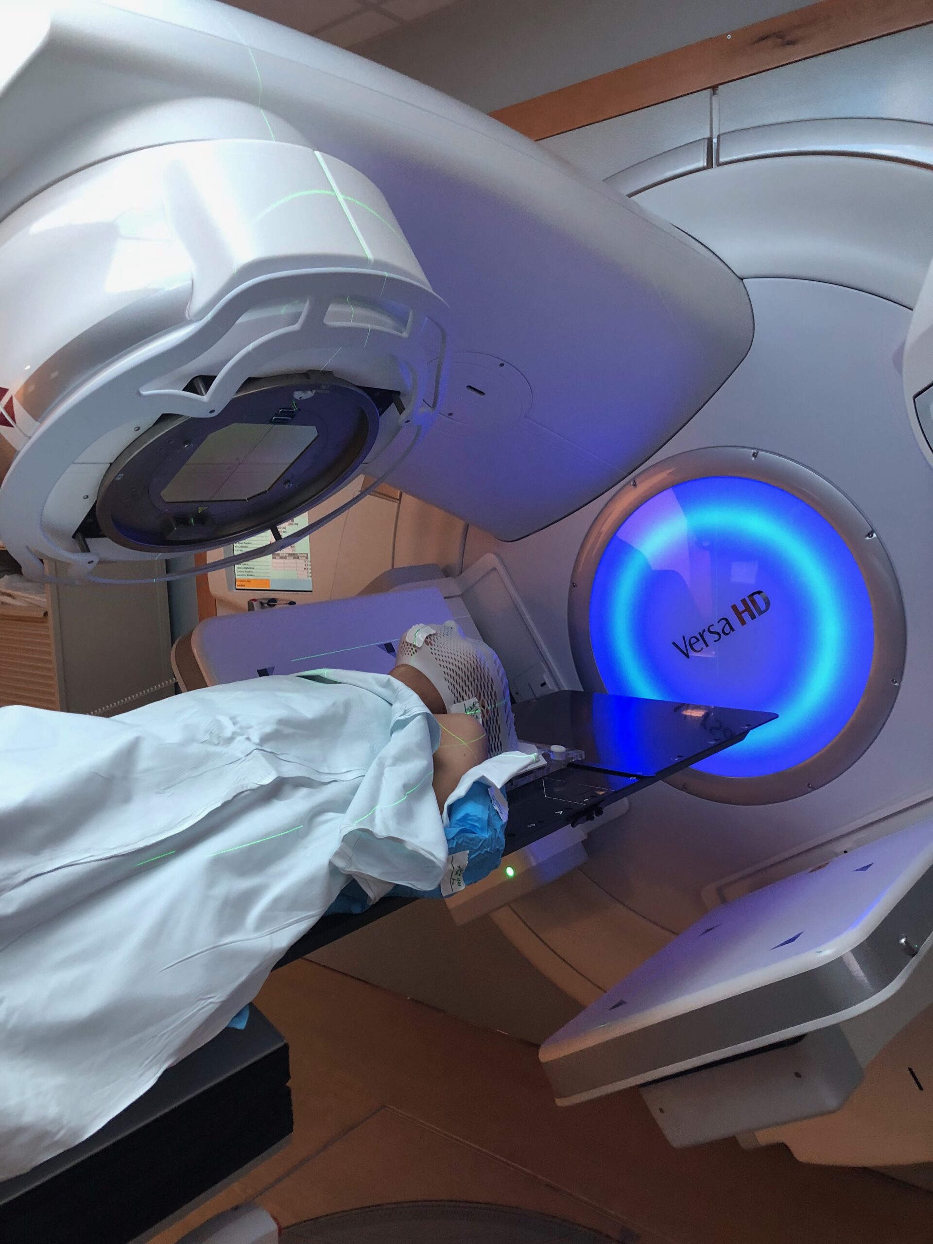
Radiation therapy for Hodgkin’s Lymphoma. Radiation therapy most commonly uses X-rays to apply high energy beams to cancer-affected areas in order to target and kill cancerous cells. However, this is a pro-inflammatory procedure and can increase the incidence of autoimmune reactions.
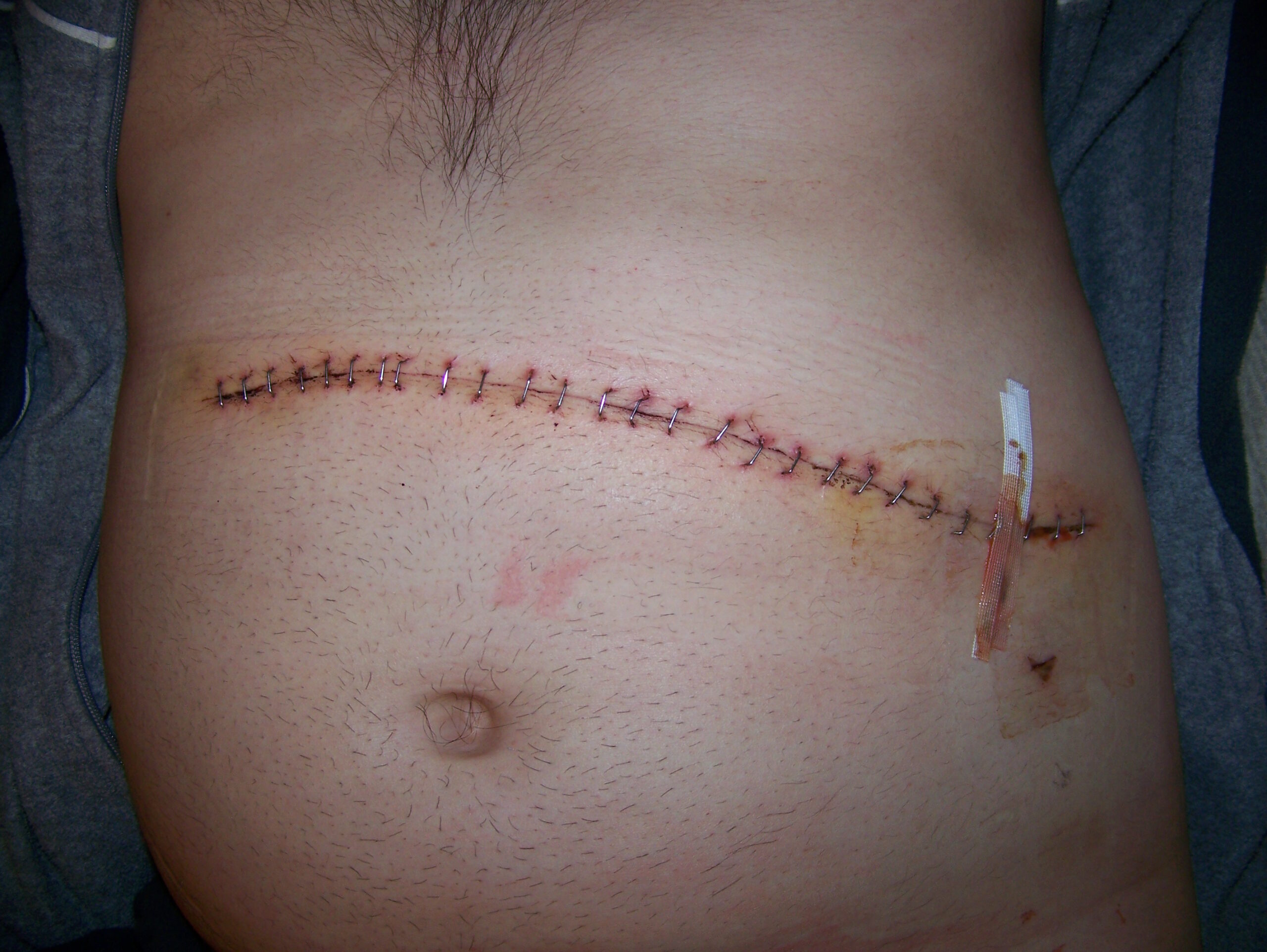
Scar following splenectomy. This procedure partially or completely removes the spleen, an organ important for immunological function.
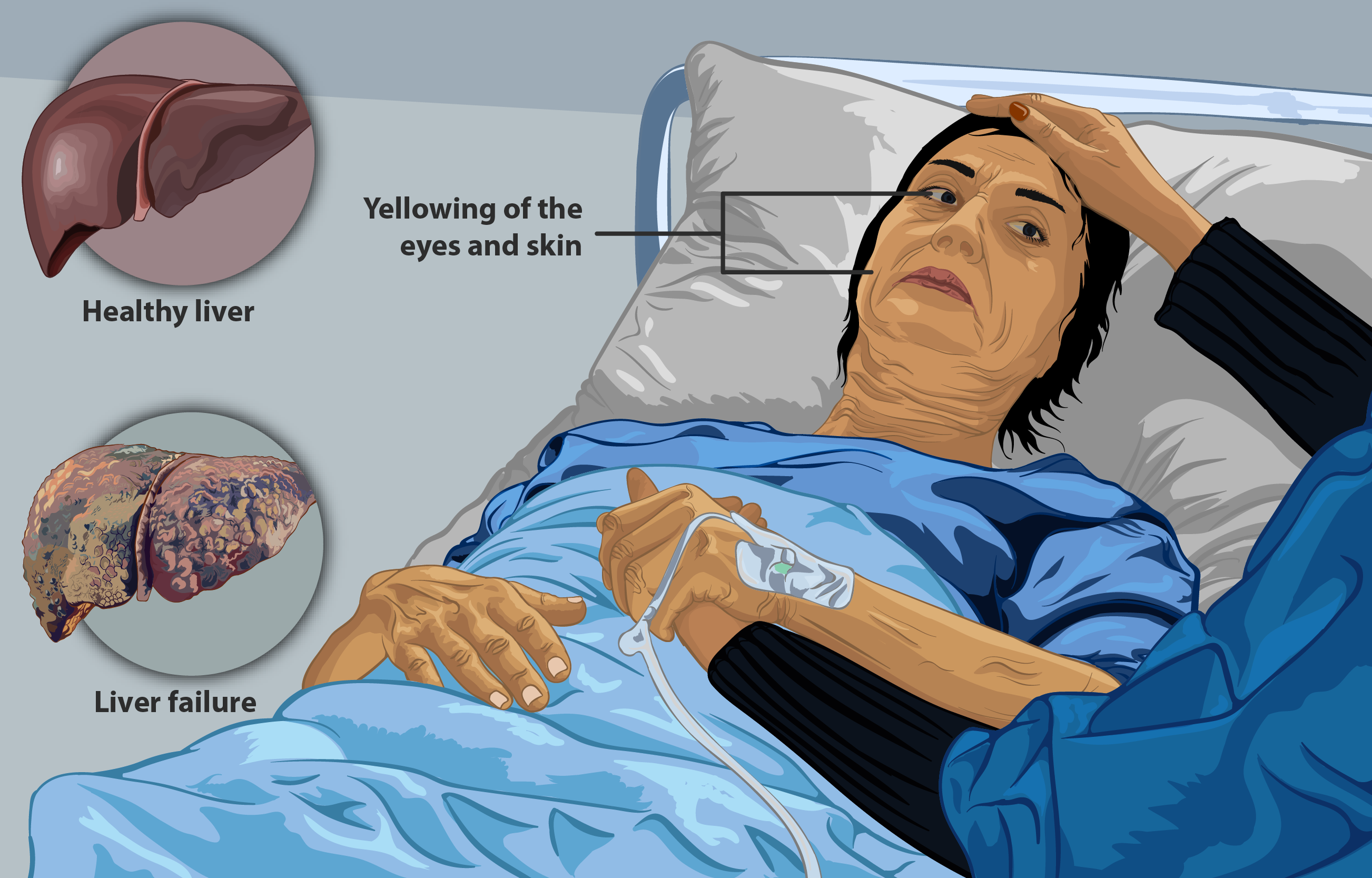
The liver is responsible for an array of essential bodily functions, including filtering blood. Early stage symptoms include jaundice (yellowing of the eyes and skin), while late stages of liver disease includes liver failure.
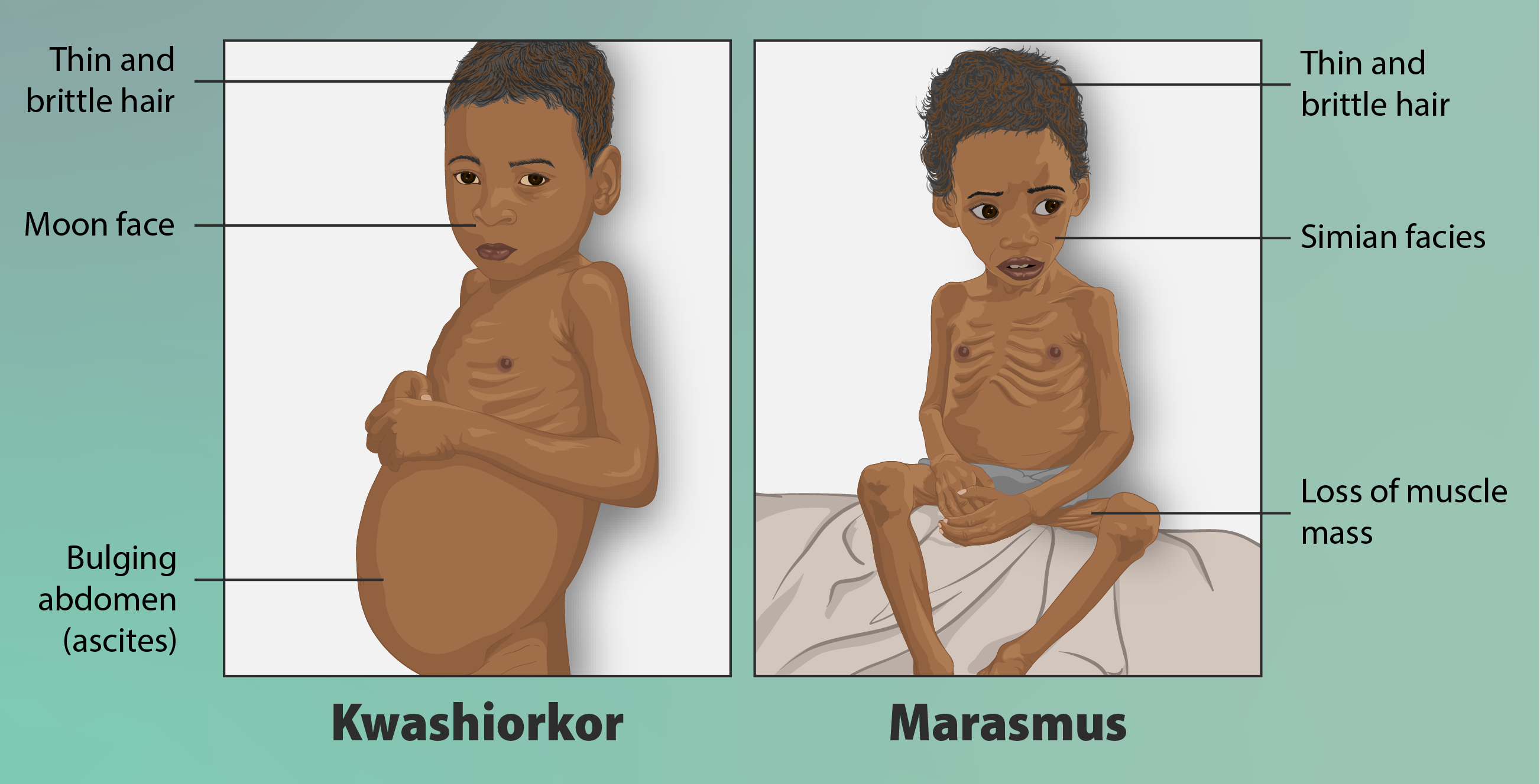
Malnutrition is a suite of conditions that consist of inadequate, imbalance, or excess energy and/or nutrient consumption. Kwashiorkor malnutrition is defined by severe protein deficiency, while marasmus malnutrition is marked by a deficiency in all macronutrients. However, obesity, an excess in energy and nutrient consumption, is also considered to be a form of malnutrition.
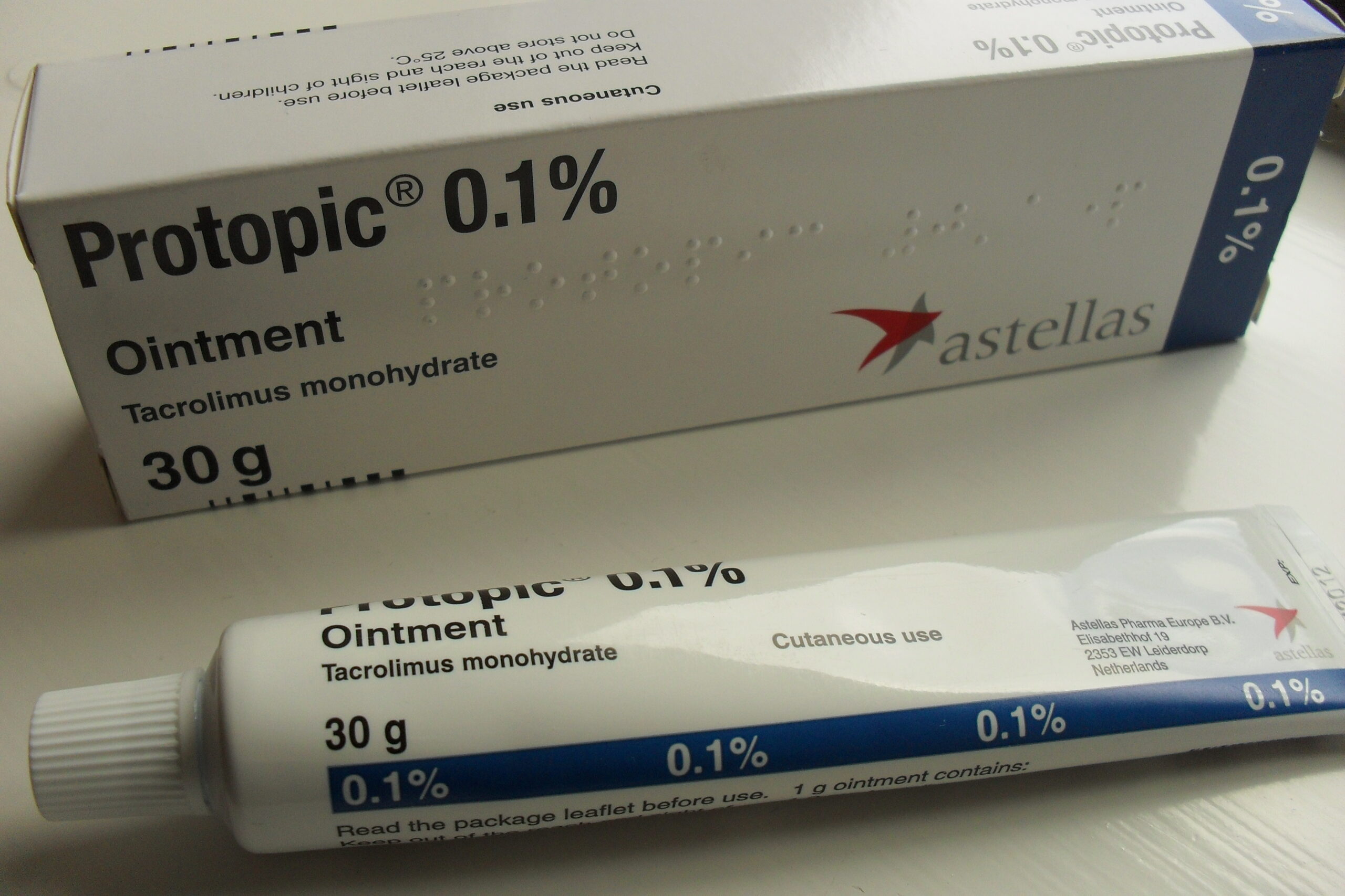
Immunosuppressant drugs are used to suppress a myriad of allergic, inflammatory, and/or autoimmune disorders. These drugs are also prescribed following organ transplant to reduce risk of rejection (for example, Tacrolimus).
Hydrocortisone is an anti-inflammatory glucocorticosteroid drug, typically applied as a topical treatment. Hydrocortisone is the synthetic version of the naturally produced hormone cortisol.


.jpg)