Chapter 2 The Inflammatory Response, Fever, Healing, Cell Proliferation, Tissue Regeneration and Repair
Chapter 2 Cellular Damage and Tissue Healing – Portiaa
Zoë Soon
Creative Commons – Simple Pictures, Images, Video Clips, and/or Gifs that help illustrate any of the following:
Chapter 2 Learning Outcomes:
By the end of this section you will be able to:
Describe Normal Defenses of the Body:
- Innate (non-specific) Defenses:
- Mechanical/Physical – skin, hair, mucus, sebum, urination, cilia, cell shedding
- Biochemical – sweat, tears & saliva (lysozymes), bile, stomach pH, cerumen, mucus, vaginal secretions, prostatic and testicular secretions,
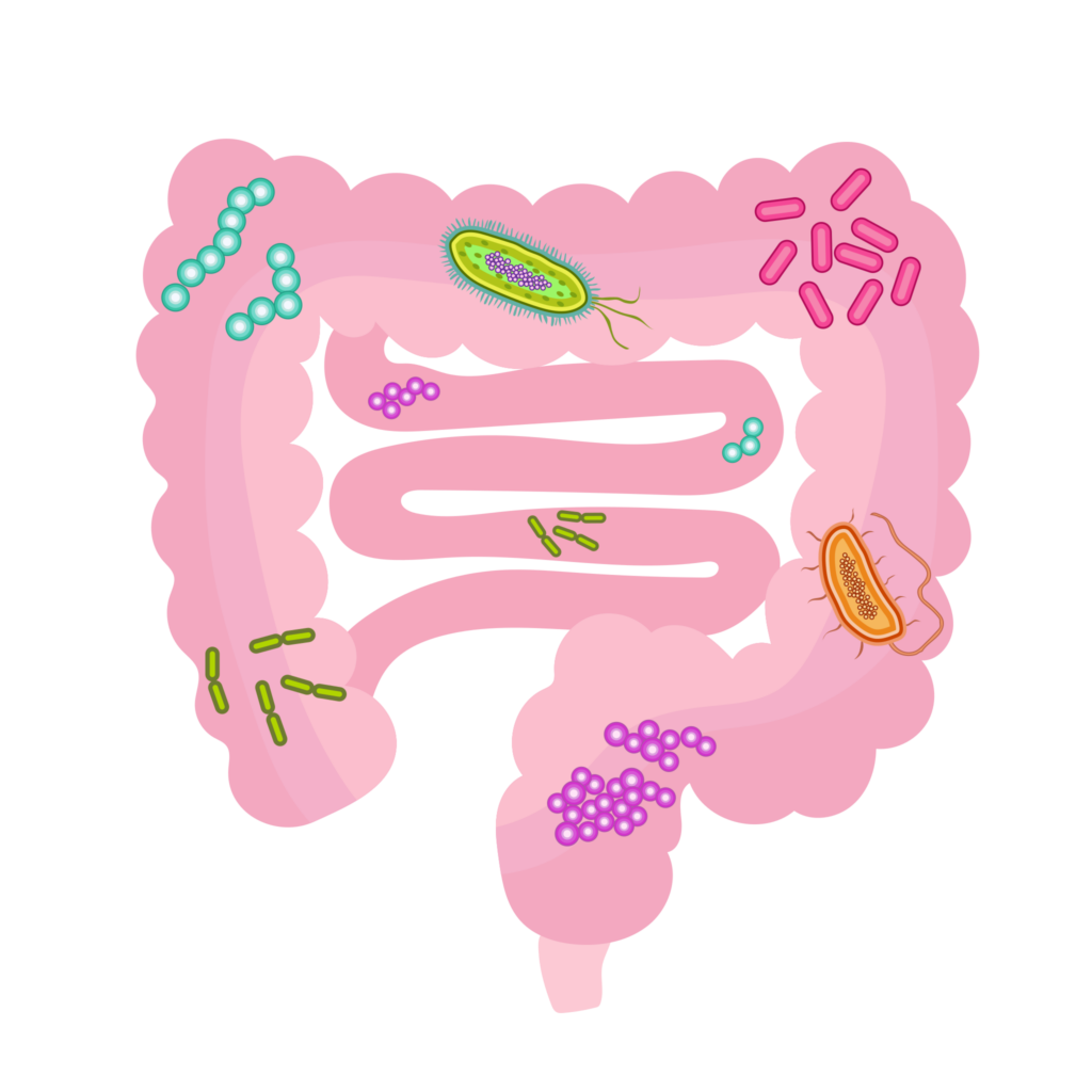
- Normal Flora
-
-
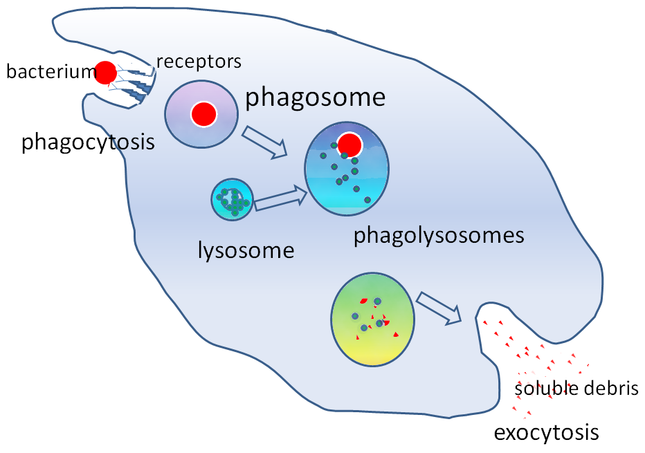
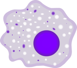
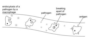
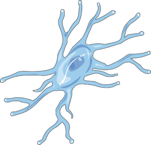
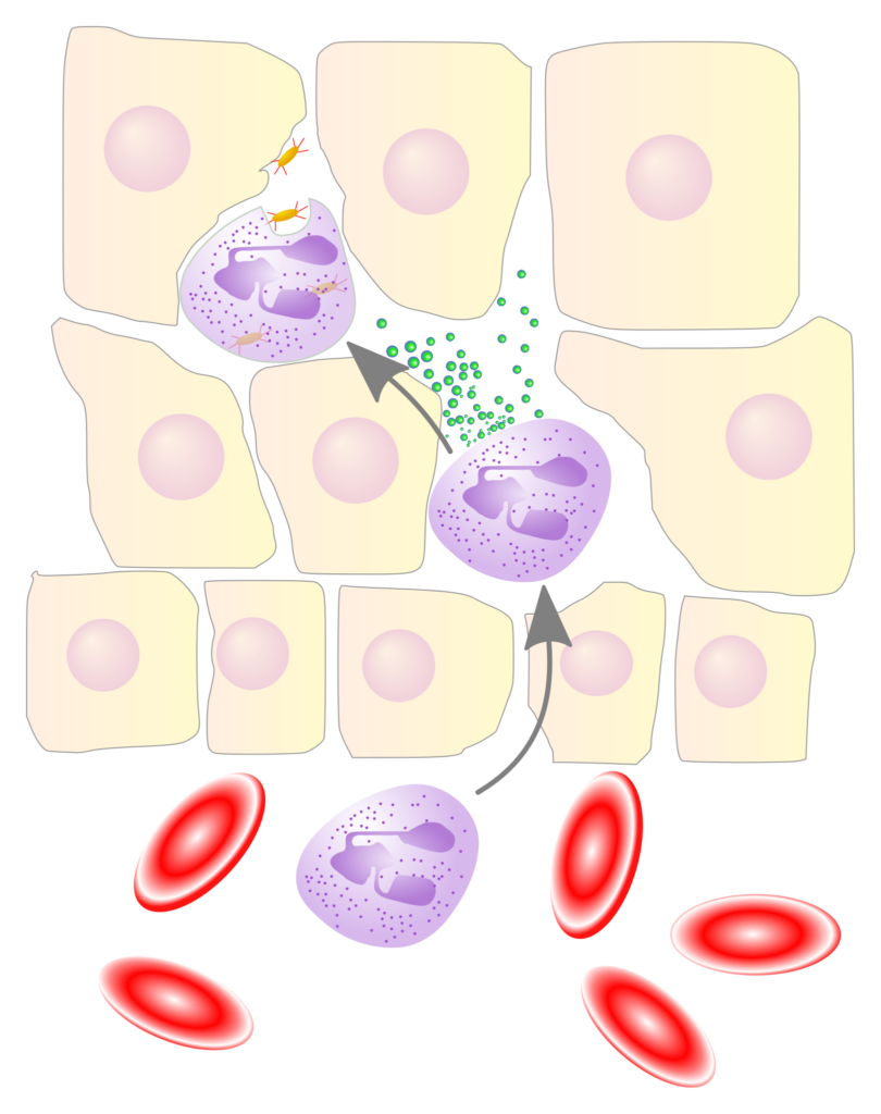
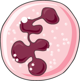
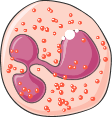
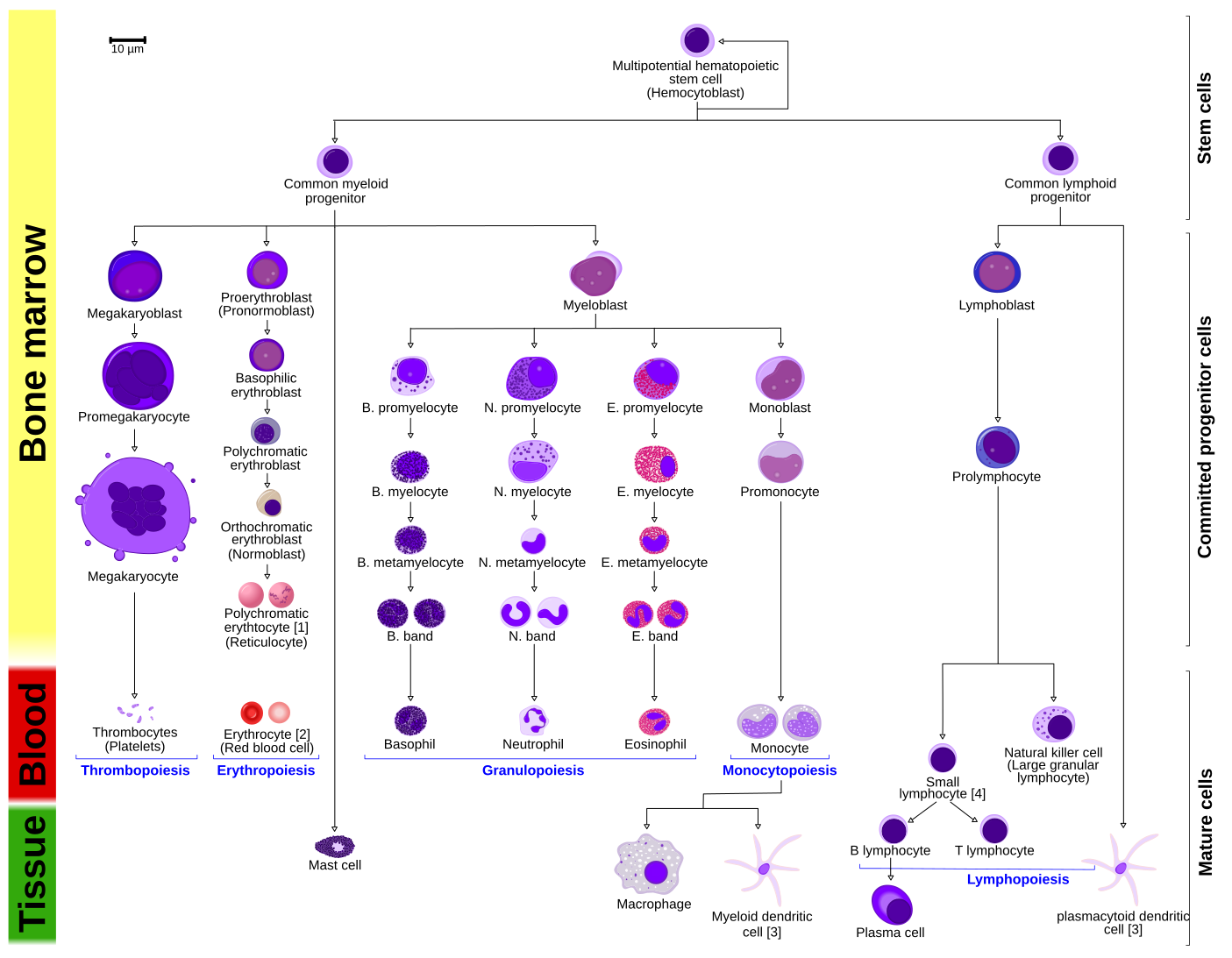
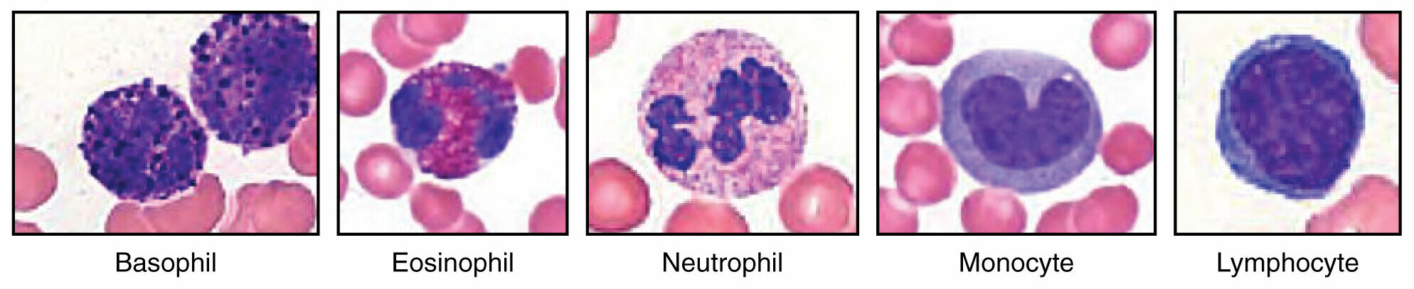
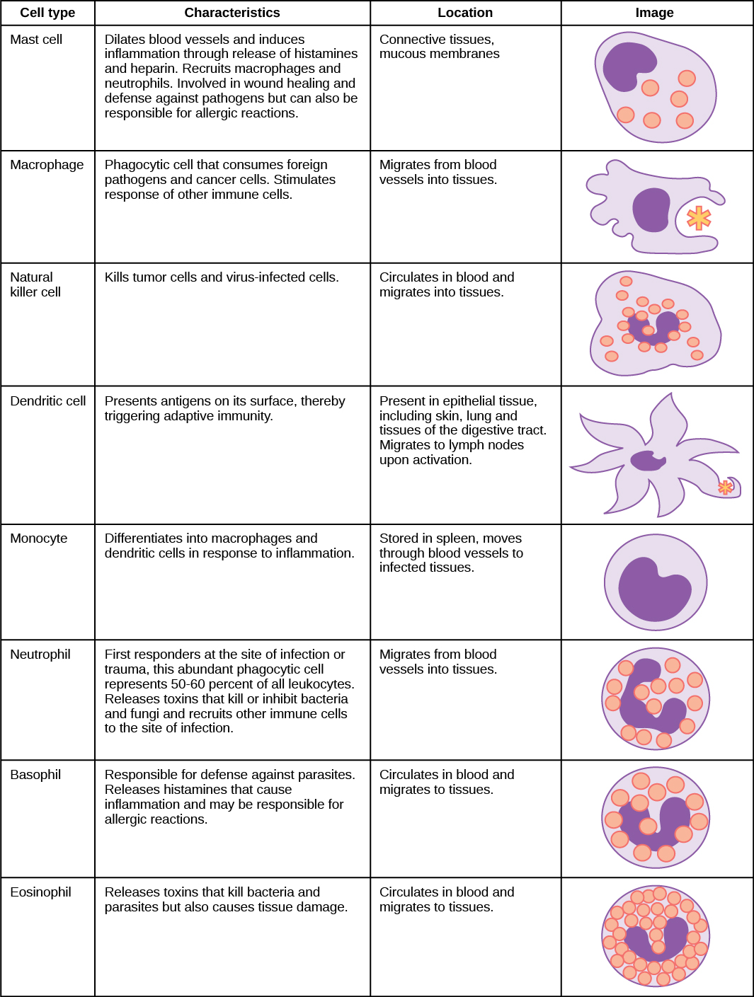
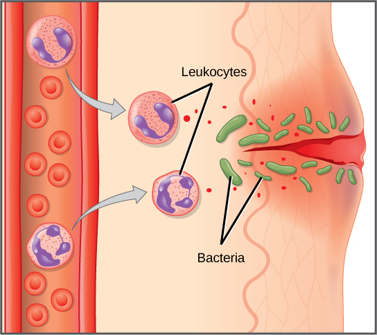
Phagocytes: (WBCs such as monocytes, fixed and free macrophages, microglia, neutrophils, eosinophils, dendritic cells) capable of diapedesis/emigration/transmigration.
-

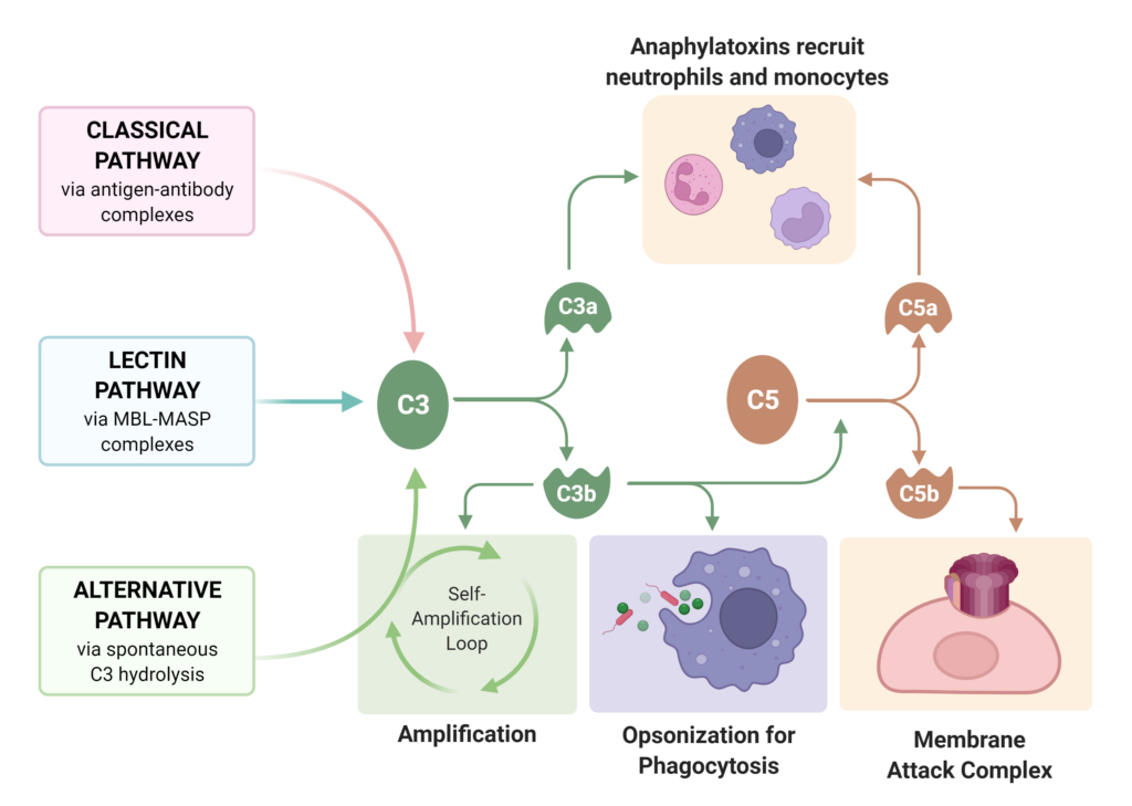
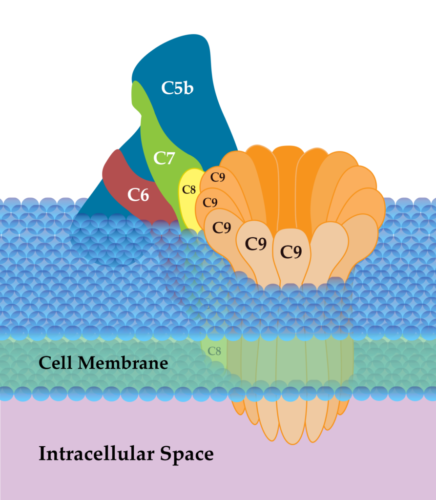
- Complement System (Classical Pathway with antibody, Lectin Pathway, and Alternative Pathway) – involving 30+ complement plasma protein cascade of activation – resulting in opsonization, MAC (Membrane Attack Complexes), stimulation of mast cells & basophils


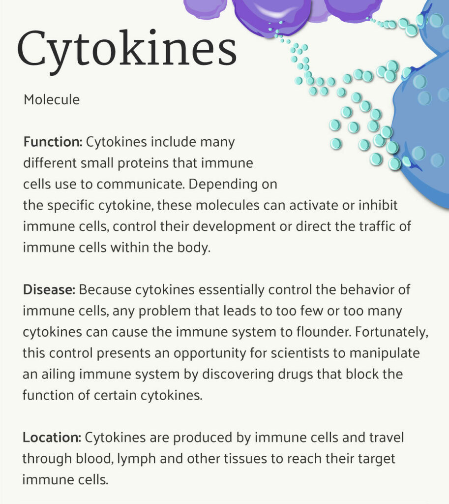
- Cytokine family: Glycoproteins produced by WBCs, fibroblasts, endothelial cells, stromal (connect tissue) cells
-
-
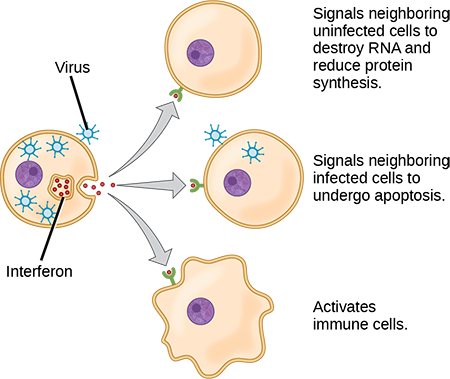
Interferons: (chemical messages that stimulate defense)
-
-
-
- Alpha Interferons – produced by virally infected host cells to attract & stimulate NK cells and stimulate AVP production in neighbouring cells.
- Beta Interferons – produced by fibroblasts to slow inflammation, and promote healing
- Gamma Interferons – produced by T & NK cells to stimulate macrophage activity
-
-
-
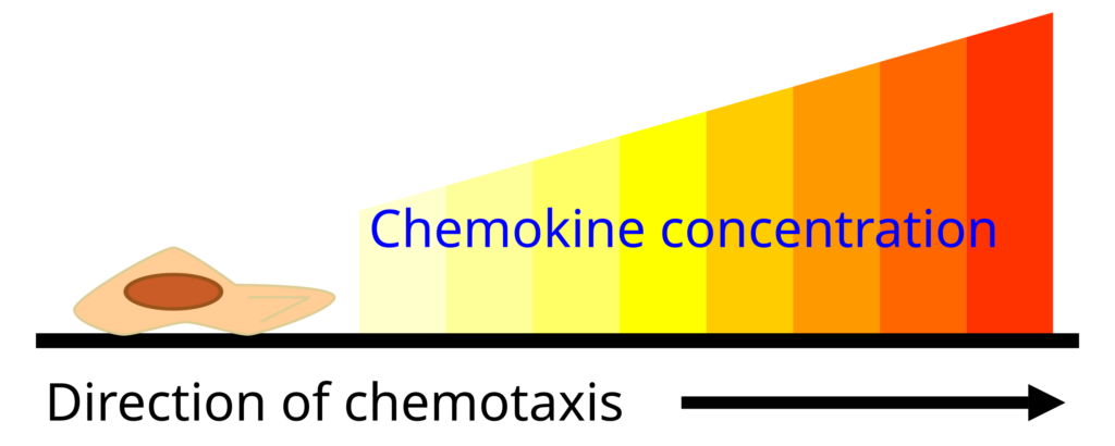
Chemokines: induce chemotaxis
-
-
-
Lymphokines: produced by T lymphocytes to: 1) attract macrophages & 2) stimulate B lymphocytes to produce antibodies
-
Interleukins: produced by helper T cells to:1. activate macrophages and stimulate fever (act as endogenous pyrogens)2. stimulate T & B cell differentiation3. Stimulate hemopoietic cells to proliferate → producing more WBCs
-
Natural Killer cells (NK Lymphocytes) – type of WBC (White Blood Cell/Leukocyte)
-
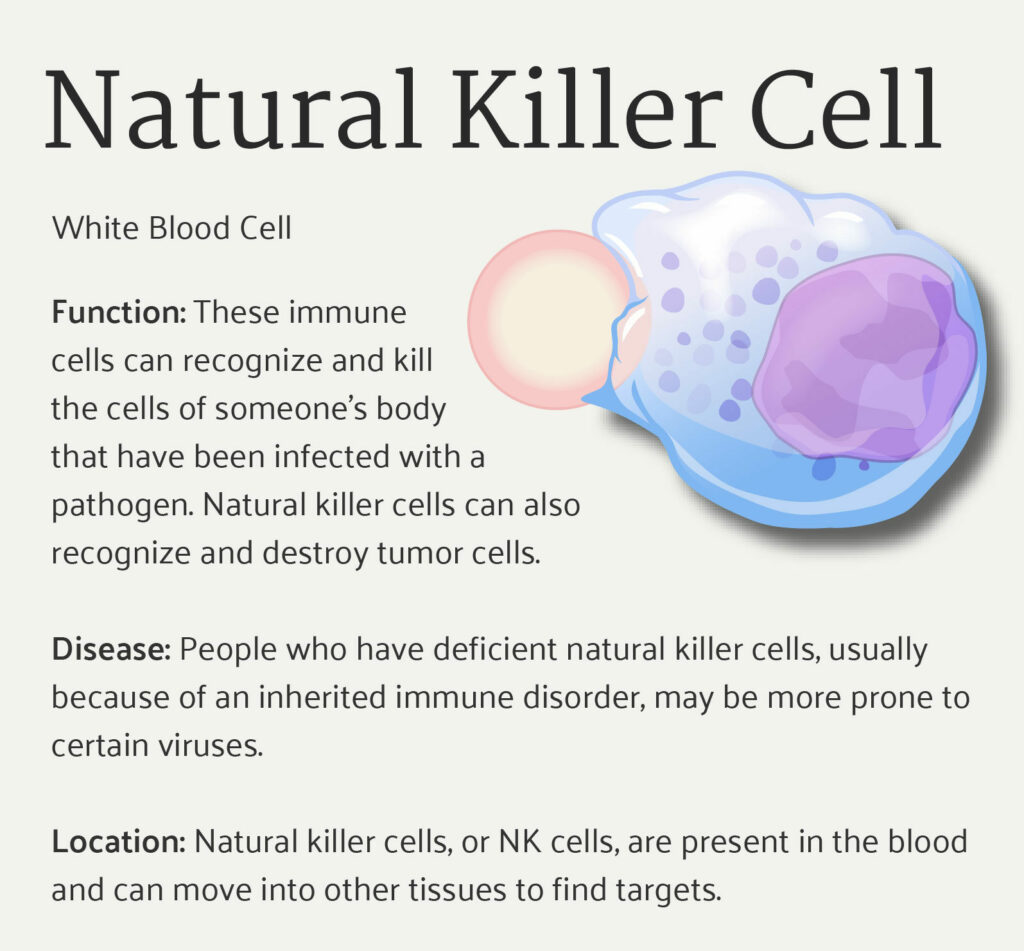
- Inflammatory Response
- Fever – speeds up WBC activity and repairs, inhibits pathogen activity
- Describe plasma components & define vocabulary words:
-
- Plasma – liquid matrix containing water, electrolytes, and plasma proteins
-
- Plasma proteins – antibodies, complement proteins, clotting factors, albumen and transporter proteins
-
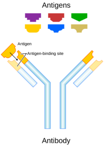
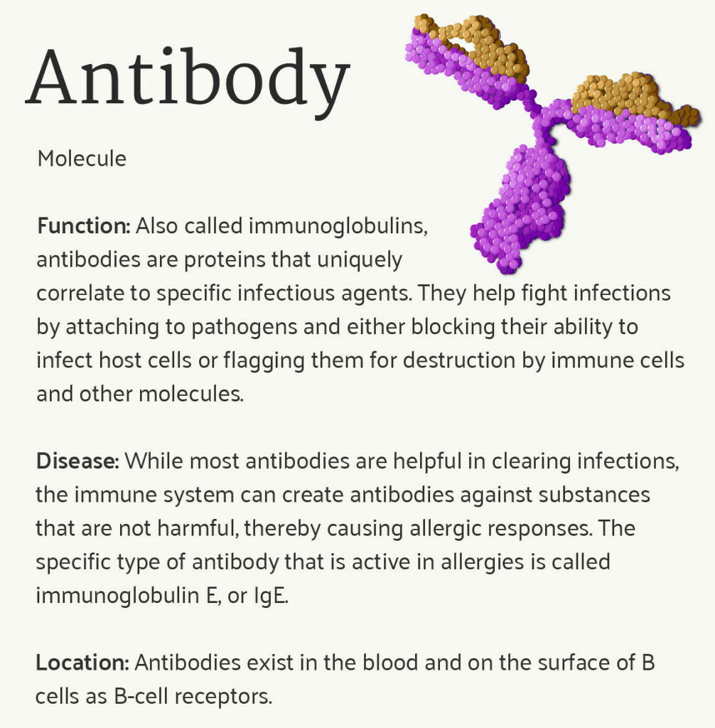
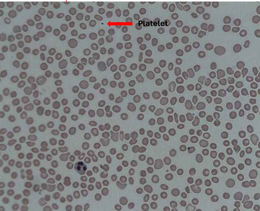
Structure of antibody molecule (protein). These molecules circulate in the blood, recognize foreign particles, and neutralize them. - Platelets/Thrombocytes – a nuclear cell fragments formed from large megakaryocytes; involved in clotting (hemostasis)
-
- Leukocytes – WBCs
- Lymphocytes: type of WBC involved in antibody production (B lymphocytes), targeted immune response (T lymphocytes), and surveillance (NK lymphocytes)
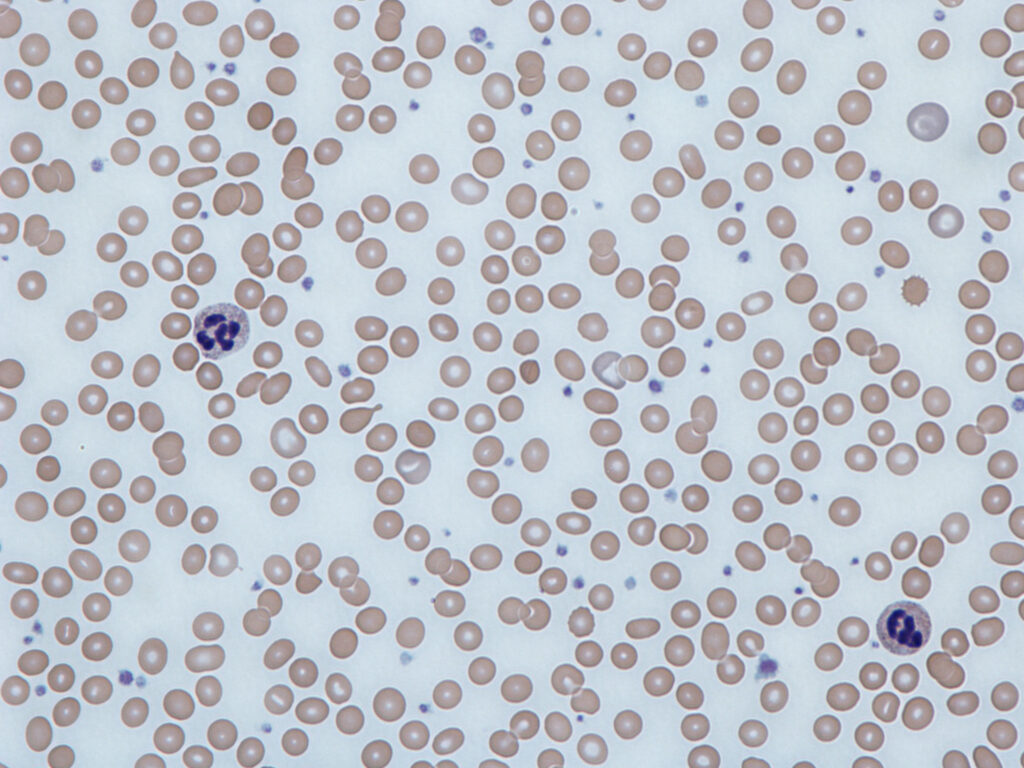
- Neutrophils: The most abundant phagocyte in the blood; contain extensive lysosomes
- Eosinophils: Destroy parasitic worms & immune complexes
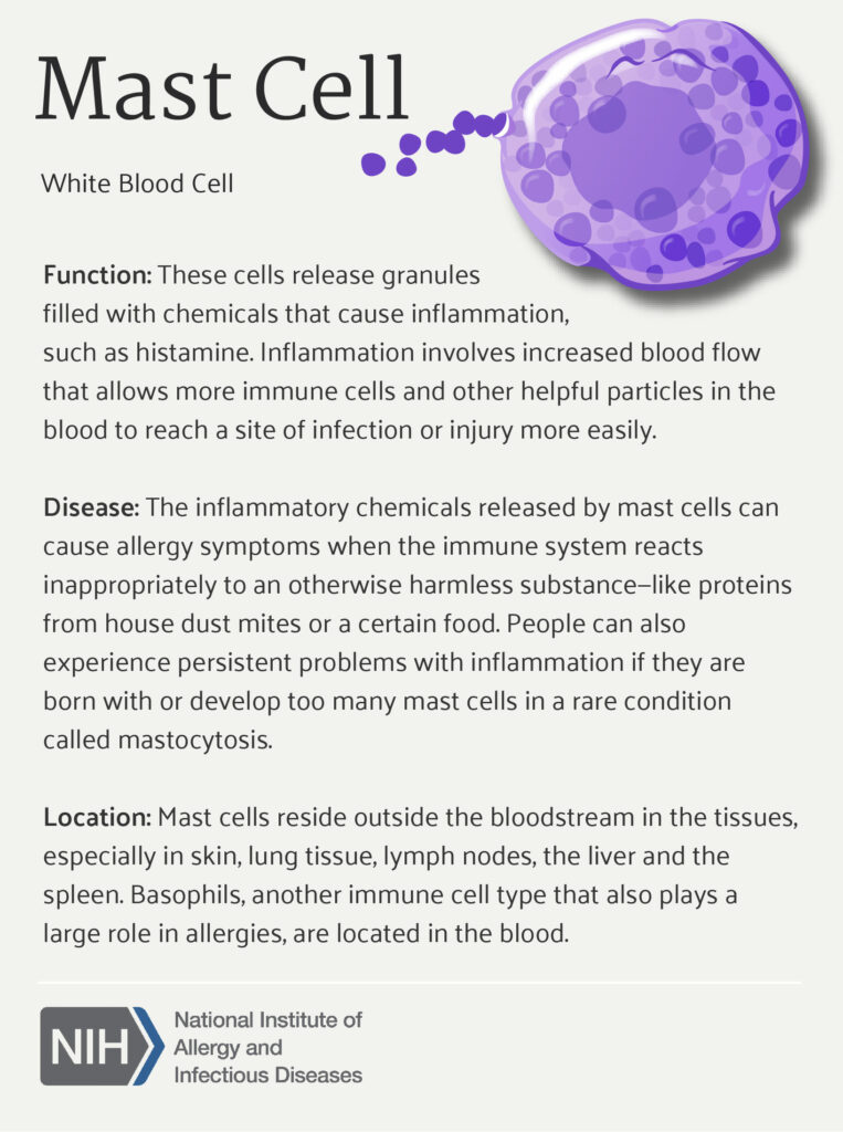
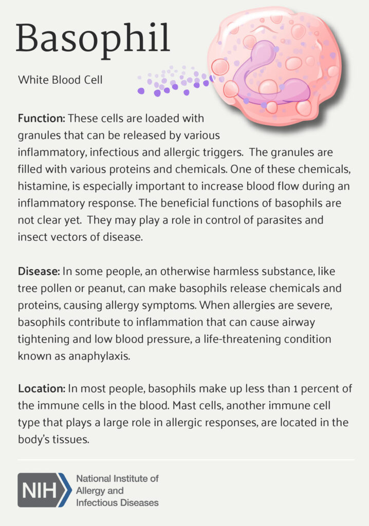
- Basophils & Mast cells: Release histamine, heparin, prostaglandins, and leukotrienes in process known as degranulation
-
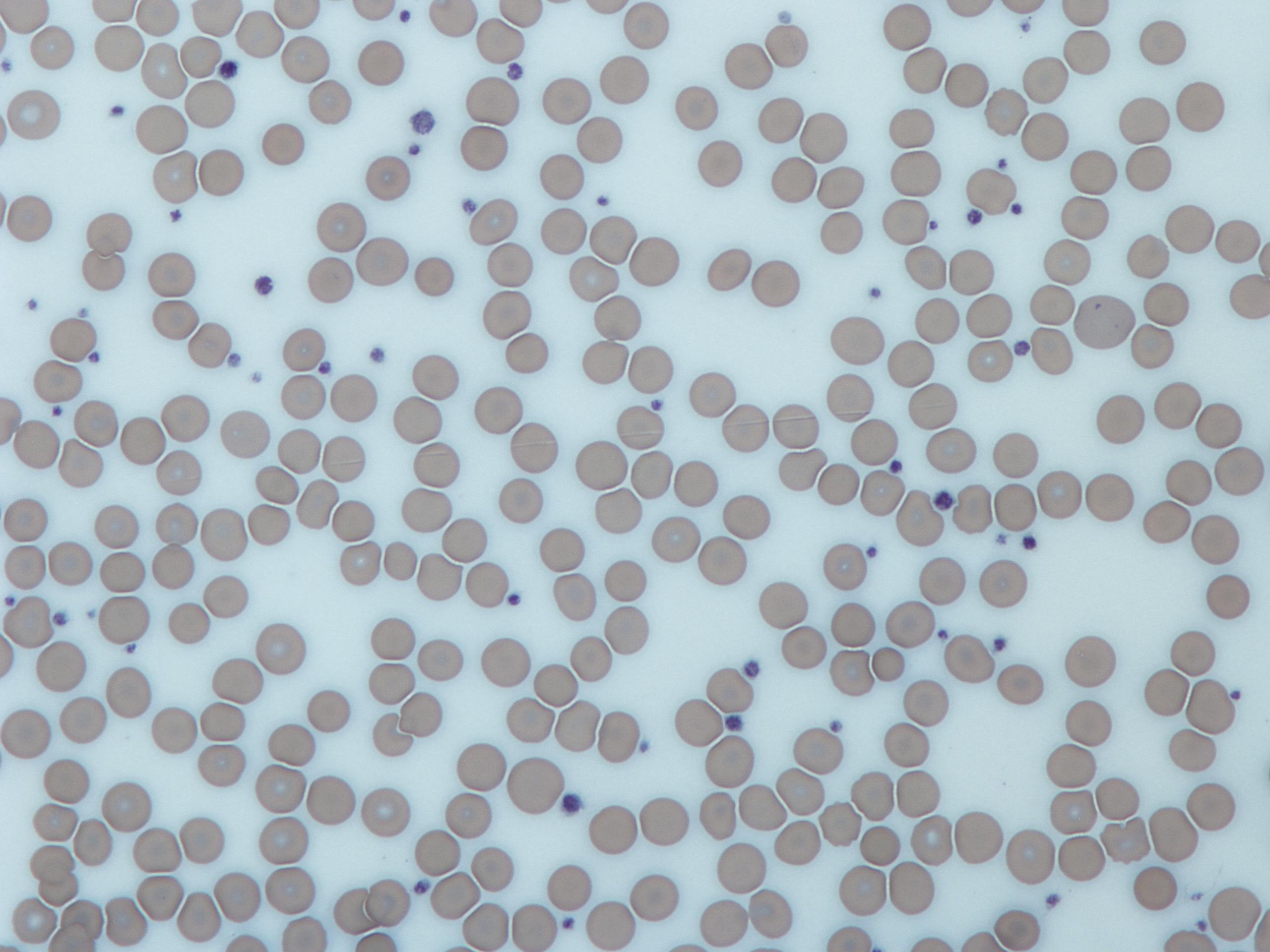
- Erythrocytes – RBCs; transport oxygen & carbon dioxide
-
- Hematocrit – % by volume of blood that is formed elements
- Anemia: reduced oxygen-carrying capacity of blood due to low levels of functional RBCs or hemoglobin.
-
-
- Polycythemia; greater than normal # of RBCs
- EPO, erythropoietin: hormone that stimulates production of RBCs
-
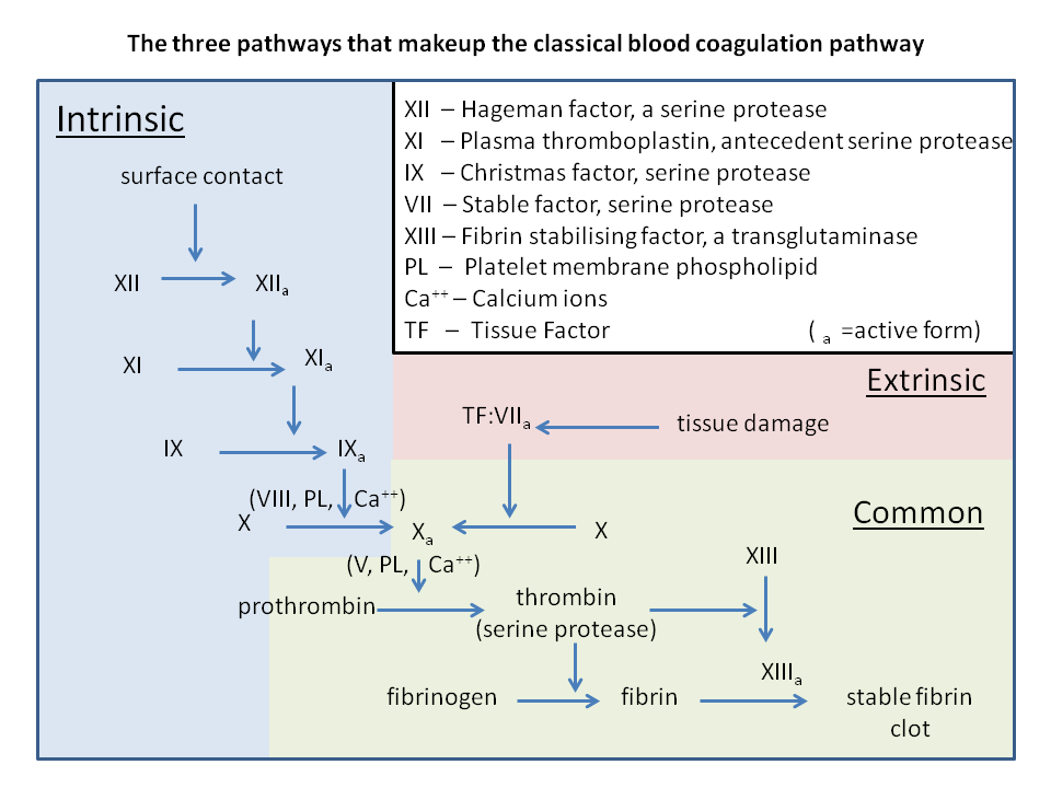
- Describe 3 stages of hemostasis: vascular spasm (role of endothelin and tunica media), platelet plug formation (extrinsic and intrinsic pathways, roles of Factor X, thrombin, clotting factors, and Ca++) and coagulation (role of fibrin)
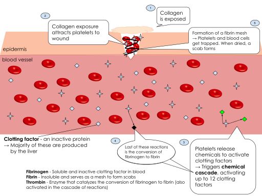
- Describe stages of healing: fibrinolysis (role of tPA, and plasmin) and regeneration (role of PDGF)
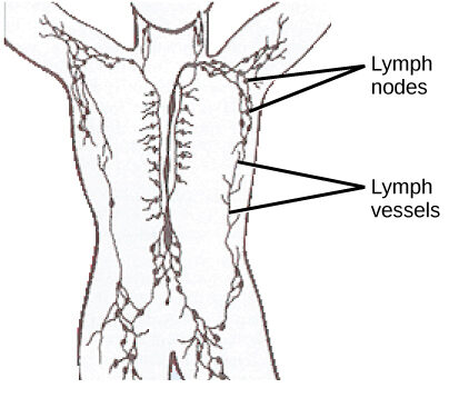
- Describe components of the Lymphatic System:
- Lymph Vessels
- Lymph Nodes
-
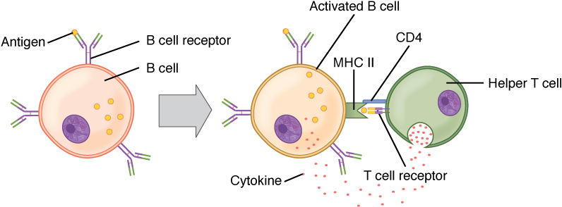
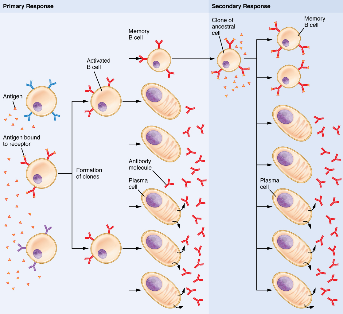
- Lymphocytes (Helper T, Cytotoxic T, Memory T, Suppressor/Regulator T, B, Memory B, plasma cells), Cell-mediated and Humoral Immunity
-
- Macrophages, Dendritic Cells
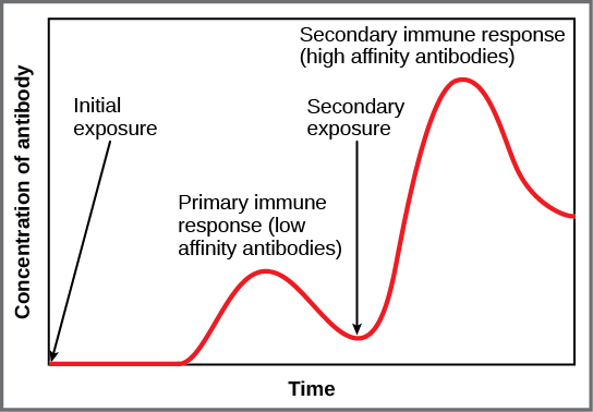
- Primary and Secondary Response, Vaccination

-
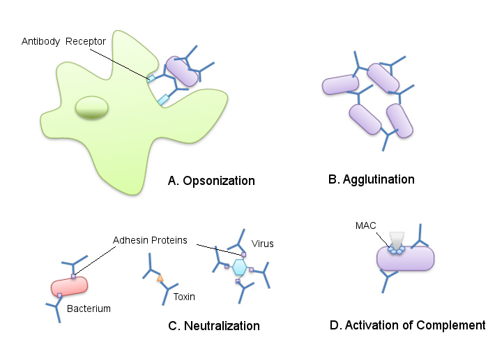
- Antibody Roles – Neutralization, Agglutination, Precipitation, Opsonization
-
- Cytotoxic T cell activity – perforin, lymphotoxin, apoptosis
-
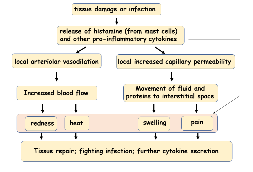
- Explain cause of Inflammation – innate (non-specific) response to tissue injury; caused by: tissue damage from cuts, sprains, chemicals, ischemia, heat, cold, infections, or foreign objects
-
-
- Stimulated by vasoactive chemicals released by mast cells: histamine, prostaglandin, leukotrienes – all induce: vasodilation, increased capillary permeability, bronchoconstriction, mucous production (stim. gland secretion), and chemotaxis of WBCs
-
-
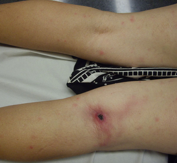
- Explain 5 signs of Inflammation:
-
Redness & warmth: due to ↑ blood flow (hyperemia) to damaged area
-
Swelling (edema): protein & fluid into interstitial space
-
Pain: increased pressure of fluid on nerves; release of chemical mediators – i.e., bradykinins, histamine (itch), prostaglandins
-
Loss of function: may develop if cells lack nutrients; edema may interfere with movement
-
- Explain 5 signs of Inflammation:
Explain 2 phases of inflammation:
- 1. Vascular: vasodilation & increased cap perm → exudate (fluid); stagnation of flow & clotting of blood occurs which aids in localizing the spread of infectious microorganisms.
-
- Histamine, Leukotrienes, Bradykinin, Prostaglandins: induces vasodilation, increased capillary permeability
- Histamine: additionally induces itch
- Prostaglandins: additionally induce pain, fever
- Bradykinogen (plasma protein): additionally induce pain when in active bradykinin form
- Histamine receptors found on nerve endings and on blood vessel walls
-
- 2. Cellular: – emigration (diapedesis) of WBCs; Production of more WBCS (e.g. neutrophils, shift to the left)
- Four types of Exudate:
-
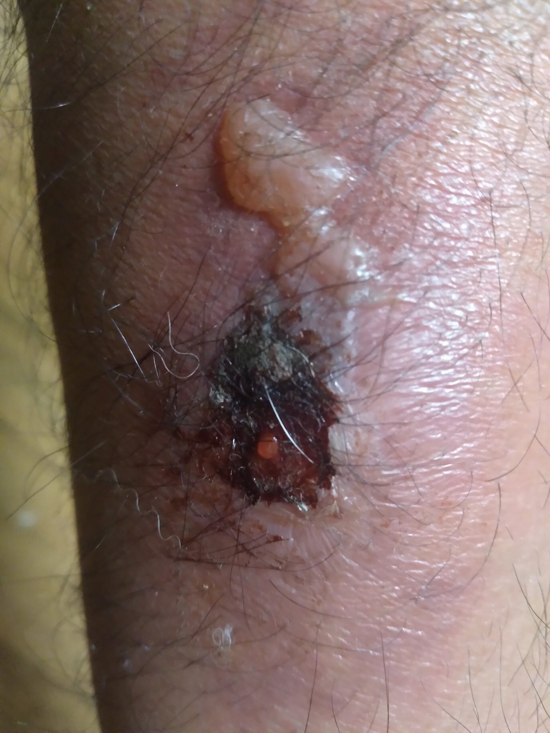
1. Serous: watery, consists primarily of fluid, some proteins, & WBCs (e.g. allergic rxns & burns);2. Fibrinous: thick, sticky, high cell & fibrin content; Increased risk of scar tissue (e.g. severe injuries, rheumatic heart disease, bacterial pneumonia)3. Purulent (“pus”): thick, yellow-green, contains more WBCs, cell debris, & microorganisms; Bacterial infection *An abscess contains purulent exudate
4. Hemorrhagic: blood from damaged blood vessels
-
Mild fever (pyrexia) – oral temp above 38ºC; (Side note: Heatstroke = 40ºC or higher)
-
Common if inflammation is extensive (can occur with heart attack, stroke, trauma, cancer)
-
Due to WBC release of endogenous pyrogens (interferons, interleukins) or (LPS= LipoPolySaccharide = slimy coat of bacteria = exogenous pyrogen)¤What is the most accurate way to take someone’s temperature? Rectal¤What is preferred method? Tympanic for elderly and Axillary for babies¤What is FUO? Fever of Unknown Origin (unknown drug rxn; undetectable infection/trauma/injury/cancer/heart attack/blood clots/inflammatory disease)¤What is a blunted/absent febrile response to infection? Indicates poorer immune response
-
-
What is Systemic Inflammatory Response Syndrome? Can be in response to burn or infection and is as follows: Enormous release of inflammatory cytokines → systemic vasodil. & cap perm → low BP → Circulatory/Septic shock (can be fatal)
-
Malaise (Feeling unwell), Fatigue, Headache, Anorexia
-
Decreased mental function (in elderly) due to cerebral hypoxia
-
-
- Describe the 4 stages of Fever
- Prodromal
- Chills
- Flush
- Defervescence (Sweating)
- Define and explain the significance of:
- Leukocytosis
- Differential Count
- Describe the 4 stages of Fever
-
-
- Plasma
- Erythrocyte Sedimentation Rate
-
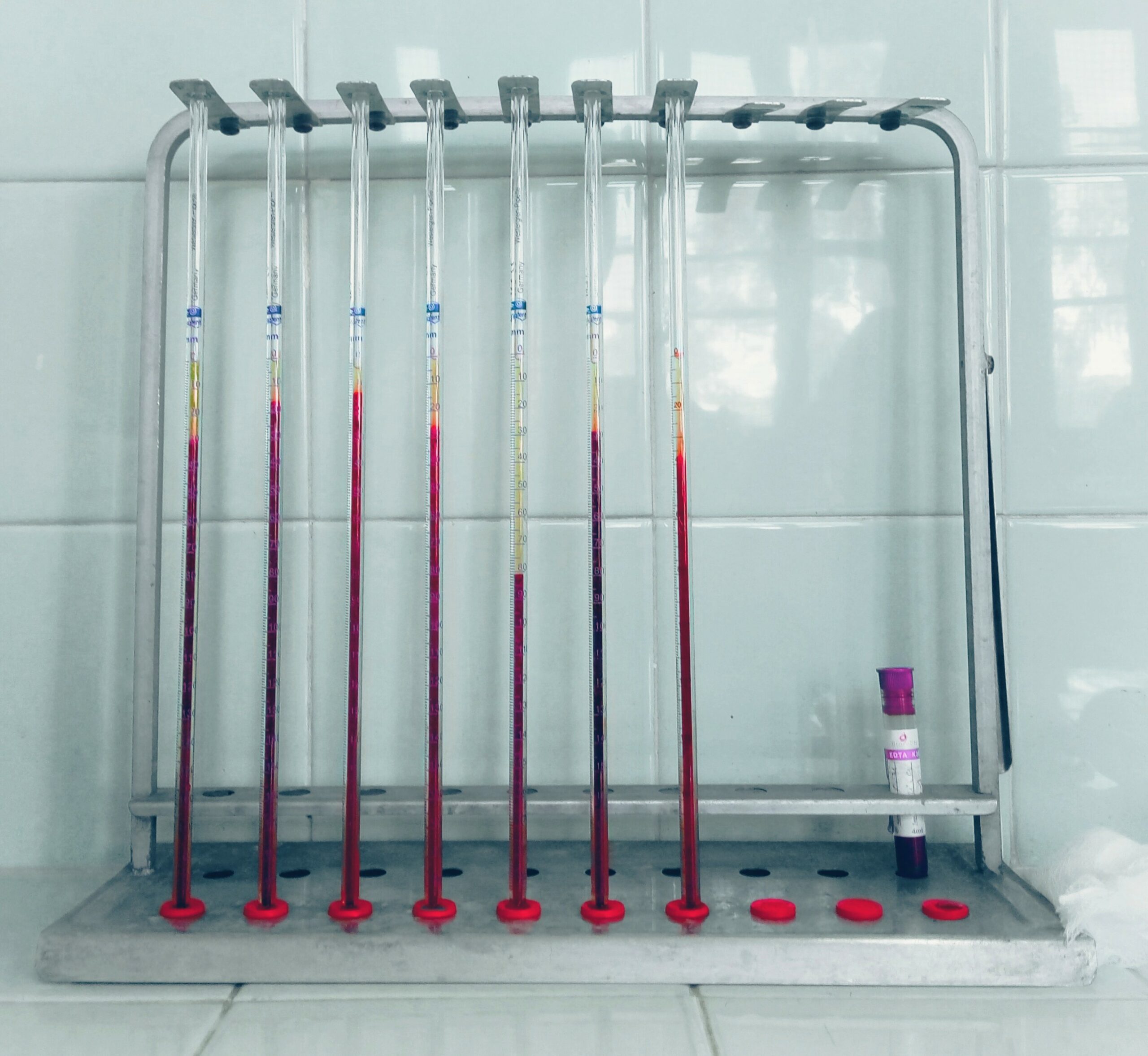
-
-
- C-reactive protein
- Neutrophilia
- Neutropenia
- Lymphocytosis
- Lymphocytopenia
- Thrombocytopenia
-
-
-
- Eosinophilia
- Scar Tissue
-

-
-
- Cellular Regeneration
- Cellular Resolution
- Cellular Replacement
- Granuloma; Granulation tissue
- Healing by 1st Intention
- Healing by 2nd Intention
- Angiogenesis
- Explain the presence of liver/heart proteins in blood
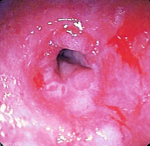
- Explain possible complications of inflammation
- Infection
- Deep ulcers
- Skeletal Muscle Spasms
- Chronic inflammation
-
-
- Explain the difference between
- ASA
- Acetaminophen
- NSAIDs
- Glucocorticoids
- Explain the difference between
-
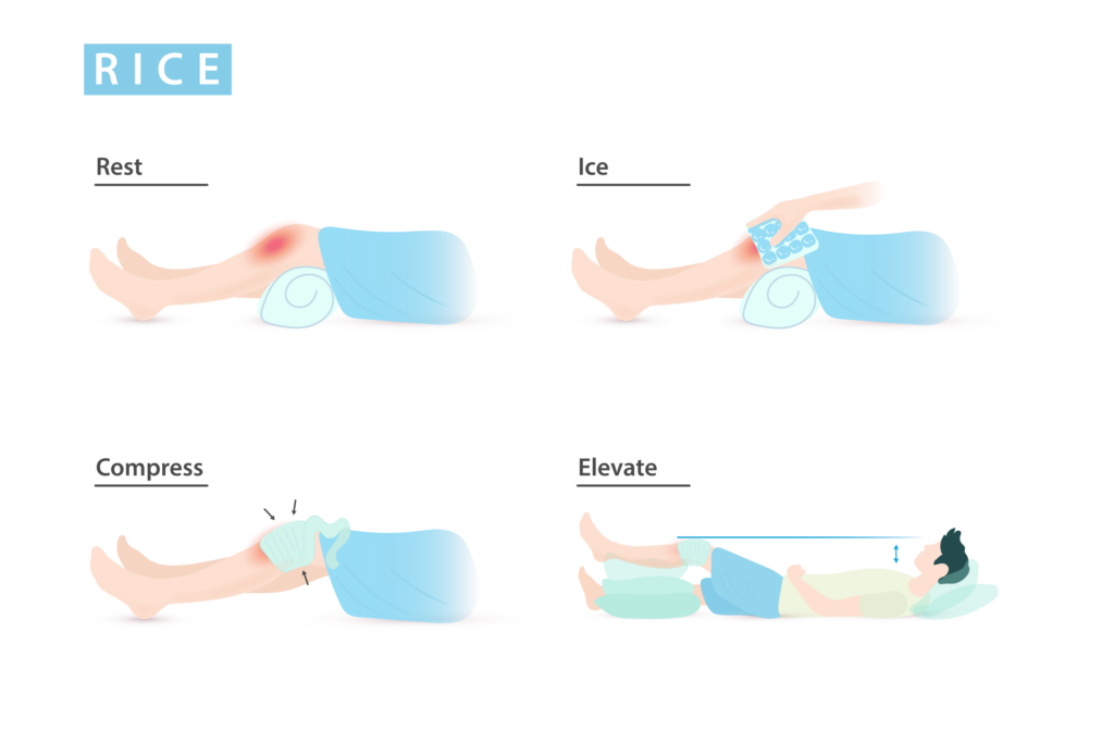
- Explain RICE (Rest, Ice, Compression, Elevation)
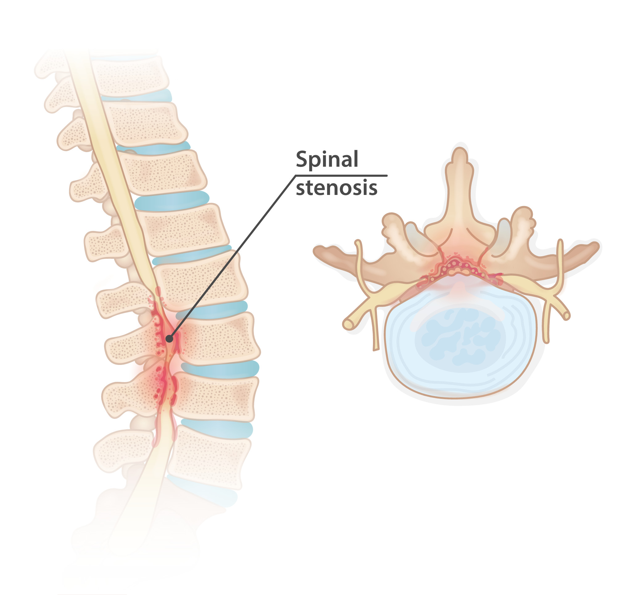
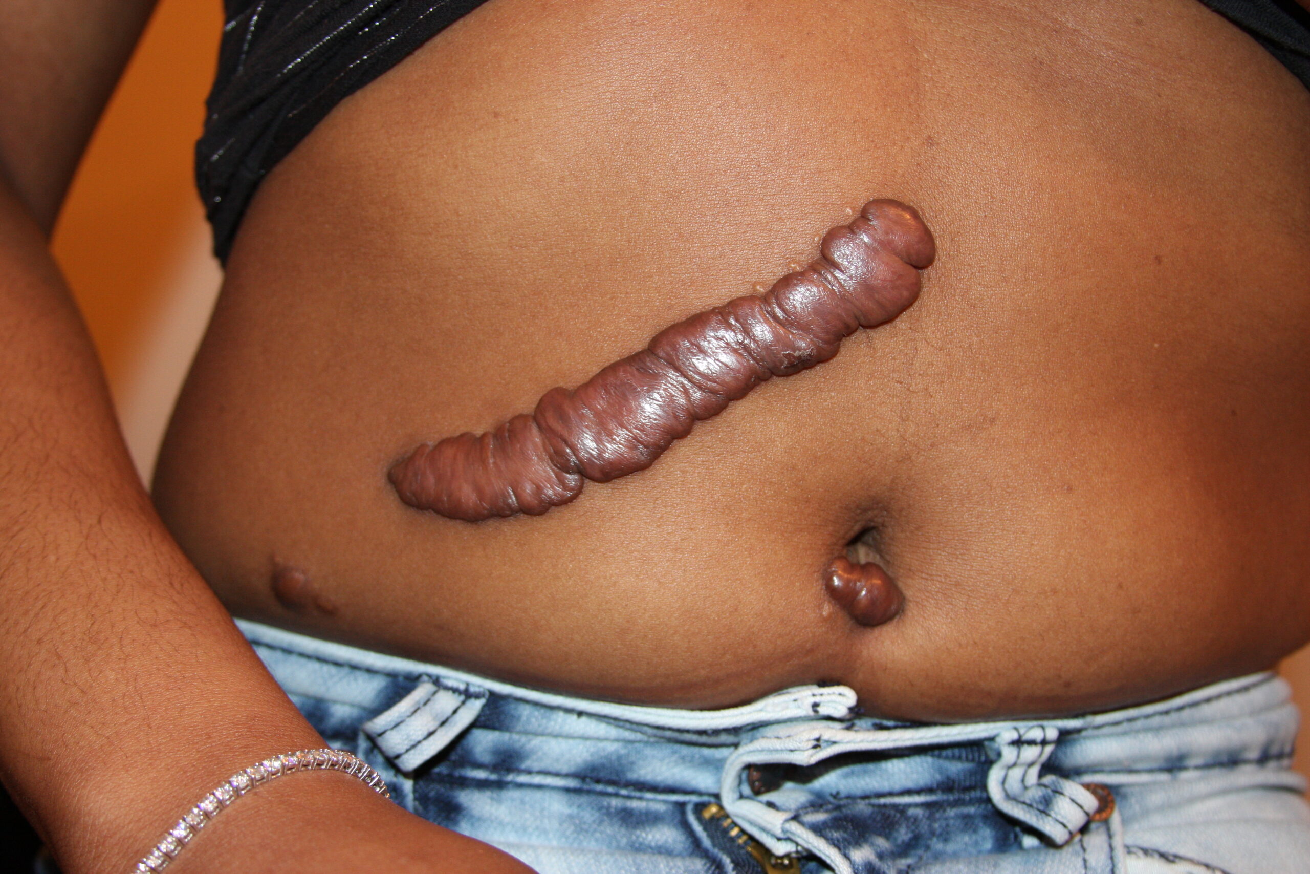
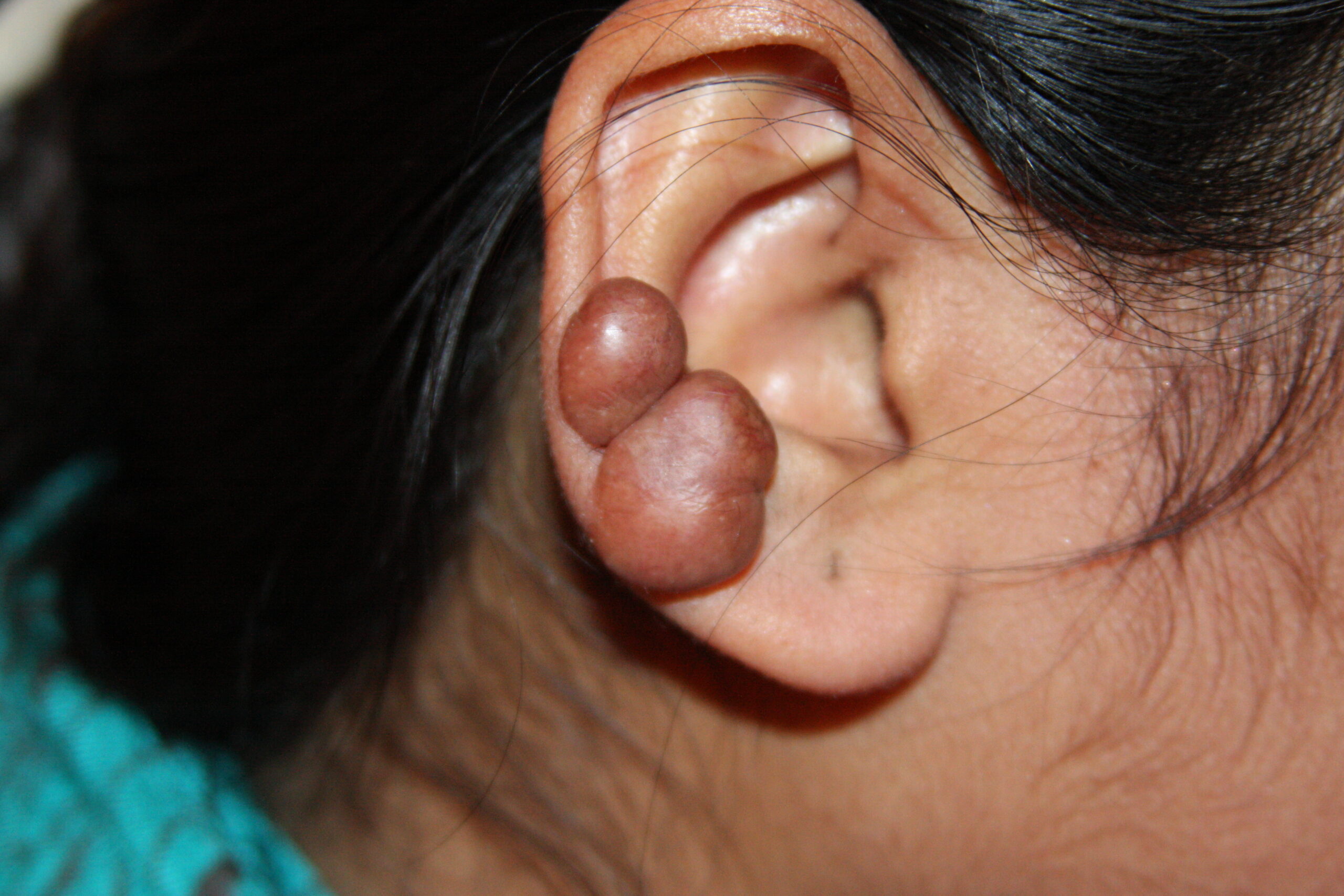
- Explain stenosis, strictures, contractures, adhesions, keloids

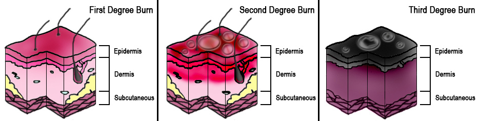
- Distinguish between 1st, 2nd, and 3rd degree burns and basic treatment strategies
- Define eschar
- Define Hypovolemic shock
Media Attributions
- Gut_microbiota © Wakana Sasaki is licensed under a CC BY (Attribution) license
- Macrophage.svg © Zahn Bariring is licensed under a CC BY-SA (Attribution ShareAlike) license
- Macrophages_and_helper_T-cells © Abutler05 adapted by Portiaa McGonigal is licensed under a CC BY-SA (Attribution ShareAlike) license
- Nervous_system_-_Microglia_3_–_Smart-Servier © Laboratoires Servier is licensed under a CC BY-SA (Attribution ShareAlike) license
- NeutrophilerAktion.svg © Mario Schubert is licensed under a Public Domain license
- Bony Heel Spur © InjuryMap - InjuryMap is licensed under a CC BY-SA (Attribution ShareAlike) license
- Complement_Overview_pathway is licensed under a CC BY-SA (Attribution ShareAlike) license
- Membrane_Attack_Complex_(Terminal_Complement_Complex_C5b-9) © SLiva2016 is licensed under a CC BY-SA (Attribution ShareAlike) license
- Mast_Cell_(30107399584) © National Institute of Allergy and Infectious Diseases (NIAID) is licensed under a CC BY (Attribution) license
- Basophil_(30439199890) © National Institue of Allergy and Infectious Diseases (NIAID) is licensed under a CC BY (Attribution) license
- cytokines © National Institute of Allergy and Infectious Disease adapted by Portiaa McGonigal is licensed under a CC BY (Attribution) license
- Chemokine_concentration_chemotaxis.svg © L Kohidai adapted by Pen1234567 is licensed under a CC BY-SA (Attribution ShareAlike) license
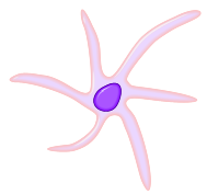
- Natural_Killer_Cell_(30439199790) © National Institute of Allergy and Infectious Disease adapted by Portiaa McGonigal is licensed under a CC BY (Attribution) license
- DK-Bcell receptor (2)
- antibody © National Institute of Allergy and Infectious Disease adapted by Portiaa McGonigal is licensed under a CC BY (Attribution) license
- Antibody © Fvasconcellos is licensed under a Public Domain license
- Iron_deficiency_anemia_blood_film © Graham Beards is licensed under a CC BY-SA (Attribution ShareAlike) license
- Tissues_Affected_In_Allergic_Inflammation © Sari Sabban is licensed under a CC BY-SA (Attribution ShareAlike) license
- Skin_Graft_on_Ankle_after_Third_Degree_Burns © Giftrapped is licensed under a CC BY-SA (Attribution ShareAlike) license
- Microglia_in_ischemic_stroke © Mary Antipova is licensed under a CC BY (Attribution) license
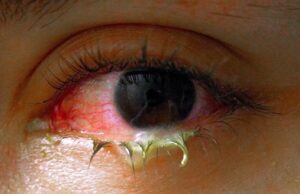
- Swollen_eye_with_conjunctivitis © Tanalai, Wikipedia Commons is licensed under a CC BY (Attribution) license
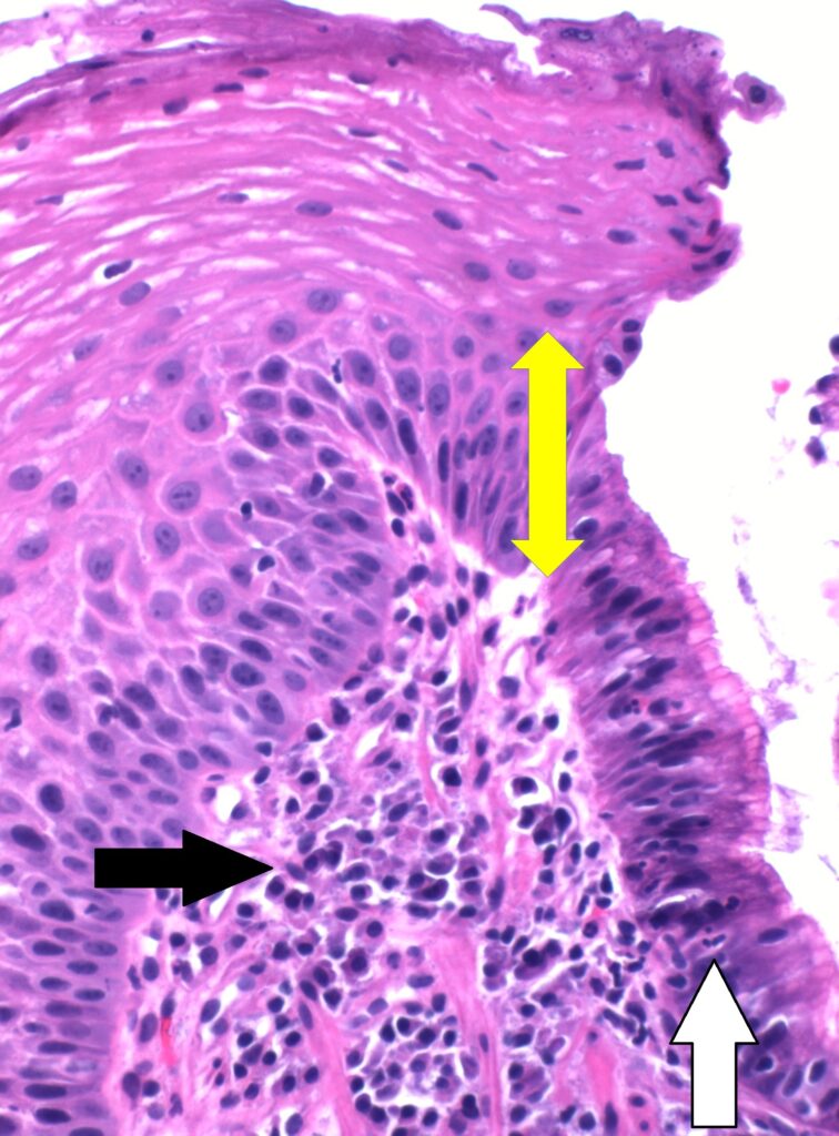
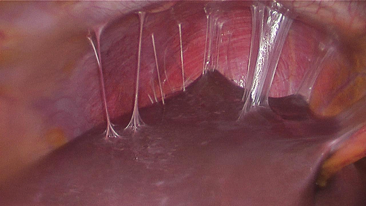
- Histopathology_of_acute_and_chronic_inflammation_of_the_gastro-esophageal_junction,_annotated © Mikael Häggström is licensed under a Public Domain license
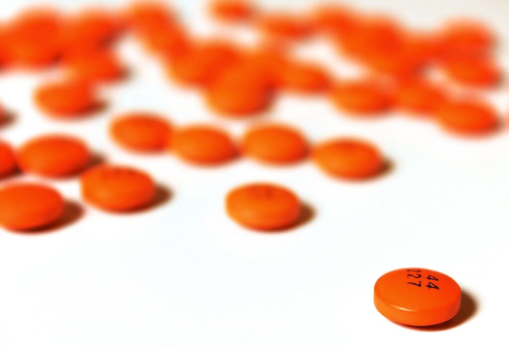
- 2048px-Orange_pills © Candy is licensed under a CC BY-SA (Attribution ShareAlike) license
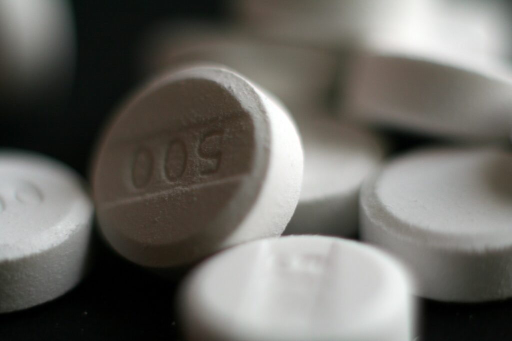
- Paracetamol_acetaminophen_500_mg_pills © Michelle Tribe is licensed under a CC BY (Attribution) license
- RICE © Injurymap is licensed under a CC BY (Attribution) license
- Peptic_stricture © Samir Grover is licensed under a Public Domain license

.png)