8 2.1. The Skeleton
The skeletal system forms the rigid internal framework of the body. It consists of the bones, cartilages, and ligaments. Bones support the weight of the body, allow for body movements, and protect internal organs. Cartilage provides flexible strength and support for body structures such as the thoracic cage, the external ear, and the trachea and larynx. At joints of the body, cartilage can also unite adjacent bones or provide cushioning between them. Ligaments are the strong connective tissue bands that hold the bones at a moveable joint together and serve to prevent excessive movements of the joint that would result in injury. Providing movement of the skeleton are the muscles of the body, which are firmly attached to the skeleton via connective tissue structures called tendons. As muscles contract, they pull on the bones to produce movements of the body. Thus, without a skeleton, you would not be able to stand, run, or even feed yourself!
Each bone of the body serves a particular function, and therefore bones vary in size, shape, and strength based on these functions. For example, the bones of the lower back and lower limb are thick and strong to support your body weight. Similarly, the size of a bony landmark that serves as a muscle attachment site on an individual bone is related to the strength of this muscle. Muscles can apply very strong pulling forces to the bones of the skeleton. To resist these forces, bones have enlarged bony landmarks at sites where powerful muscles attach. This means that not only the size of a bone, but also its shape, is related to its function. For this reason, the identification of bony landmarks is important during your study of the skeletal system.
Bones are also dynamic organs that can modify their strength and thickness in response to changes in muscle strength or body weight. Thus, muscle attachment sites on bones will thicken if you begin a workout program that increases muscle strength. Similarly, the walls of weight-bearing bones will thicken if you gain body weight or begin pounding the pavement as part of a new running regimen. In contrast, a reduction in muscle strength or body weight will cause bones to become thinner. This may happen during a prolonged hospital stay, following limb immobilization in a cast, or going into the weightlessness of outer space. Even a change in diet, such as eating only soft food due to the loss of teeth, will result in a noticeable decrease in the size and thickness of the jaw bones.
The skeletal system includes all of the bones, cartilages, and ligaments of the body that support and give shape to the body and body structures. The skeleton consists of the bones of the body. For adults, there are 206 bones in the skeleton. Younger individuals have higher numbers of bones because some bones fuse together during childhood and adolescence to form an adult bone. The primary functions of the skeleton are to provide a rigid, internal structure that can support the weight of the body against the force of gravity, and to provide a structure upon which muscles can act to produce movements of the body. The lower portion of the skeleton is specialized for stability during walking or running. In contrast, the upper skeleton has greater mobility and ranges of motion, features that allow you to lift and carry objects or turn your head and trunk.
In addition to providing for support and movements of the body, the skeleton has protective and storage functions. It protects the internal organs, including the brain, spinal cord, heart, lungs, and pelvic organs. The bones of the skeleton serve as the primary storage site for important minerals such as calcium and phosphate. The bone marrow found within bones stores fat and houses the blood-cell producing tissue of the body.
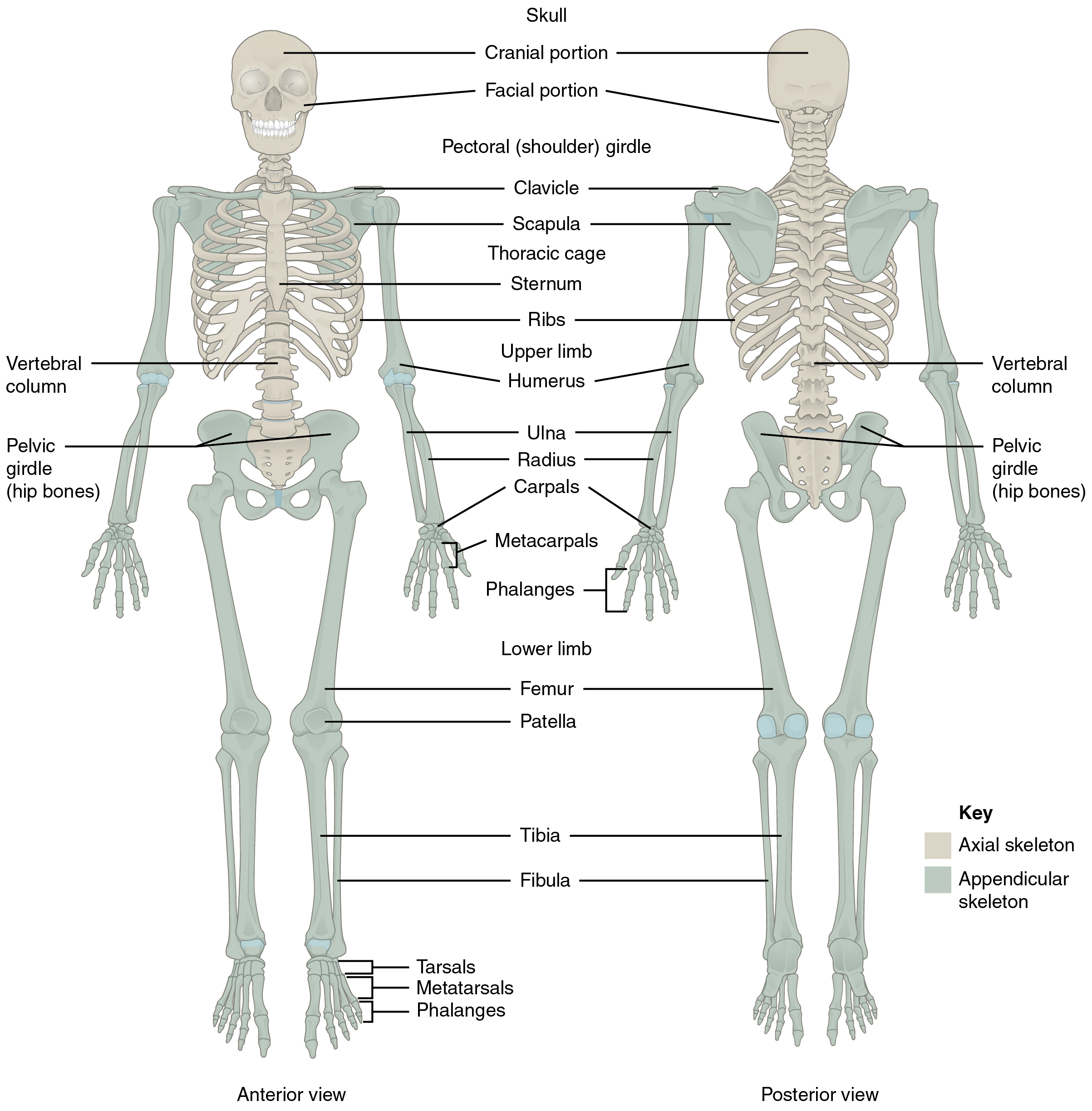
The skeleton is subdivided into two major divisions—the axial and appendicular.
The Axial Skeleton
The axial skeleton forms the vertical, central axis of the body and includes all bones of the head, neck, chest, and back (Figure 1). It serves to protect the brain, spinal cord, heart, and lungs. It also serves as the attachment site for muscles that move the head, neck, and back, and for muscles that act across the shoulder and hip joints to move their corresponding limbs.
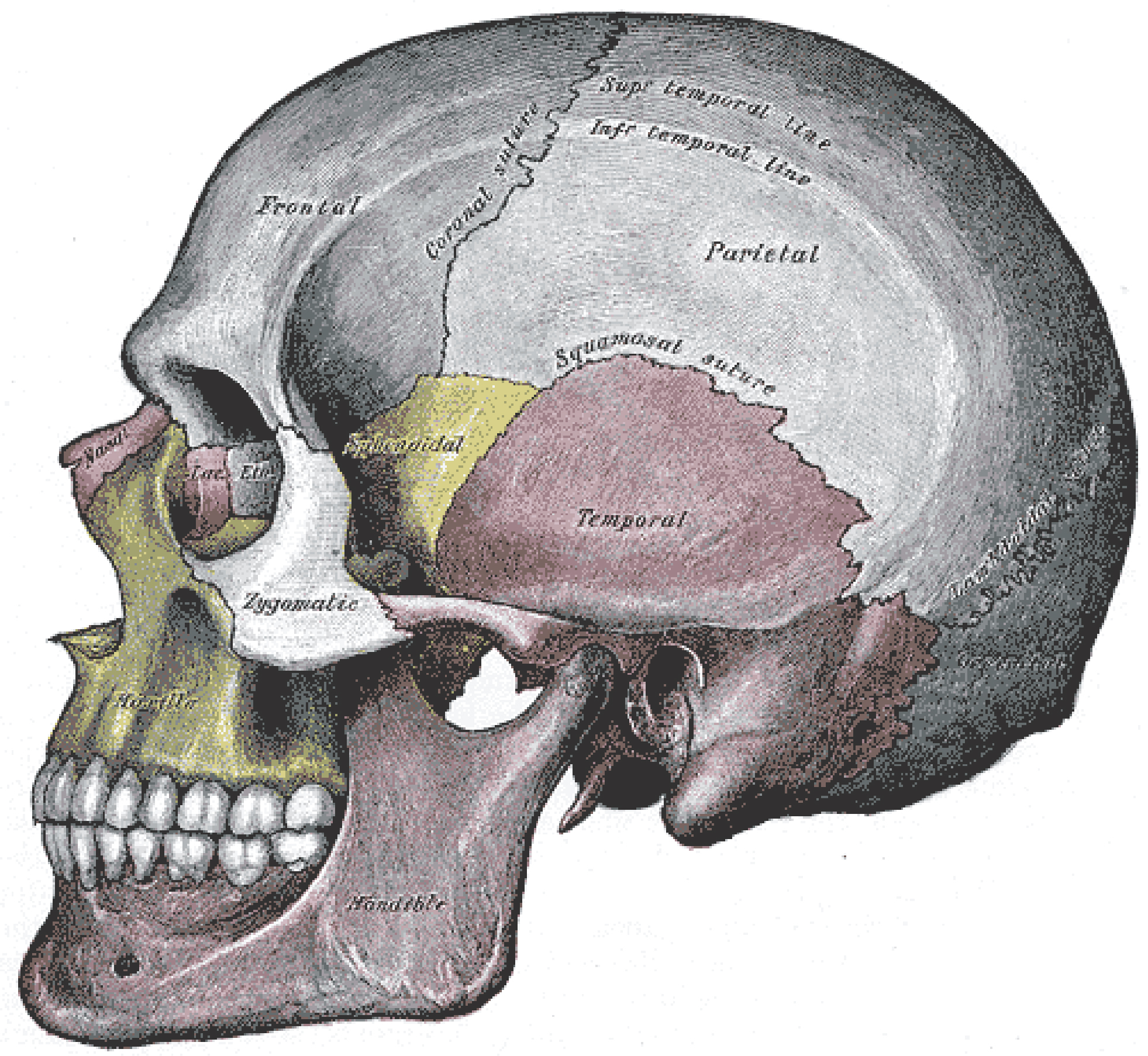
The axial skeleton of the adult consists of 80 bones, including the skull, the vertebral column, and the thoracic cage. The skull is formed by 22 bones. Also associated with the head are an additional seven bones, including the hyoid bone and the ear ossicles (three small bones found in each middle ear). The vertebral column consists of 24 bones, each called a vertebra, plus the sacrum and coccyx. The thoracic cage includes the 12 pairs of ribs, and the sternum, the flattened bone of the anterior chest.
The Appendicular Skeleton
The appendicular skeleton includes all bones of the upper and lower limbs, plus the bones that attach each limb to the axial skeleton. There are 126 bones in the appendicular skeleton of an adult.
Review
The skeletal system includes all of the bones, cartilages, and ligaments of the body. It serves to support the body, protect the brain and other internal organs, and provides a rigid structure upon which muscles can pull to generate body movements. It also stores fat and the tissue responsible for the production of blood cells. The skeleton is subdivided into two parts. The axial skeleton forms a vertical axis that includes the head, neck, back, and chest. It has 80 bones and consists of the skull, vertebral column, and thoracic cage. The adult vertebral column consists of 24 vertebrae plus the sacrum and coccyx. The thoracic cage is formed by 12 pairs of ribs and the sternum. The appendicular skeleton consists of 126 bones in the adult and includes all of the bones of the upper and lower limbs plus the bones that anchor each limb to the axial skeleton.
Glossary
- appendicular skeleton
- all bones of the upper and lower limbs, plus the girdle bones that attach each limb to the axial skeleton
- axial skeleton
- central, vertical axis of the body, including the skull, vertebral column, and thoracic cage
- coccyx
- small bone located at inferior end of the adult vertebral column that is formed by the fusion of four coccygeal vertebrae; also referred to as the “tailbone”
- ear ossicles
- three small bones located in the middle ear cavity that serve to transmit sound vibrations to the inner ear
- hyoid bone
- small, U-shaped bone located in upper neck that does not contact any other bone
- ribs
- thin, curved bones of the chest wall
- sacrum
- single bone located near the inferior end of the adult vertebral column that is formed by the fusion of five sacral vertebrae; forms the posterior portion of the pelvis
- skeleton
- bones of the body
- skull
- bony structure that forms the head, face, and jaws, and protects the brain; consists of 22 bones
- sternum
- flattened bone located at the center of the anterior chest
- thoracic cage
- consists of 12 pairs of ribs and sternum
- vertebra
- individual bone in the neck and back regions of the vertebral column
- vertebral column
- entire sequence of bones that extend from the skull to the tailbone
