16 Insect Anatomy – The Head
External Morphology – Head
The first of the three tagmata is the head.
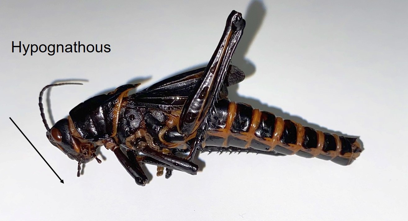
The grasshopper head is oriented with the mouthparts pointing downwards, or ventrally. Thos arrangement is called hypognathous (hypo, below/down; gnath, jaw). Other insects may have mouthparts that point forwards, or even backwards, and you’ll see examples of these later on.
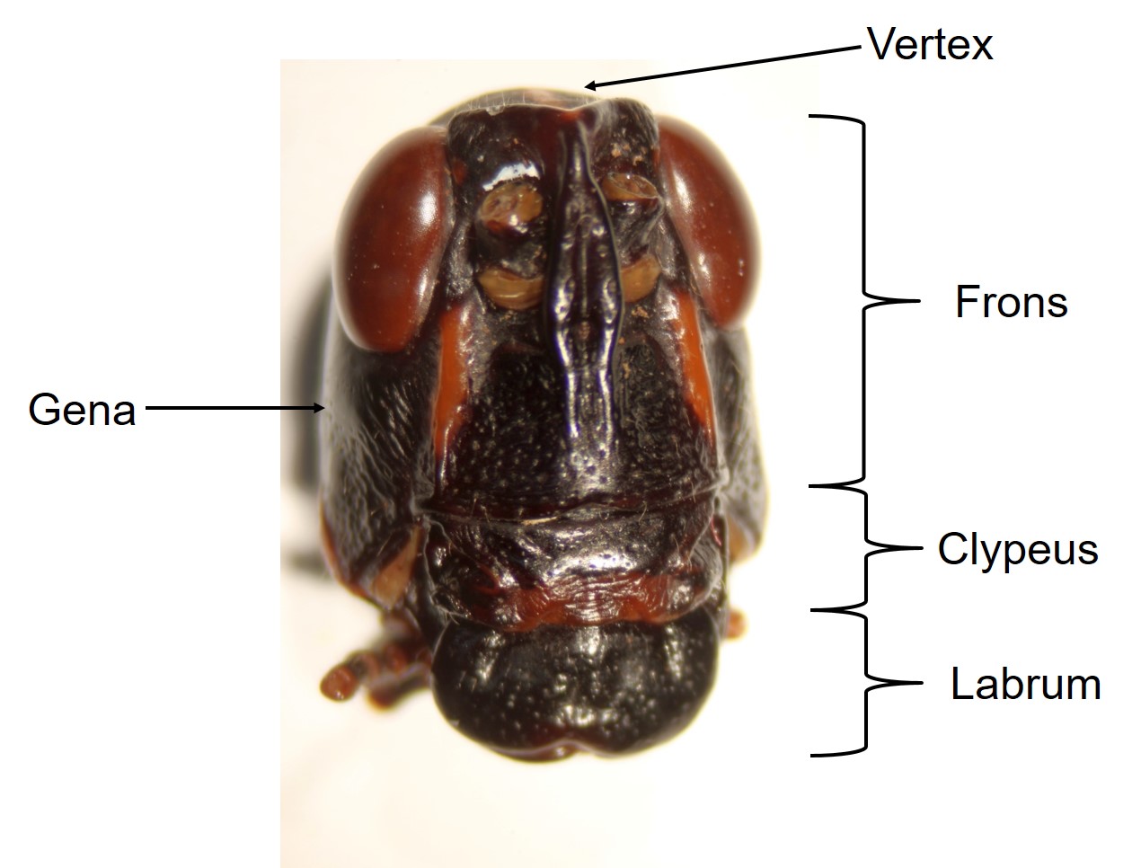
Looking more closely at the head, from the front in this next photo, you can distinguish some sclerites (antennae removed for clarity).
- Vertex – top of head
- Gena (“cheeks”) – sides of head
- Frons – front of head
- Clypeus – below frons
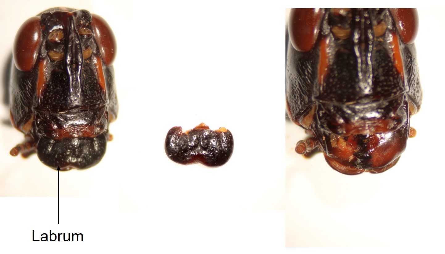
- Labrum (“upper lip”) – below clypeus
Modifications of these structures can be important for identifying insects.
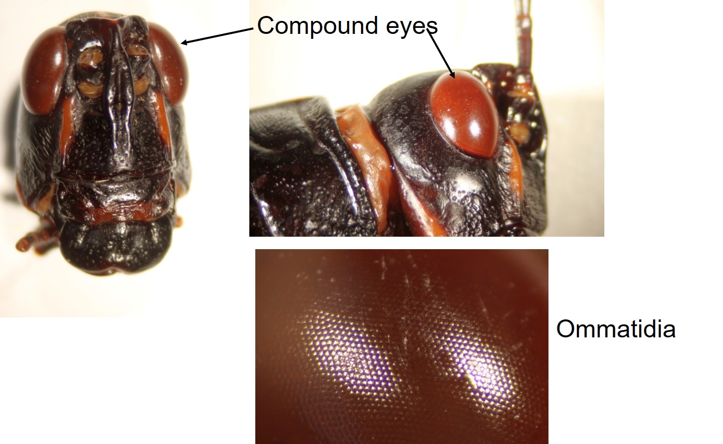
The head, as I said, is specialized for sensory functions and feeding. The major sensory structures visible include the compound eyes, which are located laterally on the grasshopper.
Zooming in on a compound eye, you can see many individual hexagonal facets, called ommatidia. Each one (ommatidium) is an image-forming structure, with the images integrated by the brain into a single image, much as our brains integrate information from our two eyes to make a single image.
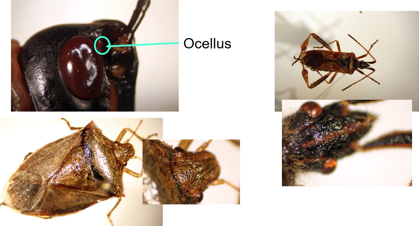
Many insects also have ocelli, which are not image-forming, but help to detect light and dark, and may assist with flight.
Ocelli are difficult to see on Romalea, but are much more obvious on some other insects. Their presence or absence, locations and number can be important identifying characteristics.
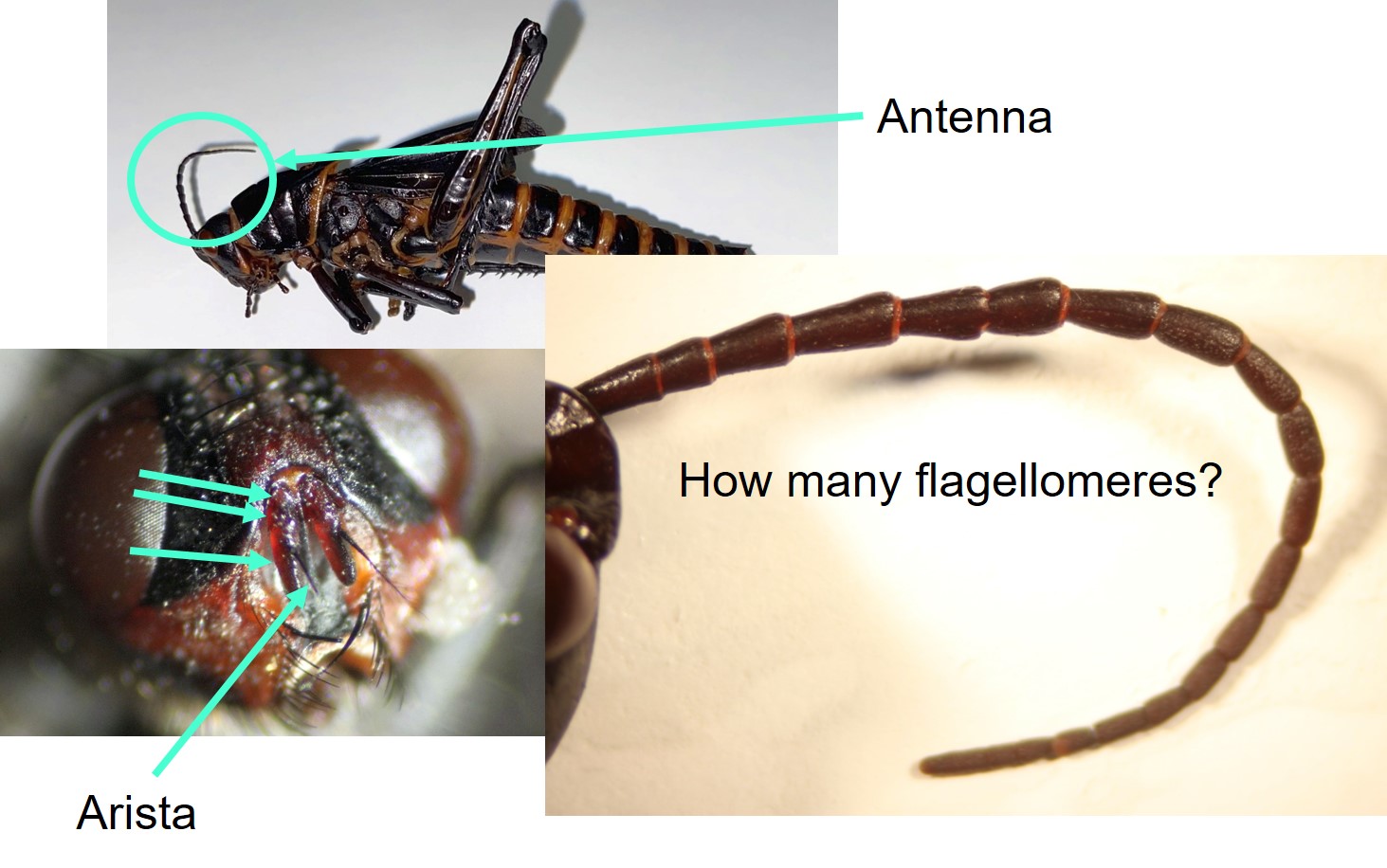
Antennae are important sensory structures, but some stages of some insects may be missing them entirely.
Looking more closely at the grasshopper’s antenna, you can see that it is segmented. These segments are called flagellomeres. Identification keys sometimes ask you to count the number of antennal segments. How many do you see on the grasshopper? It gets a bit out of focus towards the tip, but you should be able to distinguish 18.
Antennae can also come in a wide variety of shapes, sizes and lengths. For instance, this fly (bottom left image) has a three-segmented antenna, with a long bristle called an arista.
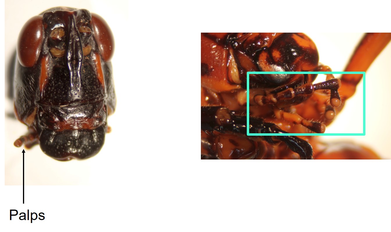
You may have noticed two pairs of structures associated with the mouthparts of the grasshopper that kind of look like short antennae. These are palps, which are also sensory structures. They are a little easier to see if we zoom in on a ventral view, and will be much easier to see as we dissect the mouthparts.
You have already seen the labrum. Remember that above the labrum is the clypeus, and above that the frons (sclerites of the head).
For the mouthparts dissection, the labrum is the first structure we remove. You will see that the shape varies in other insects.
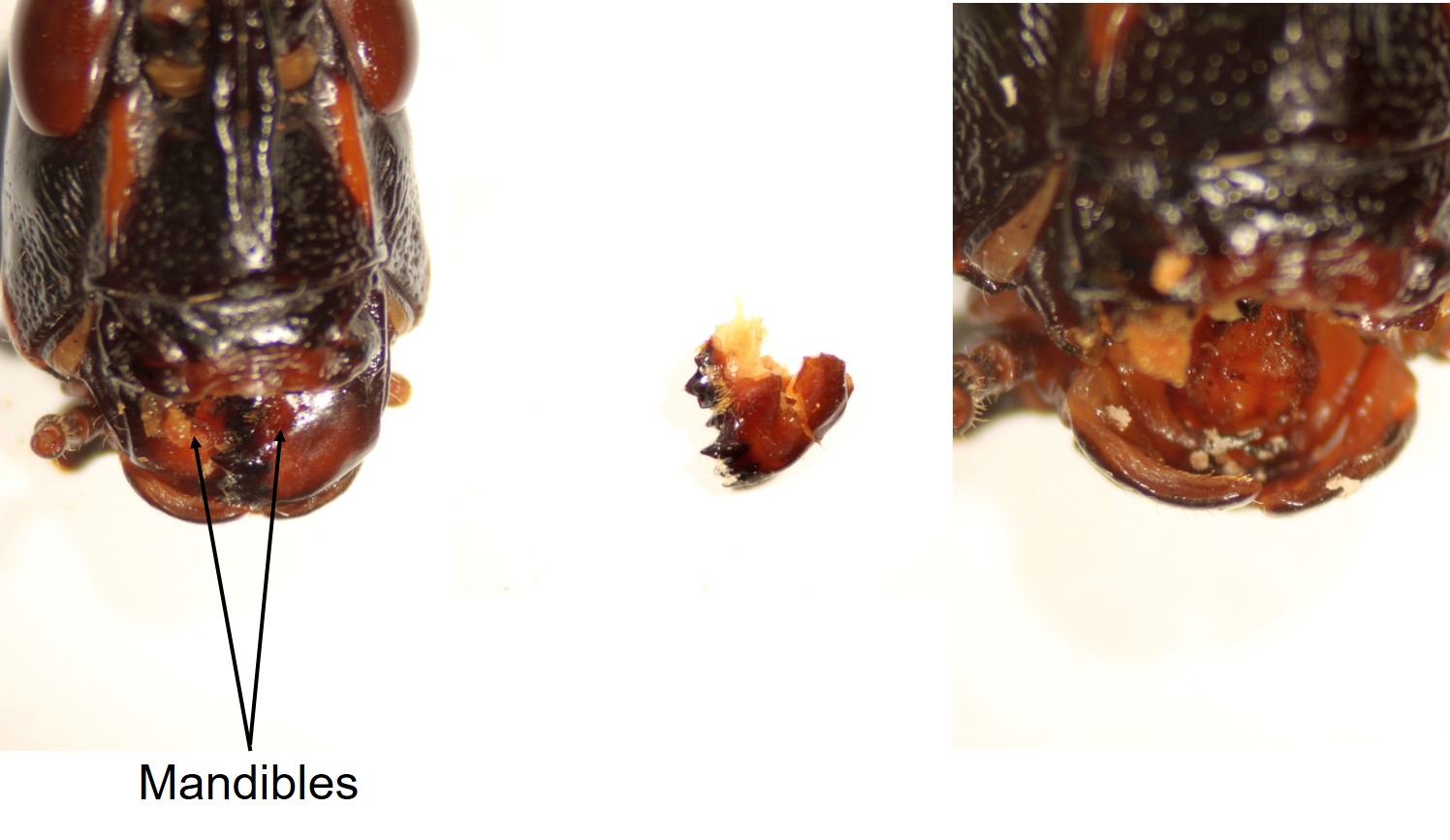
With the labrum removed, you will see some hard, toothed structures behind it.
These hard, toothed structures are the mandibles.
When one mandible is removed, you can see that it is heavily sclerotized (hardened), and that it has pronounced teeth on the inner margin. Grasshoppers eat… well… grass! So, why all the teeth? Grasses have lots of silica crystals in the leaves, as a herbivore deterrent, so grasshoppers need tough mandibles to grind up the leaves they eat. Even so, the teeth will wear down over time.
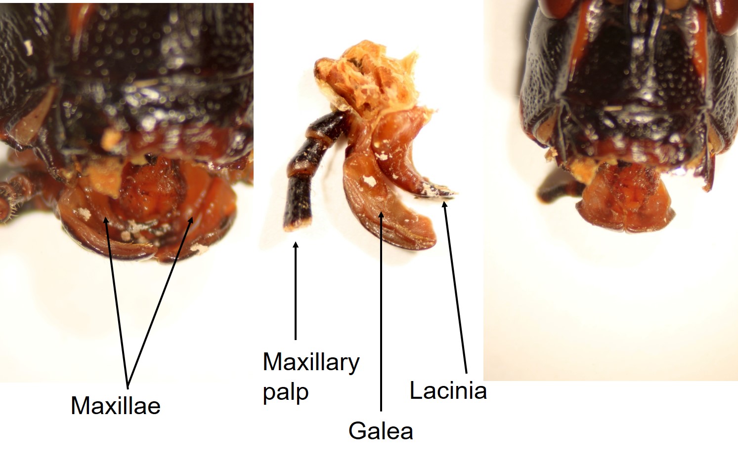
Once the mandibles are removed, you can see more mouthparts behind them – these are the maxillae.
When the maxillae are removed, you can see that each is composed of several parts. In the grasshopper, the galea is scoop-shaped, and the lacinia is pointed, with some sclerotized teeth. The maxillary palp, as you’ve already is seen, is sensory.
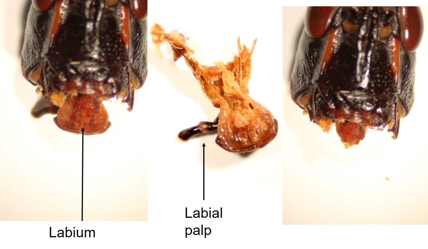
Once the maxillae are removed, you can see the remaining mouthparts towards the back of the grasshopper’s head.
At the back of the head is the labium (“lower lip”). Note the similarity in spelling between labium and labrum. Spelling it “labrium” on an exam will not get you a mark for either one!
At the top of the photo, notice the muscle fibres that attached the labium to the head of the grasshopper. The two halves of the labium are fused to form a single, median structure, again composed of multiple parts (I’m not going to cover all of those names, though!). There is also a pair of palps associated with the labium, again sensory in function.
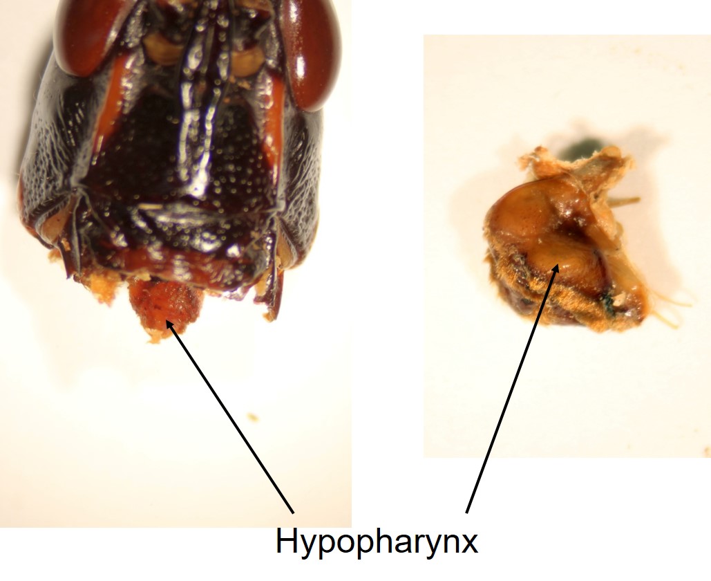
Once the labium is removed, there is only a single, central structure remaining.
The remaining structure, the hypopharynx, is in approximately the centre of all the other mouthparts you’ve seen. We have the labrum and mandibles to the front, maxillae to the sides, and labium to the back of the head.
In the grasshopper, the hypopharynx is roughly analogous to our tongue, manipulating food and guiding it towards the esophagus.
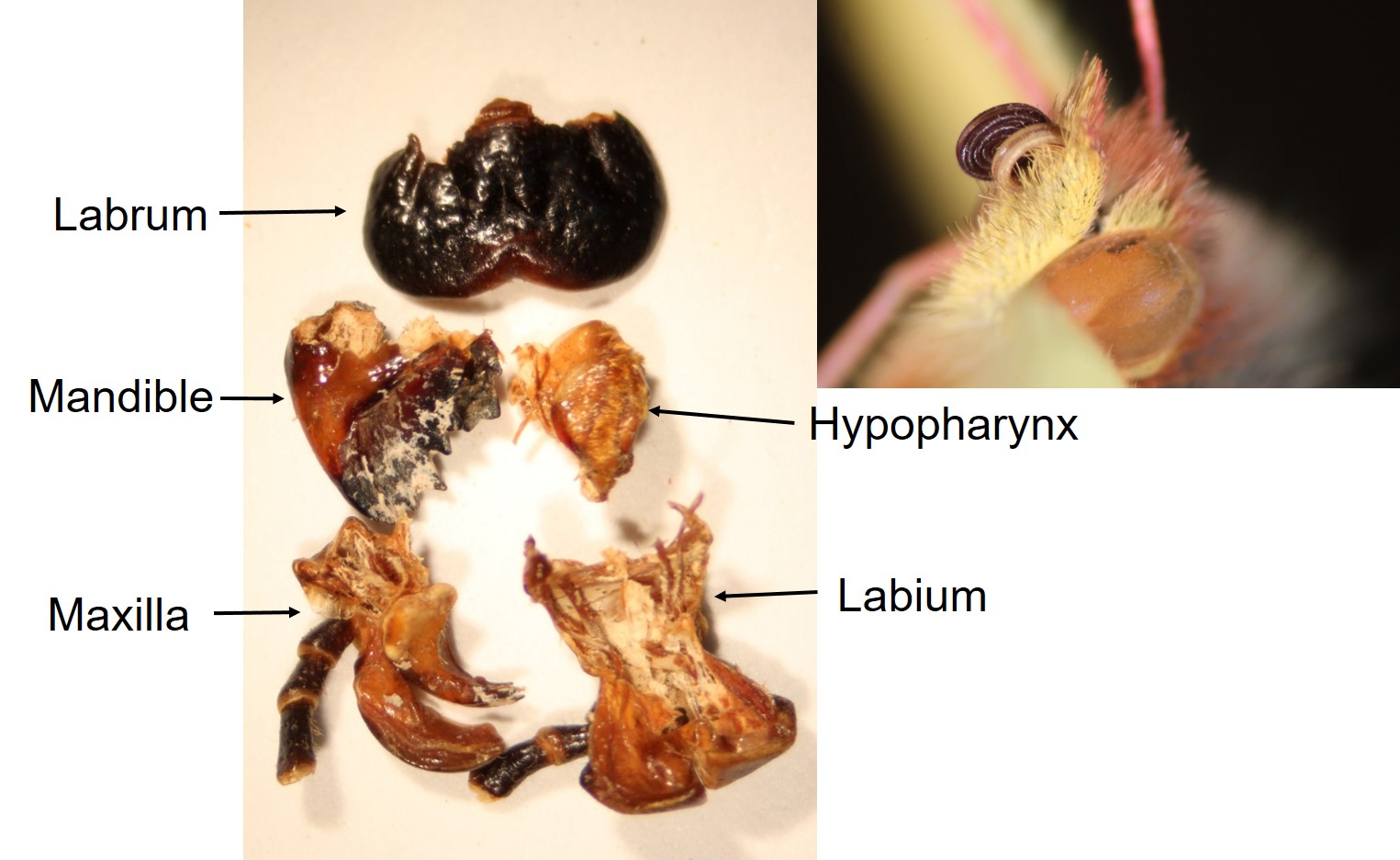
Arranged all together, here are the mouthparts, so that you can see their relative sizes and shapes in the grasshopper.
These mouthparts can all be modified into different shapes, textures, and sizes to accommodate different diets – sometimes they are radically altered! For example, in butterflies, the coiled tube that allows the butterfly to drink nectar is formed solely from the galea of the maxillae. In other insects, these structures have been modified to form a “sponge”, the labellum and associated structures, for feeding on liquids (e.g. carrion flies), or piercing and sucking structures (e.g. mosquitoes).

Feedback/Errata