22 Internal Anatomy – Reproductive Systems
Internal Anatomy – Reproductive Systems
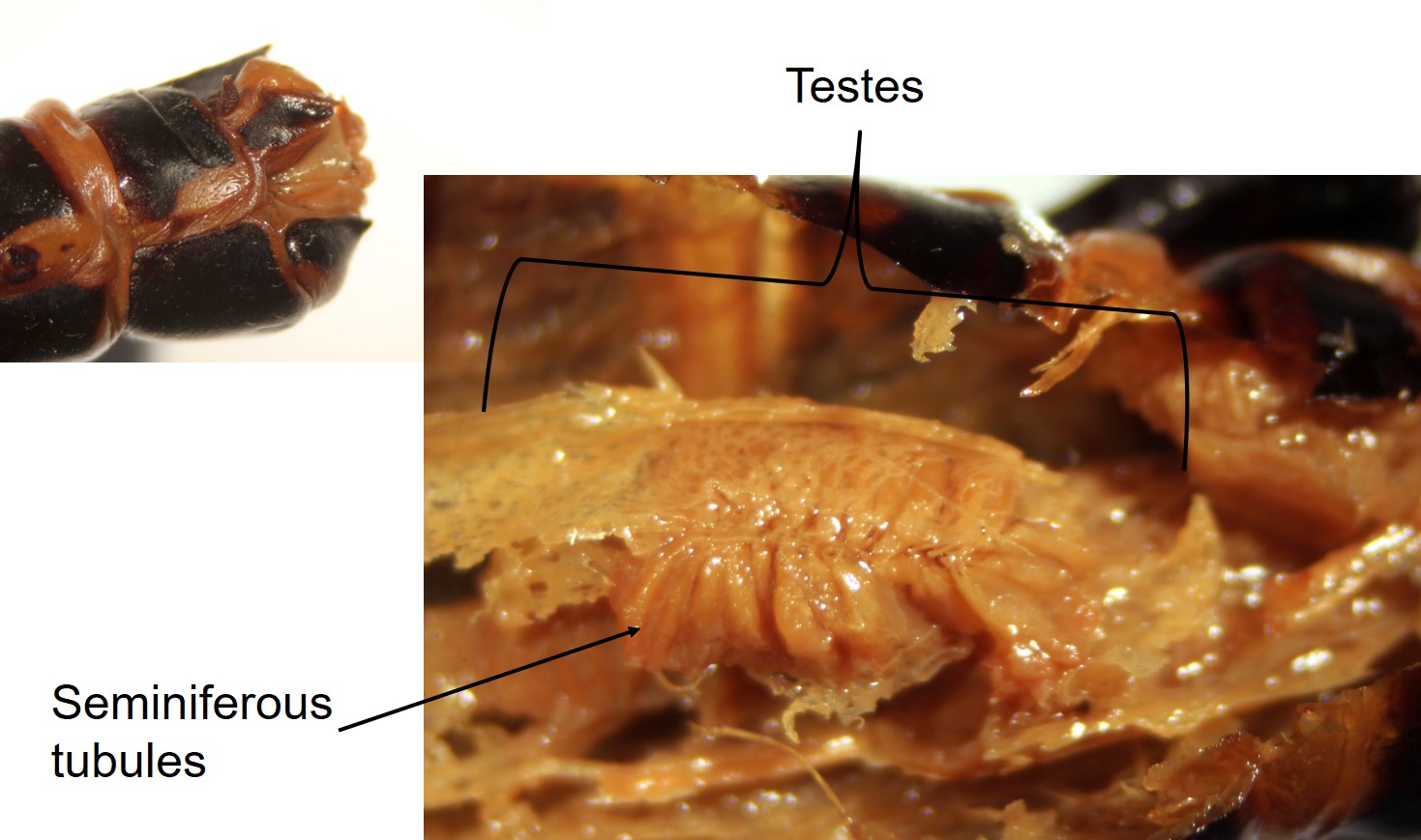
A reminder of what the male’s external genitalia look like.
Inside you can see the fused testes, with plenty of fat body covering them; the individual segments are called seminiferous tubules. These are the locations for meiosis to produce sperm cells.
Various accessory glands are also associated with the male reproductive system. These support and protect sperm, and package sperm into a spermatophore in many insects.
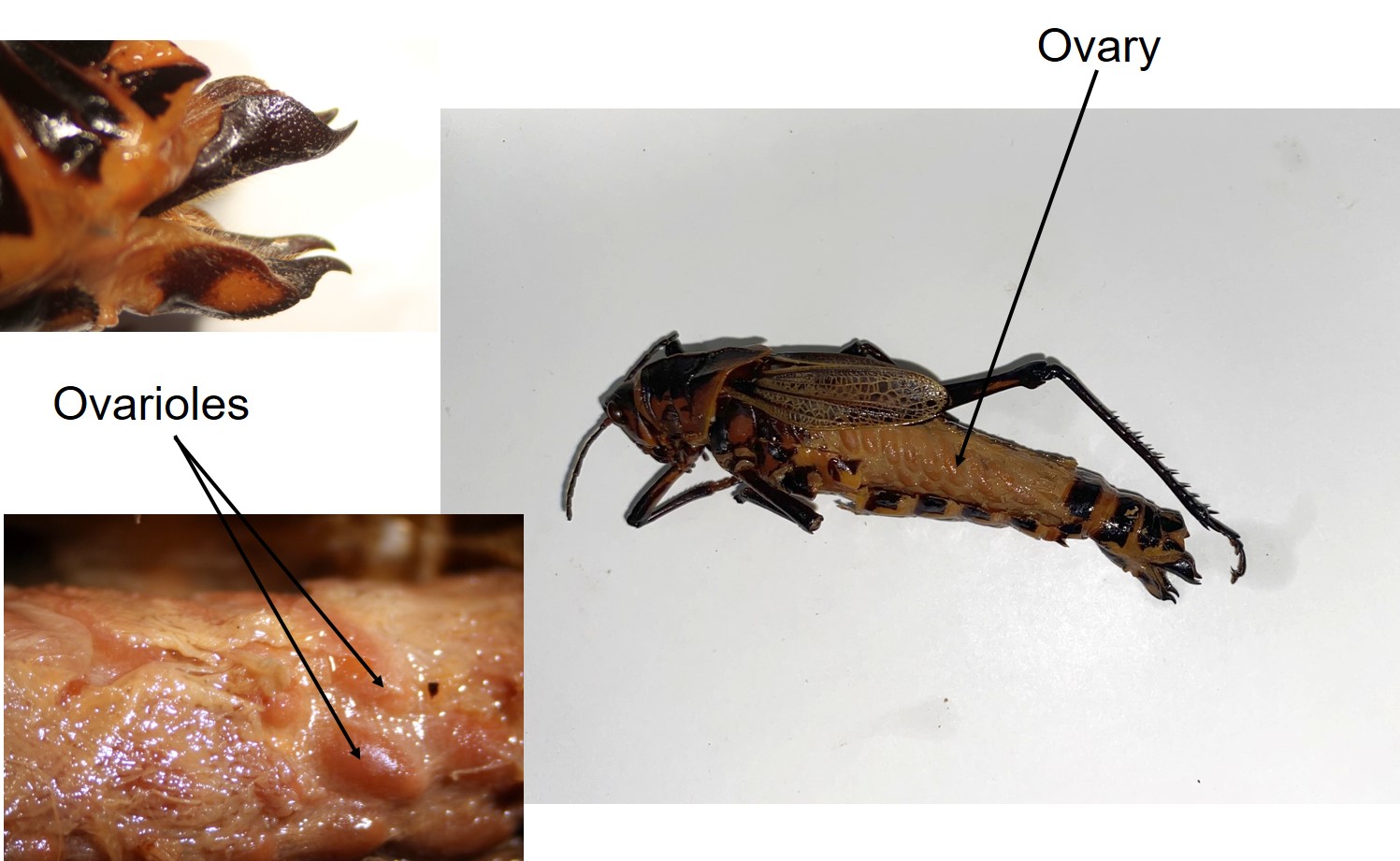
A reminder of what the female’s external genitalia look like. Note the stout ovipositor.
Following the initial incision on a mature female grasshopper, almost the only organ visible is the fused ovaries, again with plenty of fat body associated. The small sections are ovarioles, where meiosis occurs, with individual ova developing within the ovary.
Again, accessory glands are associated that protect and nourish eggs, and secrete coverings.

Feedback/Errata