23 Internal Anatomy – Digestive System
Internal Anatomy – Digestive System
Moving on to the digestive system…
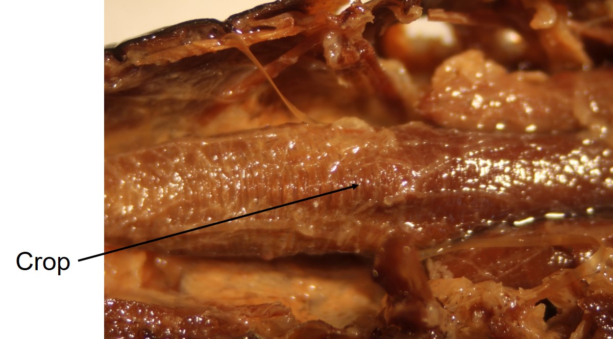
The foregut consists of a short esophagus behind the mouth, followed by large crop (shown here) for storage of ingested food.
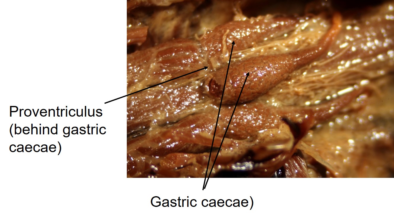
Leaving the crop, the food is passed to the muscular proventriculus. In the grasshopper, mechanical digestion continues here (the proventriculus may contain sclerotized “teeth”).
From the foregut, food passes to the midgut. At the junction of the foregut and midgut are several blind pouches called gastric caecae. These increase the surface area for secretion of enzymes, digestion, and absorption.
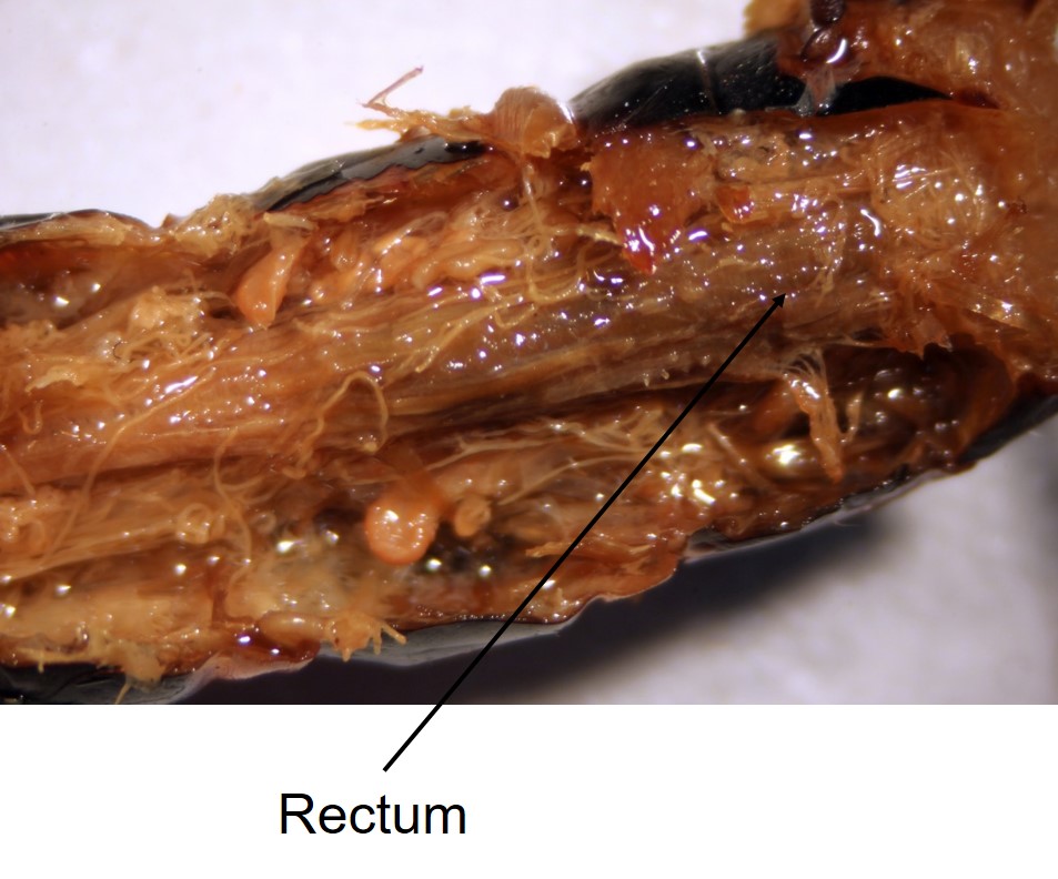
Finally, the mostly-digested food passes to the hindgut, primarily composed of a thick-walled rectum. The rectum reclaims almost all water from the feces before eliminating it. Most insects produce dry waste products, with almost all water removed. Insect feces is often called frass.
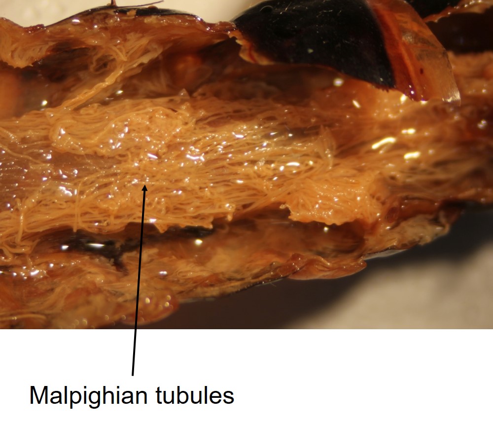
Draining into the hindgut, just posterior to the midgut, are the Malpighian tubules, which are important in osmoregulation and excretion. They are named after Marcello Malpighi, a 17th century anatomist.

Feedback/Errata