17 Alternative Cloning Methods 2 – Golden Gate Cloning and BioBRICKS
Gateway Cloning
Introduction
In this section we are going to look at the use of restriction enzymes that cut to one side of the recognition sequence rather than inside it. This type of enzyme is called type IIS. This means that the overhang generated will be different for different DNA molecules. The value of this is that you can design primers to amplify an insert that has the restriction enzyme sequence, followed by the sequence that will generate your desired overhang. You can do directional cloning easily. You still rely on restriction digest and ligation however, so the lower efficiency of this approach (compared to Gateway) is a potential drawback.
BIO Bricks is the result of a non-profit organization’s attempt to produce a set of components: promoters, coding sequences, and others (like ribosome binding sites) that can be assembled in various combinations to produce novel constructs.
Contents
Learning Outcomes
A. Golden Gate Cloning
A-1. Type IIS Restriction Enzymes
A-2. Primer design for Golden Gate Cloning
B-1. IGEM (this information comes from Labster)
B-2. Bio BRICKS
Learning Outcomes
- Be able to design primers to generate a desired overhang for cloning an insert into a vector using this method
- Outline a cloning strategy to add an insert to a Golden Gate vector
- Outline a cloning strategy using Bio Bricks and the 3A method (as done in the simulation)
- For the cloning strategies, include enough sequence to show how the cloning works to join the fragments
A. Golden Gate Cloning:
A-1. Type IIS Restriction Enzymes
Type IIS restriction enzymes differ from the types of enzymes we’ve been using so far. They do not recognize palindromic sequences. They do not cut inside the recognition sequence but to one side of it. The ones we will be focussing on generate 4 base pair 5′ overhangs. The recognition sequences are shown with the cut site indicated in brackets beside the sequence, like this: 5′-GGTCTC-3′ (1,5). This is the recognition sequence for BsaI. The first number tells you how far from the recognition sequence the enzyme cuts on the top strand, and the second number indicates the cut site on the lower strand. In this case the enzyme cuts one nucleotide 3′ to the final C of the sequence on the top strand and 5 nucleotides 5′ to the first G of the bottom strand. It looks like this:
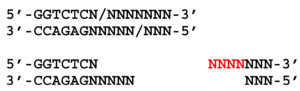
This shows the recognition sequence and the results of the cut.
The Ns indicate that the 5′ overhang could be any sequence.
This is an example of another Type IIS enzyme called BpiI. The recognition sequence for this enzyme is listed as: 5′-GAAGAC-3′ (2,6)

(where is the attribution for this image?)
Here is some practice in determining the overhang generated by this type of enzyme:
A-2. Primer design for Golden Gate Cloning
In order to do cloning with the Golden Gate method it is best to start by visualizing the product you want to generate.
- Draw out the sequence at the positions where the “joins” will be.
- Determine the overlaps that will be needed to join the sequences.
- Figure out where the BsaI (or other) restriction sites need to be placed.
- Draw out the ends ofthe PCR fragment you would get with the primers you’ve designed
- Draw what the ends will look like after the restriction digest.
- Join the ends and confirm that the ligation product matches the desired fragment.
Click here for the powerpoint slides presented in the video below (note: there are errors on slide 5 & 8 which have been corrected).
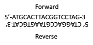
Here is a quick (I hope) practice in oligo design for golden gate cloning. Here are the oligo sequences (A); these will be annealed by the same process we are using for our CRISPR oligos. The annealed sequences are shown in B.
A.

B.
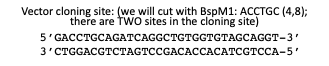
We want to clone the annealed oligos into a vector cut with a type IIS restriction enzyme. The schematic of what we are trying to do is shown in C. Note that we want the orientation as shown, with the forward oligo on the top strand and the reverse on the bottom strand. The blue squiggles drawn in represent the sequences we need to add to the oligos so that the desirded overhang is generate after annealing. The cloning site of the vector and the restriction enzyme will use to cut the vector is shown in D. Your job is to determine what overhangs are generated by the restriction enzyme, BspMI, to select the sequence that should be added to each oligo, and to determine which end it should be added to, with the aim being to clone the oligos into the vector as shown in the schematic in C.
C.
![]()
D.
B. BioBricks:
B-1. IGEM (this information comes from Labster)
“The International Genetically Engineered Machine (iGEM) Foundation is an independent, non-profit organization dedicated to the advancement of synthetic biology and the development of an open community and collaboration.”
iGEM has three main activities
- iGEM Competition – this is an international competition for students interested in the field of synthetic biology. The aim is for them to design a novel molecular machine that will address a real world problem. Teams of undergraduate students as well as high school students can enter these competitions.
- Labs Program – a program for academic labs to use the same resources as the competition teams.
- Registry of Standard Biological Parts – the group maintains a database listing a growing collection of genetic parts used for building biological devices and systems.
B-2. Bio BRICKS
The genetic parts mentioned above are the Bio BRICKs. As the name suggests, the parts can be combined to build novel constructs with unique combinations of promoter and coding sequences, somewhat like building a structure with Lego bricks. They can be used to make genetically engineered biological “machines”. [Insert examples of such constructs, what they are used for]
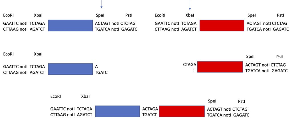
Each “part” comes in a plasmid, with an origin of replication, an antibiotic resistance gene, and the unique sequence, or “part”. Each part (coding sequence or promoter) is flanked by the same Prefix (set of restriction sites) before it and Suffix (a different set of restriction sites) after it. In the prefix is EcoRI, Not1 and XbaI. In the suffix is SpeI, NotI and PstI.
The picture below comes from Labster and shows the prefix and suffix relative to the “part”.
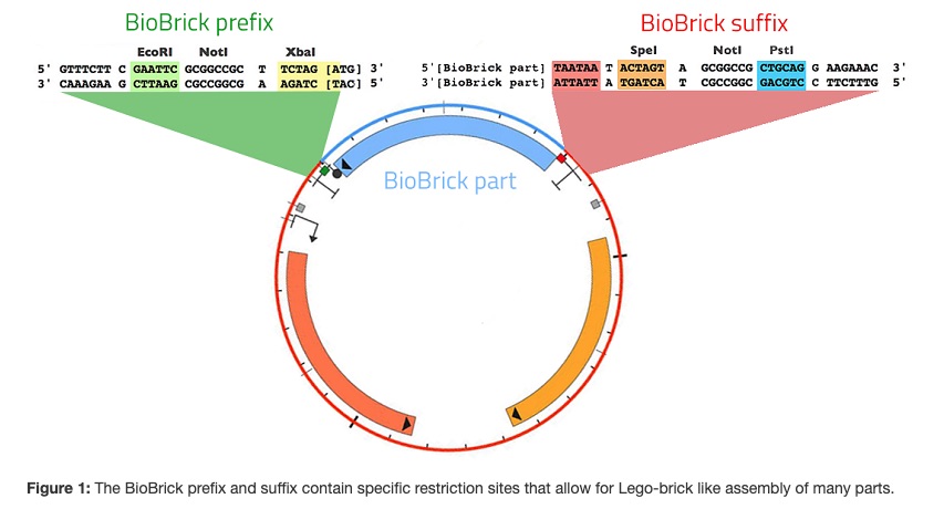
The key to being able to join different parts to assemble a novel construct is that the XbaI and SpeI restriction sites are different but leave compatible overhangs. Therefore if one part is cut with XbaI and the other with SpeI, the two pieces can be joined by ligation. When they are, the same Prefix and Suffix now flank the joined pieces. The place where the sequences are joined is now neither an XbaI nor an SpeI site, and is referred to as a “scar”.
I’ve drawn it below. Note the prefix and suffix sequences to either side of the parts, the promoter shown in blue and the coding sequence in red. When the promotor piece is cut with SpeI and the coding piece with XbaI compatible ends are formed and these can be ligated together. Notice also that the 8 nucleotides between the two pieces is the scar. One drawback to this method is that because the “join” between the two pieces is 8 nucleotides long, it will disrupt the reading frame between two coding pieces so making gene fusions is not possible. The joined pieces are now flanked by the same suffix and prefix pieces, which can be used to ligate these pieces into a vector backbone. This vector will have the ccdB gene in it and it will be cut with EcoRI and PstI. The promoter piece was cut with EcoRI and the coding piece with PstI so that the whole thing assembles into the vector backbone to make your construct. The promoter construct, the coding construct and the “backbone” construct all have different antibiotic resistance. The transformed cells are plated on the antibiotic that selects for the “backbone” or destination vector. Any of the destination vectors that were not cut and did not undergo ligation will not allow transformed cells to grow because of the ccdB gene.
Click here for the powerpoint slides presented in the video below.
