1103 Chapter 11. The Muscular System
11.1 Interactions of Skeletal Muscles, Their Fascicle Arrangement, and Their Lever Systems
Learning Objectives
By the end of this section, you will be able to:
- Describe how muscles are attached to bones to produce movement
- Explain how the location of the insertion and origin of a muscle determine the type of movement accomplished by contraction of that muscle
- Describe the principle of muscular antagonism in movement, using the forearm as an example
- Explain how synergists influence the type of movement accomplished by contraction of a muscle
- Define the following terms: level, fulcrum, resistance, effort
- Describe three kinds of levers and give an example of each type in the human body
- Calculate the force that has to be exerted by the biceps brachii to maintain the forearm in equilibrium when the hand is holding a mass of 10 kilograms
- Describe one mechanical disadvantage and two advantages of the insertion of the biceps brachii being near the elbow joint
To move the skeleton, the tension created by the contraction of the fibers in most skeletal muscles is transferred to the tendons. The tendons are strong bands of dense, regular connective tissue that connect muscles to bones. The bone connection is why this muscle tissue is called skeletal muscle.
Interactions of Skeletal Muscles in the Body
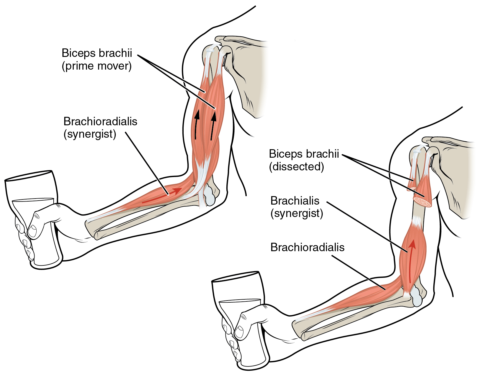
To pull on a bone, that is, to change the angle at its synovial joint, which essentially moves the skeleton, a skeletal muscle must also be attached to a fixed part of the skeleton. The moveable end of the muscle that attaches to the bone being pulled is called the muscle’s insertion, and the end of the muscle attached to a fixed (stabilized) bone is called the origin. During forearm flexion—bending the elbow—the brachioradialis assists the brachialis.
Although a number of muscles may be involved in an action, the principal muscle involved is called the prime mover, or agonist. To lift a cup, a muscle called the biceps brachii is actually the prime mover; however, because it can be assisted by the brachialis, the brachialis is called a synergist in this action (Figure 1). A synergist can also be a fixator that stabilizes the bone that is the attachment for the prime mover’s origin.
A muscle with the opposite action of the prime mover is called an antagonist. Antagonists play two important roles in muscle function: (1) they maintain body or limb position, such as holding the arm out or standing erect; and (2) they control rapid movement, as in shadow boxing without landing a punch or the ability to check the motion of a limb.
For example, to extend the knee, a group of four muscles called the quadriceps femoris in the anterior compartment of the thigh are activated (and would be called the agonists of knee extension). However, to flex the knee joint, an opposite or antagonistic set of muscles called the hamstrings is activated.
As you can see, these terms would also be reversed for the opposing action. If you consider the first action as the knee bending, the hamstrings would be called the agonists and the quadriceps femoris would then be called the antagonists. See Table 1 for a list of some agonists and antagonists.
| Agonist and Antagonist Skeletal Muscle Pairs (Table 1) | ||
|---|---|---|
| Agonist | Antagonist | Movement |
| Biceps brachii: in the anterior compartment of the arm | Triceps brachii: in the posterior compartment of the arm | The biceps brachii flexes the forearm, whereas the triceps brachii extends it. |
| Hamstrings: group of three muscles in the posterior compartment of the thigh | Quadriceps femoris: group of four muscles in the anterior compartment of the thigh | The hamstrings flex the leg, whereas the quadriceps femoris extend it. |
| Flexor digitorum superficialis and flexor digitorum profundus: in the anterior compartment of the forearm | Extensor digitorum: in the posterior compartment of the forearm | The flexor digitorum superficialis and flexor digitorum profundus flex the fingers and the hand at the wrist, whereas the extensor digitorum extends the fingers and the hand at the wrist. |
There are also skeletal muscles that do not pull against the skeleton for movements. For example, there are the muscles that produce facial expressions. The insertions and origins of facial muscles are in the skin, so that certain individual muscles contract to form a smile or frown, form sounds or words, and raise the eyebrows. There also are skeletal muscles in the tongue, and the external urinary and anal sphincters that allow for voluntary regulation of urination and defecation, respectively. In addition, the diaphragm contracts and relaxes to change the volume of the pleural cavities but it does not move the skeleton to do this.
Skeletal muscles do not work by themselves. Muscles are arranged in pairs based on their functions. For muscles attached to the bones of the skeleton, the connection determines the force, speed, and range of movement. These characteristics depend on each other and can explain the general organization of the muscular and skeletal systems.
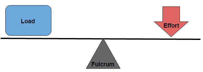
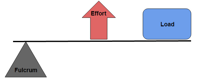
The skeleton and muscles act together to move the body. Have you ever used the back of a hammer to remove a nail from wood? The handle acts as a lever and the head of the hammer acts as a fulcrum, the fixed point that the force is applied to when you pull back or push down on the handle. The effort applied to this system is the pulling or pushing on the handle to remove the nail, which is the load, or “resistance” to the movement of the handle in the system. Our musculoskeletal system works in a similar manner, with bones being stiff levers and the articular endings of the bones—encased in synovial joints—acting as fulcrums. The load would be an object being lifted or any resistance to a movement (your head is a load when you are lifting it), and the effort, or applied force, comes from contracting skeletal muscle.
There are several types of lever systems in the body that are identified as either first-class, second-class, or third-class levers. When classifying a lever system in the human body, the “load” is located at the center of mass of the limb or structure being moved. The “effort” is applied by a muscle (or group of muscles), but its location is not the belly of the muscle being contracted; instead, it is located at the point where the muscle inserts on the structure being moved.
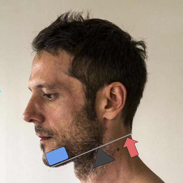
First-class levers are the most simple types of lever, where the balance depends on the distance between the effort and the load from the fulcrum, and the size of the load. In the body the best example of this is the way your head is raised off your chest. As shown in Figure 3 the posterior neck muscles act as the effort, the facial skeleton is the load, and the atlanto-occipital joint behaves as the fulcrum.
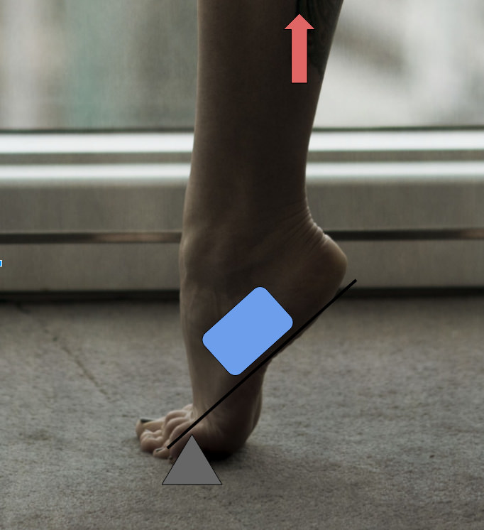
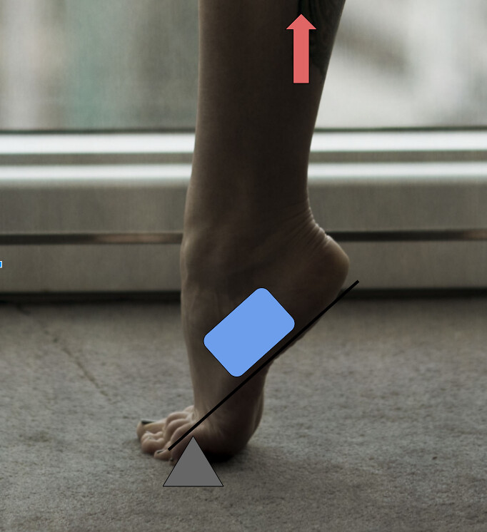
Second-class levers are levers where the load is applied between the effort and the fulcrum. The effort is closer to the load than the fulcrum, which allows a large load to be moved by a small amount of effort. However this means that the load will be moved at a slower pace, and can only be moved a short distance. Any time you stand up on your toes as shown in Figure 5, you are using a second class lever. The weight of your body acts as the load, your calf muscles are the effort, and the joints in the balls of your feet act as fulcrums.
https://www.flickr.com/photos/144136128@N02/29327887125/
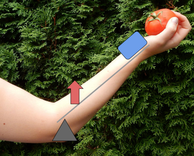
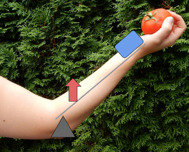
Third Class levers are the most common type of levers in your body. The effort is applied between the fulcrum and the load, which allows the load to be moved quickly over large distances. When you lift your hand by flexing your bicep you are using a third class lever. The Elbow joint acts as the fulcrum, the insertion of the biceps brachii becomes the effort, and the weight of your hand is the load being lifted (Figure 3).
