1103 Chapter 8. The Appendicular Skeleton
8.5 Development of the Appendicular Skeleton
Learning Objectives
By the end of this section, you will be able to:
- Describe the major differences between the appendicular skeleton of a baby and that of an adult
Embryologically, the appendicular skeleton arises from mesenchyme, a type of embryonic tissue that can differentiate into many types of tissues, including bone or muscle tissue. Mesenchyme gives rise to the bones of the upper and lower limbs, as well as to the pectoral and pelvic girdles. Development of the limbs begins near the end of the fourth embryonic week, with the upper limbs appearing first. Thereafter, the development of the upper and lower limbs follows similar patterns, with the lower limbs lagging behind the upper limbs by a few days.
Limb Growth
Each upper and lower limb initially develops as a small bulge called a limb bud, which appears on the lateral side of the early embryo. The upper limb bud appears near the end of the fourth week of development, with the lower limb bud appearing shortly after (Figure 1).
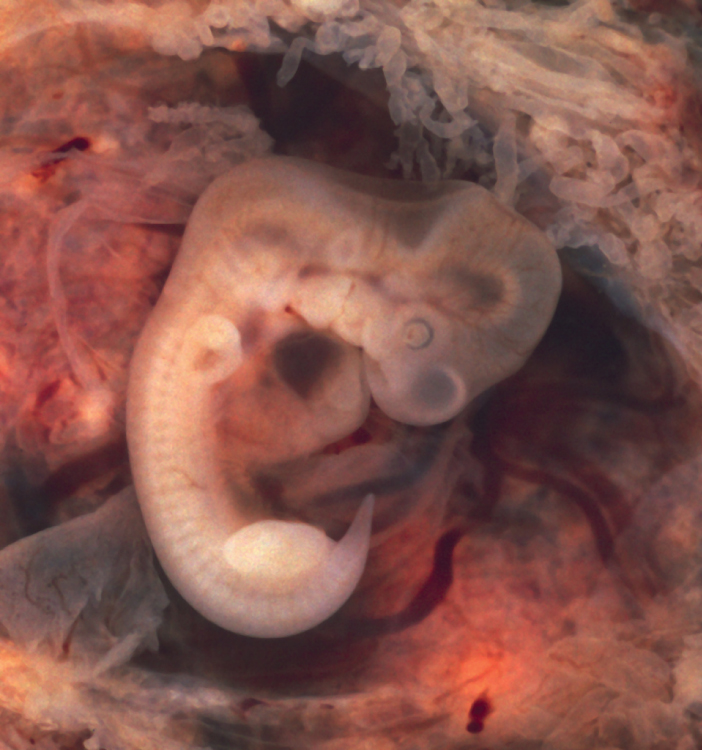
Initially, the limb buds consist of a core of mesenchyme covered by a layer of ectoderm. The ectoderm at the end of the limb bud thickens to form a narrow crest called the apical ectodermal ridge. This ridge stimulates the underlying mesenchyme to rapidly proliferate, producing the outgrowth of the developing limb. As the limb bud elongates, cells located farther from the apical ectodermal ridge slow their rates of cell division and begin to differentiate. In this way, the limb develops along a proximal-to-distal axis.
During the sixth week of development, the distal ends of the upper and lower limb buds expand and flatten into a paddle shape. This region will become the hand or foot. The wrist or ankle areas then appear as a constriction that develops at the base of the paddle. Shortly after this, a second constriction on the limb bud appears at the future site of the elbow or knee. Within the paddle, areas of tissue undergo cell death, producing separations between the growing fingers and toes. Also during the sixth week of development, mesenchyme within the limb buds begins to differentiate into hyaline cartilage that will form models of the future limb bones.
The early outgrowth of the upper and lower limb buds initially has the limbs positioned so that the regions that will become the palm of the hand or the bottom of the foot are facing medially toward the body, with the future thumb or big toe both oriented toward the head. During the seventh week of development, the upper limb rotates laterally by 90 degrees, so that the palm of the hand faces anteriorly and the thumb points laterally. In contrast, the lower limb undergoes a 90-degree medial rotation, thus bringing the big toe to the medial side of the foot.
Ossification of Appendicular Bones
All of the girdle and limb bones, except for the clavicle, develop by the process of endochondral ossification. This process begins as the mesenchyme within the limb bud differentiates into hyaline cartilage to form cartilage models for future bones. By the twelfth week, a primary ossification center will have appeared in the diaphysis (shaft) region of the long bones, initiating the process that converts the cartilage model into bone. A secondary ossification center will appear in each epiphysis (expanded end) of these bones at a later time, usually after birth. The primary and secondary ossification centers are separated by the epiphyseal plate, a layer of growing hyaline cartilage. This plate is located between the diaphysis and each epiphysis. It continues to grow and is responsible for the lengthening of the bone. The epiphyseal plate is retained for many years, until the bone reaches its final, adult size, at which time the epiphyseal plate disappears and the epiphysis fuses to the diaphysis. (Seek additional content on ossification in the chapter on bone tissue).
Small bones, such as the phalanges, will develop only one secondary ossification center and will thus have only a single epiphyseal plate. Large bones, such as the femur, will develop several secondary ossification centers, with an epiphyseal plate associated with each secondary center. Thus, ossification of the femur begins at the end of the seventh week with the appearance of the primary ossification center in the diaphysis, which rapidly expands to ossify the shaft of the bone prior to birth. Secondary ossification centers develop at later times. Ossification of the distal end of the femur, to form the condyles and epicondyles, begins shortly before birth. Secondary ossification centers also appear in the femoral head late in the first year after birth, in the greater trochanter during the fourth year, and in the lesser trochanter between the ages of 9 and 10 years. Once these areas have ossified, their fusion to the diaphysis and the disappearance of each epiphyseal plate follow a reversed sequence. Thus, the lesser trochanter is the first to fuse, doing so at the onset of puberty (around 11 years of age), followed by the greater trochanter approximately 1 year later. The femoral head fuses between the ages of 14–17 years, whereas the distal condyles of the femur are the last to fuse, between the ages of 16–19 years. Knowledge of the age at which different epiphyseal plates disappear is important when interpreting radiographs taken of children. Since the cartilage of an epiphyseal plate is less dense than bone, the plate will appear dark in a radiograph image. Thus, a normal epiphyseal plate may be mistaken for a bone fracture.
The clavicle is the one appendicular skeleton bone that does not develop via endochondral ossification. Instead, the clavicle develops through the process of intramembranous ossification. During this process, mesenchymal cells differentiate directly into bone-producing cells, which produce the clavicle directly, without first making a cartilage model. Because of this early production of bone, the clavicle is the first bone of the body to begin ossification, with ossification centers appearing during the fifth week of development. However, ossification of the clavicle is not complete until age 25.
