1103 Chapter 11. The Muscular System
11.5 Muscles of the Pectoral Girdle and Upper Limbs
Learning Objectives
By the end of this section, you will be able to:
Muscles of the shoulder and upper limb can be divided into four groups: muscles that stabilize and position the pectoral girdle, muscles that move the arm, muscles that move the forearm, and muscles that move the wrists, hands, and fingers. The pectoral girdle, or shoulder girdle, consists of the lateral ends of the clavicle and scapula, along with the proximal end of the humerus, and the muscles covering these three bones to stabilize the shoulder joint. The girdle creates a base from which the head of the humerus, in its ball-and-socket joint with the glenoid fossa of the scapula, can move the arm in multiple directions.
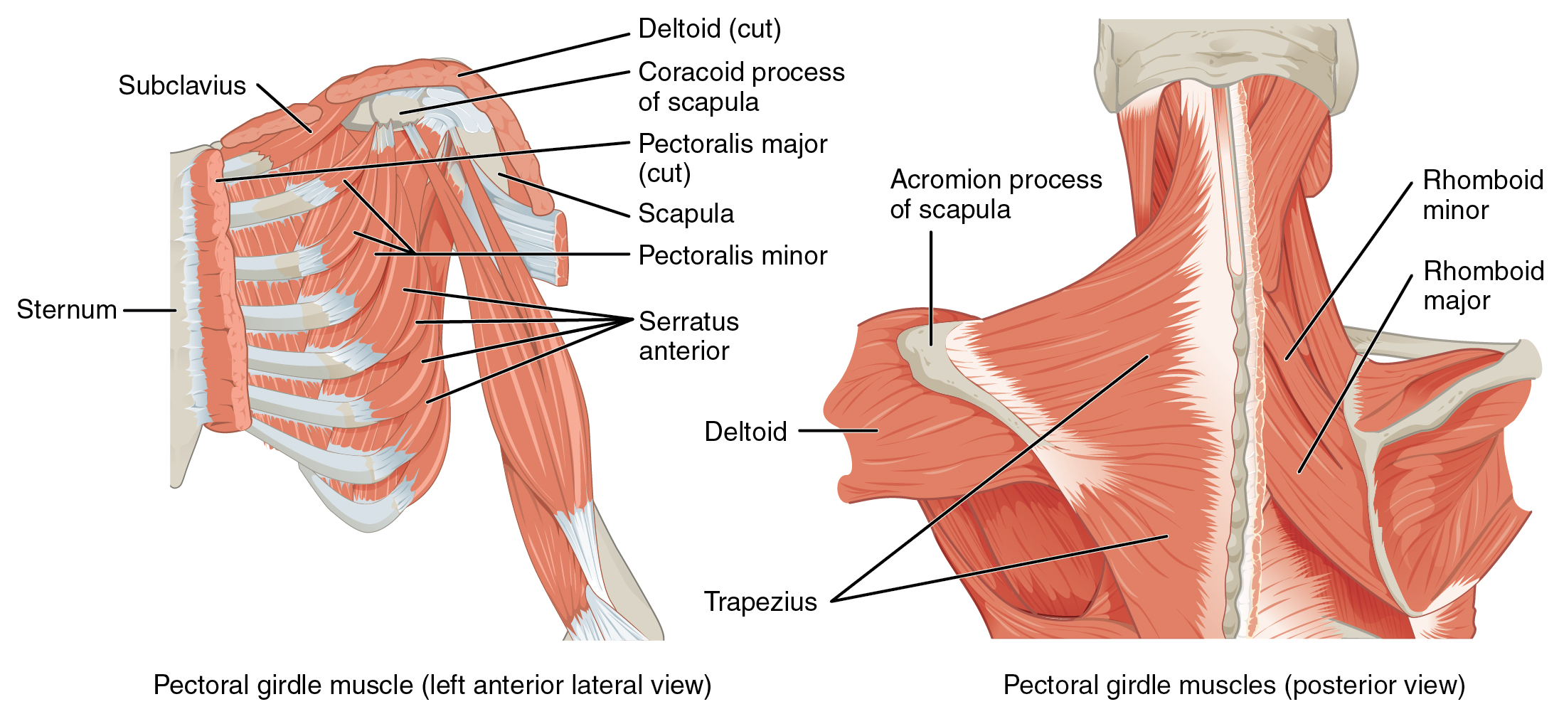
Muscles That Position the Pectoral Girdle
Muscles that position the pectoral girdle are located either on the anterior thorax or on the posterior thorax (Figure 1 and Table 8). The anterior muscles include the subclavius, pectoralis minor, and serratus anterior. The posterior muscles include the trapezius, rhomboid major, and rhomboid minor. When the rhomboids are contracted, your scapula moves medially, which can pull the shoulder and upper limb posteriorly.
| Muscles that Position the Pectoral Girdle (Table 8) | ||||||
|---|---|---|---|---|---|---|
| Position in the thorax | Movement | Target | Target motion direction | Prime mover | Origin | Insertion |
| Anterior thorax | Stabilizes clavicle during movement by depressing it | Clavicle | Depression | Subclavius | First rib | Inferior surface of clavicle |
| Anterior thorax | Rotates shoulder anteriorly (throwing motion); assists with inhalation | Scapula; ribs | Scapula: depresses; ribs: elevates | Pectoralis minor | Anterior surfaces of certain ribs (2–4 or 3–5) | Coracoid process of scapula |
| Anterior thorax | Moves arm from side of body to front of body; assists with inhalation | Scapula; ribs | Scapula: protracts; ribs: elevates | Serratus anterior | Muscle slips from certain ribs (1–8 or 1–9) | Anterior surface of vertebral border of scapula |
| Posterior thorax | Elevates shoulders (shrugging); pulls shoulder blades together; tilts head backwards | Scapula; cervical spine | Scapula: rotests inferiorly, retracts, elevates, and depresses; spine: extends | Trapezius | Skull; vertebral column | Acromion and spine of scapula; clavicle |
| Posterior thorax | Stabilizes scapula during pectoral girdle movement | Scapula | Retracts; rotates inferiorly | Rhomboid major | Thoracic vertebrae (T2–T5) | Medial border of scapula |
| Posterior thorax | Stabilizes scapula during pectoral girdle movement | Scapula | Retracts; rotates inferiorly | Rhomboid minor | Cervical and thoracic vertebrae (C7 and T1) | Medial border of scapula |
Muscles That Move the Humerus
Similar to the muscles that position the pectoral girdle, muscles that cross the shoulder joint and move the humerus bone of the arm include both axial and scapular muscles (Figure 2 and Figure 3). The two axial muscles are the pectoralis major and the latissimus dorsi. The pectoralis major is thick and fan-shaped, covering much of the superior portion of the anterior thorax. The broad, triangular latissimus dorsi is located on the inferior part of the back, where it inserts into a thick connective tissue shealth called an aponeurosis.
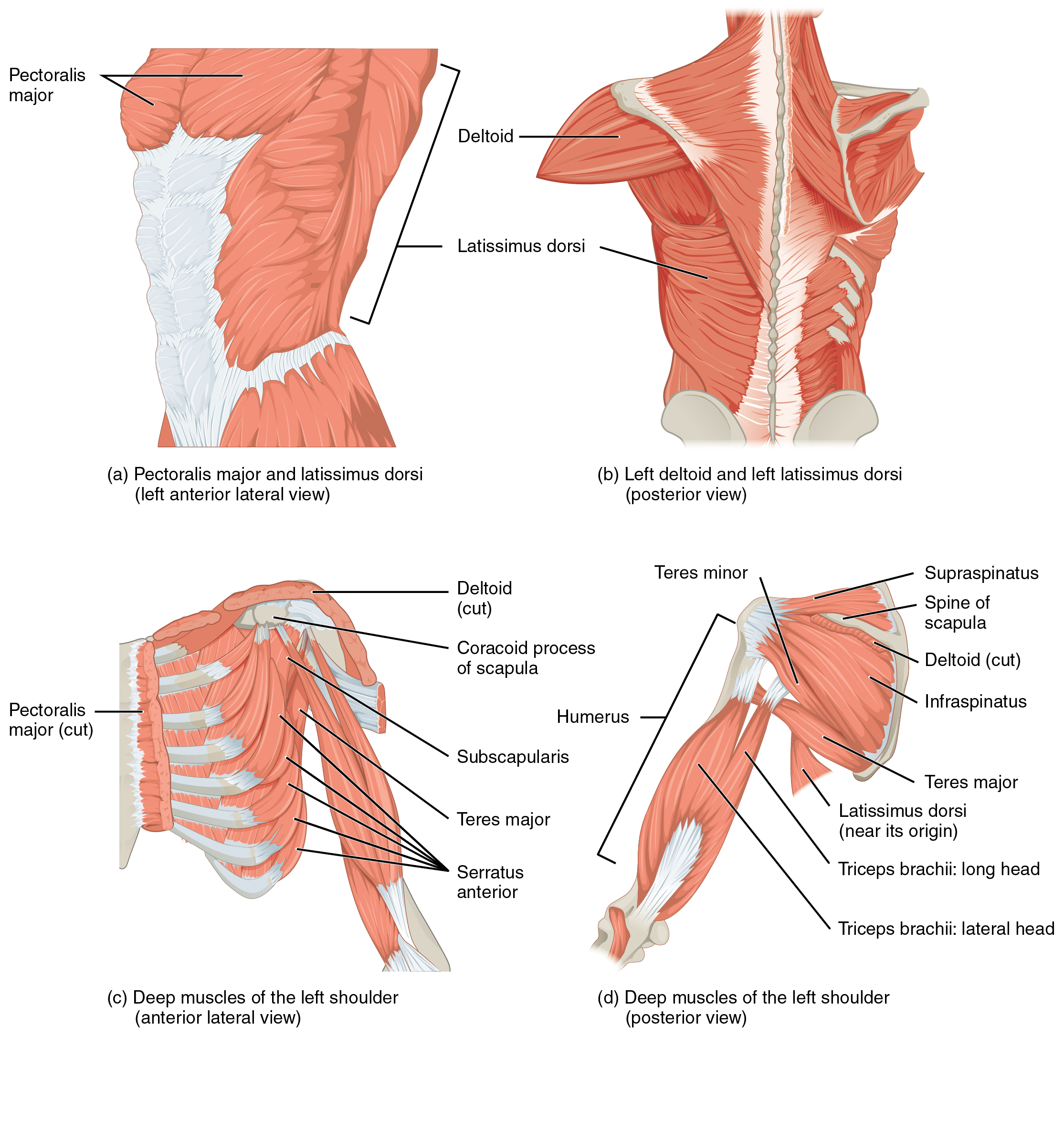
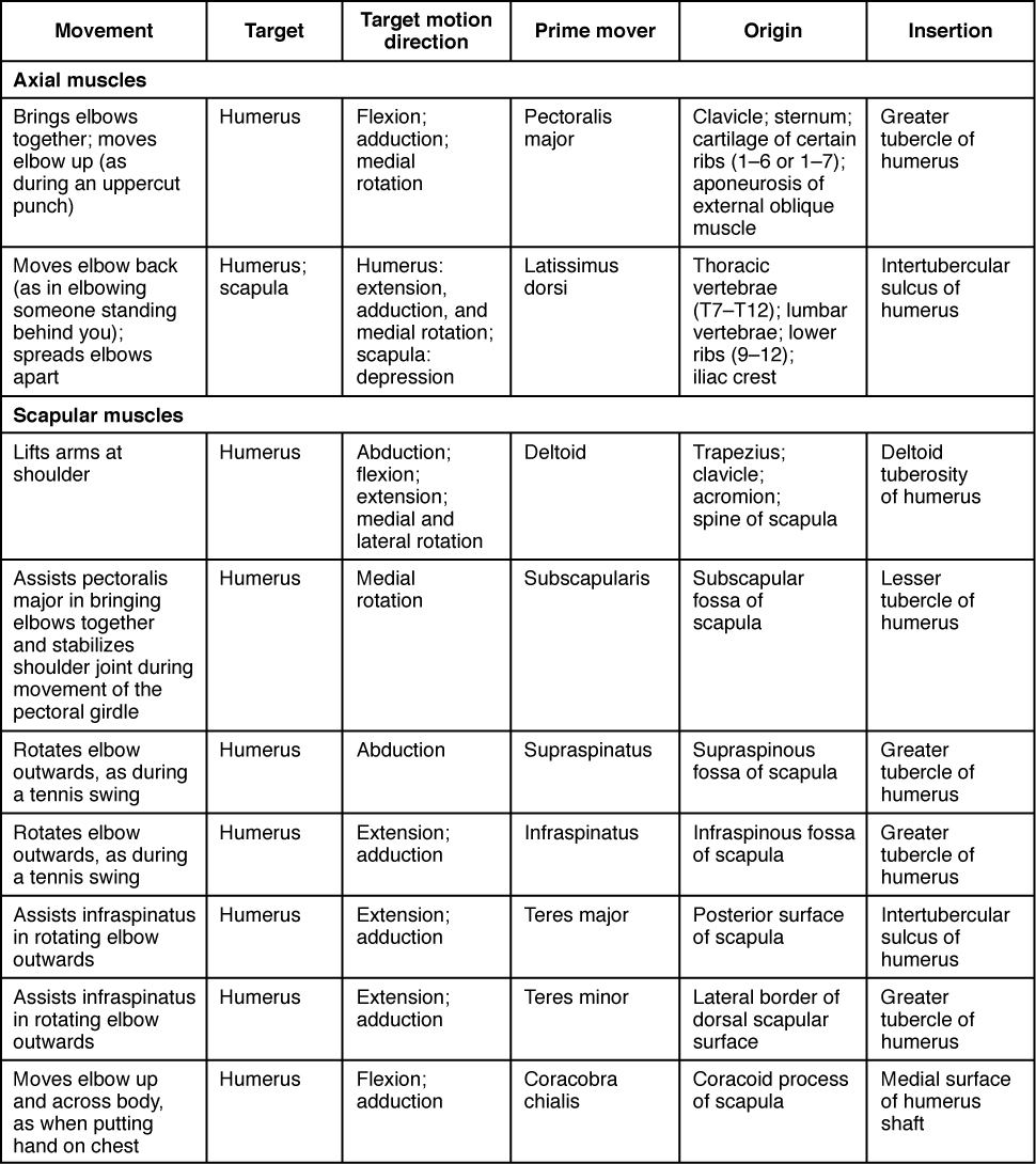
The rest of the shoulder muscles originate on the scapula. The anatomical and ligamental structure of the shoulder joint and the arrangements of the muscles covering it, allows the arm to carry out different types of movements. The deltoid, the thick muscle that creates the rounded lines of the shoulder is the major abductor of the arm, but it also facilitates flexing and medial rotation, as well as extension and lateral rotation. The subscapularis originates on the anterior scapula and medially rotates the arm. Named for their locations, the supraspinatus (superior to the spine of the scapula) and the infraspinatus (inferior to the spine of the scapula) abduct the arm, and laterally rotate the arm, respectively. The thick and flat teres major is inferior to the teres minor and extends the arm, and assists in adduction and medial rotation of it. The long teres minor laterally rotates and extends the arm. Finally, the coracobrachialis flexes and adducts the arm.
The tendons of the deep subscapularis, supraspinatus, infraspinatus, and teres minor connect the scapula to the humerus, forming the rotator cuff (musculotendinous cuff), the circle of tendons around the shoulder joint. When baseball pitchers undergo shoulder surgery it is usually on the rotator cuff, which becomes pinched and inflamed, and may tear away from the bone due to the repetitive motion of bring the arm overhead to throw a fast pitch.
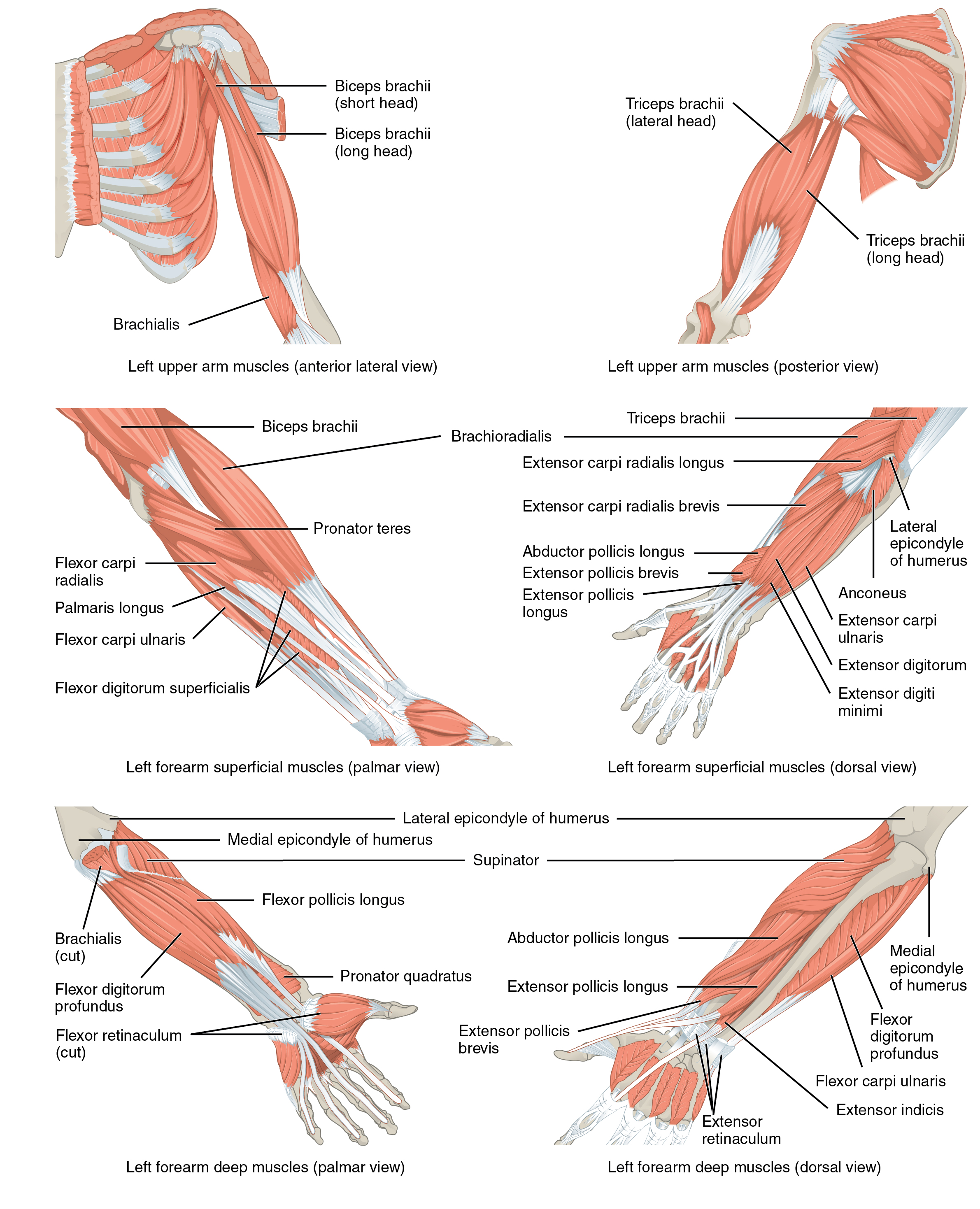
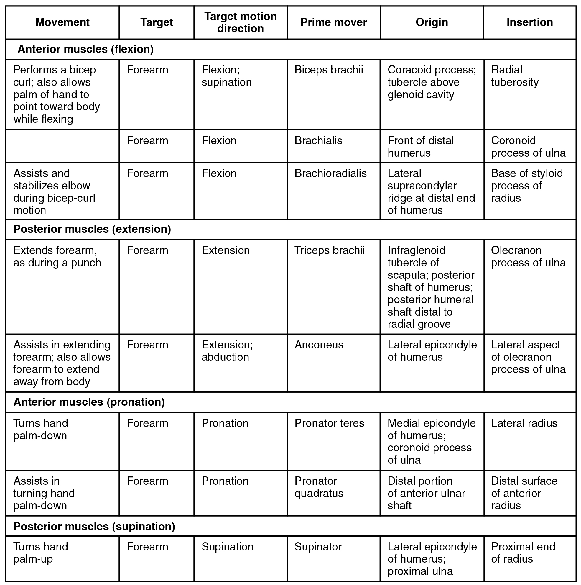
Muscles That Move the Forearm
The forearm, made of the radius and ulna bones, has four main types of action at the hinge of the elbow joint: flexion, extension, pronation, and supination. The forearm flexors include the biceps brachii, brachialis, and brachioradialis. The extensors are the triceps brachii and anconeus. The pronators are the pronator teres and the pronator quadratus, and the supinator is the only one that turns the forearm anteriorly. When the forearm faces anteriorly, it is supinated. When the forearm faces posteriorly, it is pronated.
The biceps brachii, brachialis, and brachioradialis flex the forearm. The two-headed biceps brachii crosses the shoulder and elbow joints to flex the forearm, also taking part in supinating the forearm at the radioulnar joints and flexing the arm at the shoulder joint. Deep to the biceps brachii, the brachialis provides additional power in flexing the forearm. Finally, the brachioradialis can flex the forearm quickly or help lift a load slowly. These muscles and their associated blood vessels and nerves form the anterior compartment of the arm (anterior flexor compartment of the arm) (Figure 4 and Figure 5).