1103 Chapter 11. The Muscular System
11.3 Axial Muscles of the Head, Neck, and Back
Learning Objectives
By the end of this section, you will be able to:
The skeletal muscles are divided into axial (muscles of the trunk and head) and appendicular (muscles of the arms and legs) categories. This system reflects the bones of the skeleton system, which are also arranged in this manner. The axial muscles are grouped based on location, function, or both. Some of the axial muscles may seem to blur the boundaries because they cross over to the appendicular skeleton. The first grouping of the axial muscles you will review includes the muscles of the head and neck, then you will review the muscles of the vertebral column, and finally you will review the oblique and rectus muscles.
Muscles That Move the Head
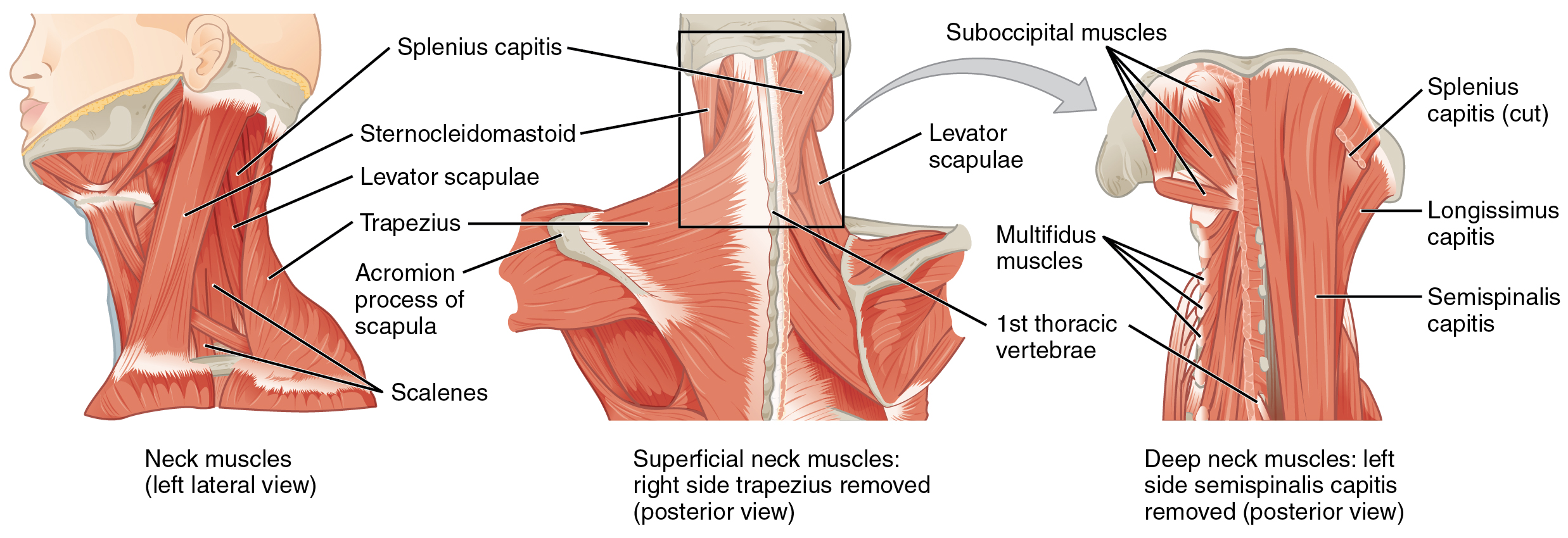
The head, attached to the top of the vertebral column, is balanced, moved, and rotated by the neck muscles (Table 5). When these muscles act unilaterally, the head rotates. When they contract bilaterally, the head flexes or extends. The major muscle that laterally flexes and rotates the head is the sternocleidomastoid. In addition, both muscles working together are the flexors of the head. Place your fingers on both sides of the neck and turn your head to the left and to the right. You will feel the movement originate there. This muscle divides the neck into anterior and posterior triangles when viewed from the side (Figure 8).
| Muscles That Move the Head (Table 5) | |||||
|---|---|---|---|---|---|
| Movement | Target | Target motion direction | Prime mover | Origin | Insertion |
| Rotates and tilts head to the side; tilts head forward | Skull; vertebrae | Individually: rotates head to opposite side; bilaterally: flexion | Sternocleidomastoid | Sternum; clavicle | Temporal bone (mastoid process); occipital bone |
| Rotates and tilts head backward | Skull; vertebrae | Individually: laterally flexes and rotates head to same side; bilaterally: extension | Semispinalis capitis | Transverse and articular processes of cervical and thoracic vertebra | Occipital bone |
| Rotates and tilts head to the side; tilts head backward | Skull; vertebrae | Individually: laterally flexes and rotates head to same side; bilaterally: extension | Splenius capitis | Spinous processes of cervical and thoracic vertebra | Temporal bone (mastoid process); occipital bone |
| Rotates and tilts head to the side; tilts head backward | Skull; vertebrae | Individually: laterally flexes and rotates head to same side; bilaterally: extension | Longissimus capitis | Transverse and articular processes of cervical and thoracic vertebra | Temporal bone (mastoid process) |
Muscles of the Posterior Neck and the Back
The posterior muscles of the neck are primarily concerned with head movements, like extension. The back muscles stabilize and move the vertebral column, and are grouped according to the lengths and direction of the fascicles.
The splenius muscles originate at the midline and run laterally and superiorly to their insertions. From the sides and the back of the neck, the splenius capitis inserts onto the head region, and the splenius cervicis extends onto the cervical region. These muscles can extend the head, laterally flex it, and rotate it (Figure 9).
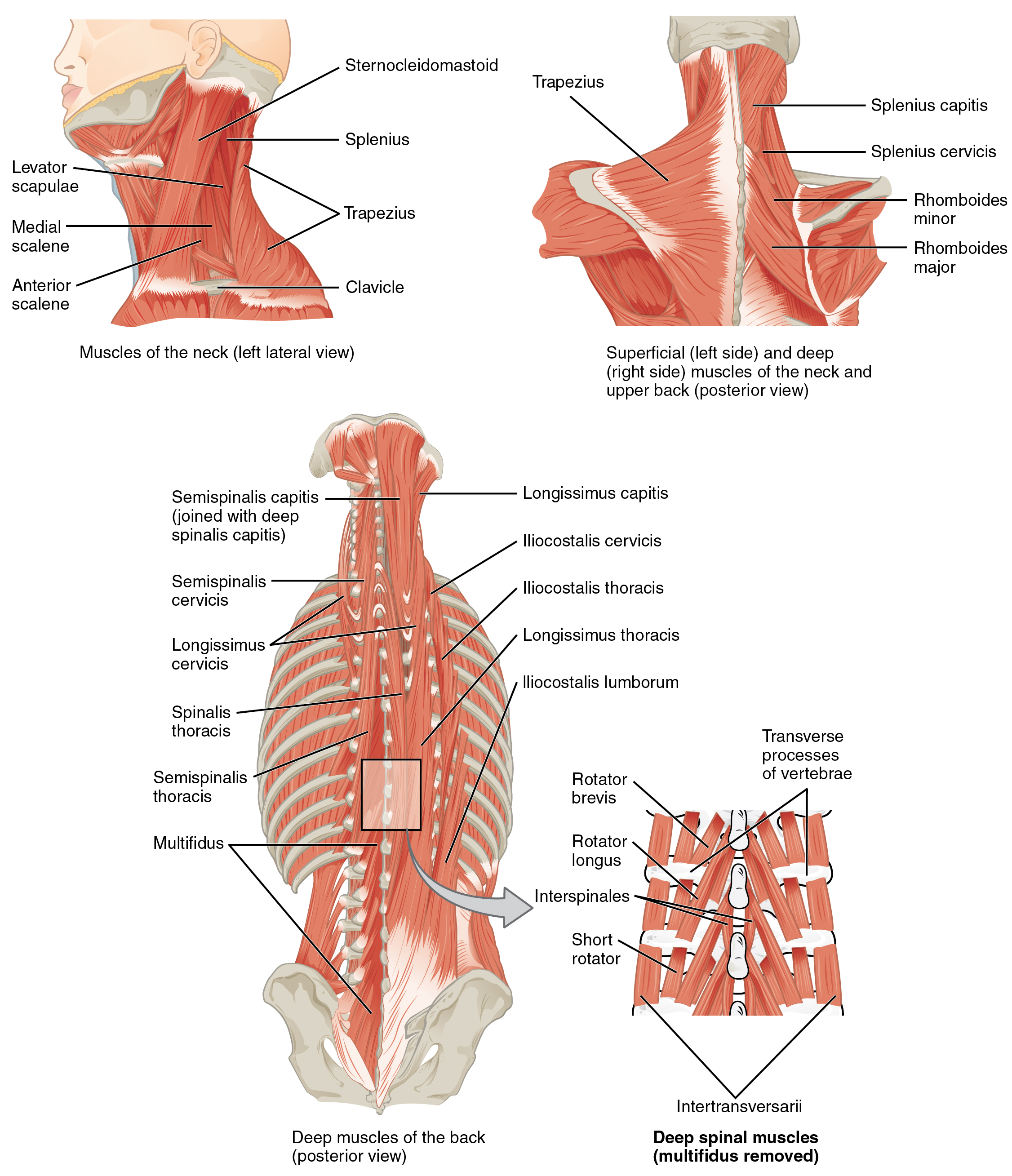
The erector spinae group forms the majority of the muscle mass of the back and it is the primary extensor of the vertebral column. It controls flexion, lateral flexion, and rotation of the vertebral column, and maintains the lumbar curve. The erector spinae comprises the iliocostalis (laterally placed) group, the longissimus (intermediately placed) group, and the spinalis (medially placed) group.
The iliocostalis group includes the iliocostalis cervicis, associated with the cervical region; the iliocostalis thoracis, associated with the thoracic region; and the iliocostalis lumborum, associated with the lumbar region. The three muscles of the longissimus group are the longissimus capitis, associated with the head region; the longissimus cervicis, associated with the cervical region; and the longissimus thoracis, associated with the thoracic region. The third group, the spinalis group, comprises the spinalis capitis (head region), the spinalis cervicis (cervical region), and the spinalis thoracis (thoracic region).
The transversospinales muscles run from the transverse processes to the spinous processes of the vertebrae. Similar to the erector spinae muscles, the semispinalis muscles in this group are named for the areas of the body with which they are associated. The semispinalis muscles include the semispinalis capitis, the semispinalis cervicis, and the semispinalis thoracis. The multifidus muscle of the lumbar region helps extend and laterally flex the vertebral column.
Important in the stabilization of the vertebral column is the segmental muscle group, which includes the interspinales and intertransversarii muscles. These muscles bring together the spinous and transverse processes of each consecutive vertebra. Finally, the scalene muscles work together to flex, laterally flex, and rotate the head. They also contribute to deep inhalation. The scalene muscles include the anterior scalene muscle (anterior to the middle scalene), the middle scalene muscle (the longest, intermediate between the anterior and posterior scalenes), and the posterior scalene muscle (the smallest, posterior to the middle scalene).
