Chapter 2: Patient Assessment
2.8 Head-to-Toe Assessment: Cardiovascular Assessment
Critical Thinking Exercises: Questions, Answers, and Sources / References
Critical thinking questions are in bold type, and the answers are italicized. Additional resources or references are provided below.
- A client has +4 edema to bilateral feet and ankles. Identify two strategies to assist in maintaining skin integrity.
Edematous tissue is at risk for breakdown. Suggest good hygiene, use of lotions, attention to skin integrity on the feet, and elevate the feet to promote venous return and reduce edema. Consult with an interdisciplinary team to determine other contributing factors (intrinsic and extrinsic) that may need to be addressed.
Sources:
Registered Nurses’ Association of Ontario (RNAO). (n.d.). Nursing best practice guidelines: Skin integrity. https://bpgmobile.rnao.ca/content/risk-and-related-interventions
Registered Nurses’ Association of Ontario (RNAO). (n.d.). Nursing best practice guidelines: Risk assessment tools. https://bpgmobile.rnao.ca/content/management-patient-nutritional-risk-and-pressure-ulcer-risk
2. A client has just had a femoral popliteal bypass. Which peripheral pulses should be included in the assessment specific to determining arterial perfusion of the affected leg?
Assessment of pulses distal to a vascular surgery will help to determine patency of vascular grafts. After a femoral popliteal bypass surgery, the dorsalis pedis and posterior tibialis pulses should be assessed.
Sources:
Berti-Hearn, L., & Elliott, B. (2018). A closer look at lower extremity peripheral arterial disease. Nursing, 48(1), 34-41. https://www.nursingcenter.com/cearticle?an=00152193-201801000-00009&Journal_ID=54016&Issue_ID=4468826
Stanford Health Care. (n.d.). What is femoral popliteal bypass surgery? https://stanfordhealthcare.org/medical-treatments/f/femoral-popliteal-bypass.html
Sample Learning Activity
- On the following diagram, identify with an X where the following pulses are located:
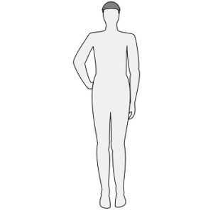
- Carotid
- Femoral
- Dorsalis pedis
- Posterior tibialis
- Radial
- Brachial
2. Have students work in partners and/or with a manikin equipped with pulses to palpate each of the pulses noted above. Integrate the learning of how to use a doppler for peripheral pulse assessment. Obtain a pulse assessment flow sheet or clinical record, and have students document their findings using the document’s rating scale. The faculty member and/or student partners will give feedback on documentation.
Attribution: Figure 2.1 Male body silouhette- front by nicubunu is in the Public Domain.
Media Attributions
- Front view of a male body silhouette © nicubunu is licensed under a Public Domain license
