Chapter 2: Biology and Human Potential
The Nervous and Endocrine Systems
The Nervous System: Connecting Sensation and Movement
As we consider the human genotype, we will start by providing an overview of the nervous system (see Figure 2.7), those structures which transmit information regarding external and internal stimulation and coordinate behavior.
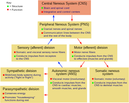
Figure 2.7 Overview of human nervous system
The central nervous system , consisting of the brain and spinal cord, organizes and interprets information received from the peripheral nervous system and initiates responding. The somatic division of the peripheral nervous system responds to sensory information originating outside the body and stimulates the skin, joints, and skeletal muscles. This type of behavior is often considered voluntary. The autonomic nervous system governs the activity of the smooth muscles and glands internal to the body involved in circulation, respiration, and digestion (see Figure 2.8). This type of activity is often considered involuntary. The sympathetic division results in arousal under stressful or dangerous conditions as the body is prepared for “fight or flight.” The parasympathetic division calms the body upon removal of the stress or danger.

Figure 2.8 The autonomic nervous system
Making the Physical Connections: The Neuron
Even very simple animals require some way of connecting environmental input with behavioral output. Specialized nerve cells called neurons are required to respond to external and internal stimulation (i.e., sensory neurons) and carry information to parts of the body capable of responding (i.e., motor neurons). A third type of cell referred to as an interneuron connects nerve cells to each other. Nervous systems consist of these types of specialized neurons and range in size from a few hundred nerve cells in worms to approximately 100 billion nerve cells in humans. Neurons are capable of transmitting information electrically and chemically. Figure 2.9 portrays the major parts of a neuron. Dendrites are small branches which can connect to nearby neurons. A single axon can extend in length up to about a meter in humans and connect to the dendrites of more distant neurons. For example, a neuron could connect the spinal cord to a foot.

Figure 2.9 The neuron
Nerve cells “fire” (i.e., achieve their electrical action potential) according to an all-or-none principle. That is, either the cell is totally activated or not at all. Increasing the intensity of stimulation does not increase the likelihood of a nerve responding. Rather, it increases the nerve’s rate of firing (i.e., frequency over time). For example, as a lamp becomes brighter, this does not increase the likelihood of a receptor cell in your eye firing. Rather, it increases the frequency with which the receptor cell fires. Nerves can fire at rates as high as a thousand times per second.
Making Chemical Connections: Neurotransmitters
The chemical exchange between neurons occurs at synapses, the small spaces separating the dendrites and axon endings (see Figure 2.10).

Figure 2.10 The synapse
The first nerve cell releases chemical neurotransmitters that can bind with receptors in the second neuron. The exchange can result in excitation or inhibition, depending upon the type of receptor activated. Figure 2.11 lists the major neurotransmitters along with their roles in the body.

Figure 2.11 The major neurotransmitters
Psychoactive drugs can affect mood, thought, and behavior. Most achieve these effects by impacting upon neurotransmitters and synaptic connections. In Chapter 11 (Maladaptive Behavior), we will consider the use of psychoactive drugs in the treatment of depression and schizophrenia.
The Brain
For literal and figurative reasons, it is tempting to refer to the human brain as evolution’s crowning achievement. After all, the brain sits atop our nervous system and enables our most complex overt and covert behaviors. Your thoughts, your feelings, all the complex things you do, would not be possible without this organ housed inside your skull on top of your head.
The human brain is similar in construction to the brains of other mammals but much larger in comparison to the size of our bodies. Without the increase in brain size occurring during human evolution it would not matter if we inherited the physical structures necessary to speak and create tools. This potential would never be realized. Manhattan would still look the same as it did 400 years ago. We are now using our remarkable brain to study itself. The United States government declared the 1990s as the “Decade of the Brain” and much progress has been made in understanding how the brain operates. President Barack Obama of the United States declared “The BRAIN (Brain Research through Advancing Innovative Neurotechnologies) Initiative” in 2013, hoping to advance this knowledge.
There are many ways of describing the brain in terms of its structure (i.e., anatomy) or function (i.e., parts operating together in producing a specific effect). Figure 2.12 shows the major parts of the human brain. The prefrontal cortex is involved in the higher human cognitive functions including attention, perception, thinking, memory, language and consciousness.
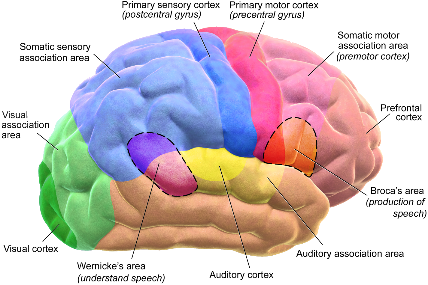
Figure 2.12 Human brain
The brain is an adaptive organ connecting sensation with movement. Besides the primary somatosensory area in the parietal lobe, sensory areas include the occipital lobe for vision and temporal lobe for hearing. Besides the primary motor area at the rear of the frontal lobe, motor areas include the brain stem and spinal cord. The rest of the cortex is referred to as association areas and is dedicated to perception and cognition. It is the size and structure of this area which expanded enormously as humans evolved and enabled us to not only survive but to transform the human condition.
A human brain weighs about three pounds and feels “squishy” (something like gelatin). The cerebral cortex covers most of the brain and is comprised of nerve fibers folded in such a manner (called convolutions ) to increase the amount of surface area in the total space. There are two symmetrical halves often referred to as the left brain (or hemisphere) and right brain (or hemisphere). The two halves are connected by the corpus callosum, a thick network of nerve fibers enabling the two sides to communicate. The left side of the brain connects to the right side of the body and vice versa. Certain activities appear more characteristic of one side than the other (see Figure 2.13). These distinctions are referred to as lateralization . Despite the different emphases, both sides usually act in concert in carrying out these activities (Toga & Thompson, 2003).

Figure 2.13 Brain lateralization
Most of the expansion in the size of the human brain occurred in the frontal lobe. This part of the brain is involved in self-control, described in Chapter 1, and in abstract thought and problem-solving, described in Chapter 7. The small occipital lobe is dedicated to vision, described in Chapter 3. At the borders of the frontal and parietal lobes is a deep fissure (the central sulcus ) where large strips of neural tissue dedicated to sensation (the primary somatosensory cortex) and movement (the primary motor cortex) meet. The temporal lobe is primarily involved with memory and language, described in Chapter 6. The parietal lobe is involved with sensation originating in the skin, muscles, and joints.
The Endocrine System: Hormonal Regulation
The endocrine system consists of ductless glands that secrete hormones (chemical messengers) into the blood stream to maintain homeostasis. It exists in all animals having a nervous system. Like the nervous system, the endocrine system enables communication between different parts of the body.
The endocrine system maintains homeostasis through a series of feedback loops, the most important of which are controlled by the hypothalamus interacting with the pituitary gland. Often, the hypothalamus stimulates the pituitary gland to secrete an activating hormone to another gland. If a signal is transmitted to a gland, indicating low blood levels of its hormone, it secretes additional amounts into the blood stream. Once the optimal level is restored, the gland stops secreting the hormone. In this way the endocrine system plays its critical role in metabolism, growth, sexual development, reproduction, and responding to stress. Figure 2.14 shows the locations of the major glands.

Figure 2.14 Major gland locations
The pituitary gland connects to the base of the hypothalamus and is often referred to as the master gland since it secretes several different hormones impacting upon other glands involved in maintaining homeostasis. Hormones secreted by the pituitary control growth, blood pressure, water balance, temperature regulation, and pain relief. The pineal gland is located at the base of the cortex between the two hemispheres and next to the thalamus. It influences the sleep-wake cycle by secreting the hormone melatonin when stimulated by light. The thyroid gland is located in the neck by the larynx (voice box) and affects metabolism by controlling the rate at which energy is expended. It is one of the glands under the control of the pituitary which secretes thyroid-stimulating hormone (TSH). The pituitary in turn is controlled by the hypothalamus through the release of thyrotropin-releasing hormone (TRH). Humans usually have four parathyroid glands located on the rear surface of the thyroid gland. These control the amount of calcium in the blood and bones. The thymus is located below the thyroid gland in the middle of the chest. It is an important part of the immune system. Damage, such as through contracting the HIV virus, can result in increased susceptibility to infection (e.g., AIDS). The spleen lies toward the bottom of your rib cage and is involved in the removal of red blood cells. The adrenal glands are located on top of the kidneys and through the release of epinephrine (adrenalin) are significantly involved in the body’s “fight-or-flight” response in reaction to danger. The sex glands (ovaries for the female and testes for the male) secrete hormones controlling the development of the reproductive sex organs and secondary sex characteristics (e.g., pubic hair) during puberty.
Attributions
Figure 2.7 “Human nervous system” is licensed under CC BY-SA 3.0
Figure 2.8 “Autonomic nervous system” by Geo-Science-International is licensed under CC BY 1.0
Figure 2.9 “The neuron” is licensed under CC BY-SA 4.0
Figure 2.10 “Synapse” by Dwindrim is licensed under CC BY-SA 1.0
Figure 2.11 “Neurotransmitters” is licensed under CC BY-SA 3.0
Figure 2.12 “Human brain” by Bruce Blaus is licensed under CC BY 3.0
Figure 2.13 “Brain lateralization” is licensed under CC BY-SA 3.0
Figure 2.14 “Major endocrine glands” by US Government is in the Public Domain, CC0
structures that transmit information regarding external and internal stimulation and coordinate behavior.
the brain and spinal cord
responds to sensory information originating outside the body and stimulates the skin, joints, and skeletal muscles; the resulting behavior is often considered voluntary.
governs the activity of the smooth muscles and glands internal to the body involved in circulation, respiration, and digestion; resulting behavior is often considered involuntary
activation arouses body under stressful or dangerous conditions to prepare for “fight or flight.”
calms the body upon removal of the stress or danger
cells that respond to external and internal stimulation (i.e., sensory neurons) and carry information to parts of the body capable of responding (i.e., motor neurons)
connect nerve cells to each other
small branches of a neuron that can connect to nearby neurons
long branches of a neuron that connect to the dendrites of more distant neurons
neural activity occurring after a threshold is reached; cells “fire” according to an all-or-none principle
a nerve cell is activated totally or not at all
small spaces separating the dendrites and axon endings where chemical exchange between neurons occur
chemicals released by nerve cells that can bind with receptors in the second neuron; major neurotransmitters include dopamine (alertness) , norepinephrine (attention and concentration), and serotonin (pleasure and anxiety)
neural process stimulating a nerve cell to transmit information
neural process decreasing the ability for a nerve cell to transmit information
affect mood, thought, and behavior; most achieve these effects by impacting upon neurotransmitters and synaptic connections
part of brain involved in human cognitive functions including attention, perception, thinking, memory, language and consciousness.
part of brain involved with sensation originating in the skin, muscles, and joints
part of brain involved in vision
part of brain involved in hearing, memory, and language
part of brain involved in self-control
nerve fibers folded in such a manner as to increase the amount of surface area in the total space
thick network of nerve fibers enabling the two sides of the brain to communicate
the left side of the brain connects to the right side of the body and vice versa; certain activities appear more characteristic of one side than the other
a deep fissure at the borders of the frontal and parietal lobes where large strips of neural tissue dedicated to sensation (the primary somatosensory cortex) and movement (the primary motor cortex) meet
consists of ductless glands that secrete hormones (chemical messengers) into the blood stream to maintain homeostasis; enables communication between different parts of the body playing critical roles in metabolism, growth, sexual development, reproduction, and responding to stress
secreted by glands and carried in the blood as chemical messengers activating other glands and parts of the body
stimulates the pituitary gland to secrete an activating hormone to another gland to maintain homeostasis
often referred to as the master gland since it secretes several different hormones impacting upon other glands involved in maintaining homeostasis; controlled by the hypothalamus through the release of thyrotropin-releasing hormone (TRH)
influences the sleep-wake cycle by secreting the hormone melatonin when stimulated by light
affects metabolism by controlling the rate at which energy is expended; one of the glands under the control of the pituitary gland
located on the rear surface of the thyroid gland; control the amount of calcium in the blood and bones
an important part of the immune system, located below the thyroid gland in the middle of the chest
lies toward the bottom of your rib cage and is involved in the removal of red blood cells
involved in the body’s “fight-or-flight” response through the release of epinephrine (adrenalin)in reaction to danger; located on top of the kidneys
ovaries for the female and testes for the male; secrete hormones controlling the development of the reproductive sex organs and secondary sex characteristics during puberty
