Chapter 2: Rashes of the Newborn
Potentially Concerning Skin Changes in Newborns
Blisters
Blisters can occur in neonates or a variety of reasons including infection, genetic blistering diseases (see Epidermolysis bullosa), and infiltration of the skin with mast cells. Appropriate testing to rule out infection is necessary and proper wound care is crucial to prevent secondary bacterial infection.
Neonatal Herpes Simplex
Neonatal herpes simplex usually presents with vesicles and occurs due to HSV exposure during vaginal delivery. Vesicles are seen most commonly on the presenting part of the baby such as the crown of the head. Neonatal HSV is more likely if the mother is experiencing her first episode of HSV, so she might not have a history of genital herpes. The rash may be present from birth if it is acquired in utero but typically starts at least 5 days after birth. Infection may be complicated by encephalitis, and mortality is ~50% in these cases if not treated with IV acyclovir.
Neonatal Lupus
Neonatal lupus is seen in babies born to mothers with anti-Ro, anti-La, or U1RNP antibodies. The antibodies can cross the placenta and cause changes in the baby. Skin findings include annular plaques with fine scale especially on the head and neck and concentrated around the eyes. The lesions typically first appear by 2 months of age and worsen after sun exposure. While skin changes will self-resolve, babies with neonatal lupus are at risk for heart block, cytopenias, and liver function changes.
Collodion Membrane
Collodion membrane is the name given to a parchment or plastic wrap-like membrane of skin that wraps some newborns. It can cause ectropion and/or eclabium. It may be the first sign of an ichthyosis, but also can be self-resolving. Treatment is with moisturizers, and possibly incubator, to help preserve skin function. The membrane will slough spontaneously and should not be removed.
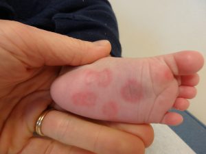
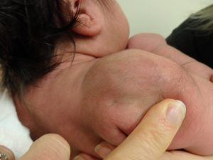
Subcutaneous Fat Necrosis of the Newborn
Subcutaneous fat necrosis of the newborn occurs due to crystal formation in fat cells in newborn fat. It is seen most often in newborns who have required cooling and presents with tender red-brown nodules. Babies with extensive subcutaneous fat necrosis should not be given Vitamin D and should be followed for possible development of hypercalcemia.
Blueberry Muffin Baby
Blueberry muffin baby describes the clinical finding of widespread red to purple papules and nodules in a newborn baby. There is a wide range of conditions that lead to the finding of blueberry muffin baby. The most common of these are congenital infections, but different forms of anemia and hematologic malignancy are among other potential causes. Evaluation for underlying cause of the nodules is imperative.
Selected causes of blueberry muffin baby:
