Chapter 7: Vascular Conditions
Vascular Malformations: Venous, Arteriovenous and Lymphatic Malformations
What are they?
Vascular malformations represent localized anomalous vessels and are categorized by the predominant vessel type. There are capillary malformations (discussed above, most commonly a port wine stain), venous, arteriovenous and lymphatic malformations. Another way to think of these is as slow flow (capillary, venous, and lymphatic malformation) or fast flow (arteriovenous malformations) and this can be seen by doppler ultrasound. Vascular malformations are congenital lesions that are typically present at birth and persist throughout life with either proportionate growth or a slow increase in size over time.
What do they look like?
Venous malformations are soft, blue papules or plaques that are compressible and fill with dependency. These can involve underlying muscle, bone and joints.
Primary lymphedema presents as fluid accumulation most commonly in the lower extremities.
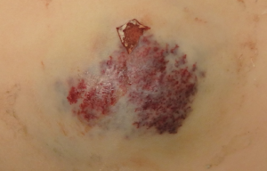
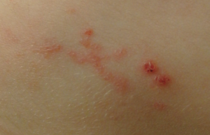
Microcystic lymphatic malformation, also known as lymphangioma circumscriptum, presents as clusters of clear or hemorrhagic vesicles. They are most common on the proximal limbs and chest but can occur anywhere including the oral cavity.
Macrocystic lymphatic malformation are most common on the neck, axilla and trunk and present as a large, soft, translucent mass underlying the skin. They often enlarge if the child has an infection.
Arteriovenous malformation (AVM) represent direct communications between arteries and veins which results in a fast flow shunt. These are rare vascular malformations, and unlike the other types only 40% are present at birth and the remainder appear later in life. The most common location is cephalic. They can cause complications such as skin necrosis or even high output cardiac failure.
There are many syndromes that are associated with vascular malformations. If a patient has multiple or large vascular malformations this should prompt a thorough work-up and involvement of a multidisciplinary team.
How are they treated?
Treatment of a vascular malformation depends on the size, location and other patient factors. Possible treatments include close observation, surgical excision, laser therapy, embolization, sclerotherapy, or oral medications such as mTOR inhibitors.

Note: compare to hemangioma on arm
