Chapter 7: Vascular Conditions
Vascular Tumors: Infantile Hemangioma
What is it?
Infantile hemangiomas (IH) are common, benign vascular tumors. They occur in approximately 5% of infants. Risk factors for IH include female sex, prematurity and low birth weight, placental insufficiency, multiple gestations and advanced maternal age. The cause of infantile hemangioma is incompletely understood and likely involves several mechanisms. It is thought that hypoxia plays a key role in initiation the growth of IH. Most are not present at birth, but appear by 3-4 weeks of age and grow rapidly within the first 3 months. The majority of growth happens by 6-9 months, followed by growth arrest. Spontaneous gradual involution starts around 1 year of age and continues until the child reaches 9 years old. There are several syndromes and specific complications that are associated with IH discussed below.
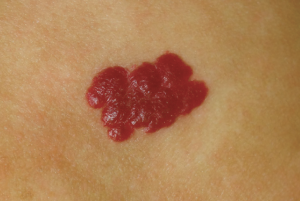
What does it look like?
At birth, precursor lesions including pale areas, pink macules or bruise-like patches may be noted. More mature hemangiomas may be superficial, deep variants or mixed lesions with features of both. Superficial IH are bright red plaques with a finely lobulated surface leading to the name “strawberry hemangioma”. Deep IH present as ill-defined blue masses which may have minimal or no overlying skin changes. Mixed lesions have a bright red superficial component overlying a deeper blue nodule. Hemangiomas, especially large or genital lesions, may develop central ulceration. Infantile hemangioma can be focal or segmental. The distribution and size of IH is important because of the risk of associated syndromes. Large, segmental IH especially on the face have a higher risk of PHACES syndrome (Posterior fossa malformations, Hemangioma, Arterial anomalies, Cardiac anomalies and aortic coarctation, Eye abnormalities, Sternal clefting and Supraumbilical raphe).
- IH located in the midline lumbosacral area are a marker of occult spinal dysraphism and large IH on the lower body have a risk of LUMBAR syndrome (Lower body/lumbosacral hemangioma and Lipomas, Urogenital anomalies and Ulceration of hemangioma, Myelopathy, Bony deformities, Anorectal and arterial anomalies, and Renal anomalies).
- IH that occur in a “beard” distribution over the mandible, chin and neck have a risk of airway involvement.
- Patients with multifocal (>5) IH are at risk of having visceral hemangiomas, most commonly in the liver.
How is it treated?
Since many will regress spontaneously, not all infantile hemangioma require treatment and active non-intervention with close follow-up may be appropriate for small, non-ulcerated IH on the trunk or extremities. Small, superficial IH in more cosmetically sensitive areas may be treated with topical betablockers such timolol. Standard therapy for complex IH is oral propranolol. Propranolol works quickly to halt growth, but in some instances oral corticosteroids may be required. More information on timolol and propranolol can be found in Chapter 16.
Indications for treatment include:
- Location in cosmetically sensitive areas and may result in deformity (such as on the face, and especially the nose, lip and ear),
- Potential to interfere with function (such as periorbital interfering with vision, around the mouth that impacts feeding, or airway), and
- Large, deep or ulcerated IH.
Patients at risk of PHACES or LUMBAR syndrome should be referred for multidisciplinary care including general pediatrics, dermatology, neurology and cardiology. Infants with beard IH should be referred to ENT to rule out airway involvement. Infants with multiple (>5) IH should be have an abdominal ultrasound to rule out visceral hemangiomas.
Though hemanigiomas do typically regress, they may not disappear. Sometimes the residual skin changes are treated with laser or surgery when children reach school age.
Hover over image for caption.
