Chapter 10: Inflammatory Skin Conditions
Vasculitis
What is it?
Vasculitis is an inflammatory process involving the blood vessels. It is classified according to the size of the affected vessels and the type of inflammatory process causing the problem.
What causes it?
The most common cause of cutaneous small vessel vasculitis (leukocytoclastic vascultitis) is a hypersensitivity reaction following an infection or exposure to a new medication. It may also occur due to an underlying malignancy or autoimmune condition such as lupus or rheumatoid arthritis. Often, a trigger is not identified. An inflammatory response targets the blood vessels and causes leakage of blood into the skin.
What does it look like?
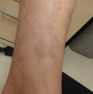
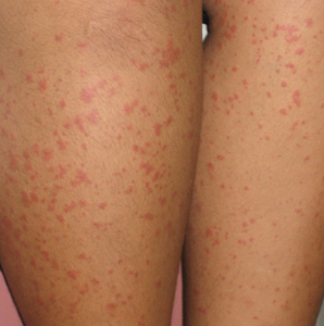
Cutaneous vasculitis is most commonly seen on the lower extremities due to gravity. It presents with non-blanchable violaceous macules and papules often described as palpable purpura. They range from pinpoint to several millimeters in diameter and may be associated with mild edema of the ankles.
How is it diagnosed?
The diagnosis of vasculitis is often made clinically, but may be confirmed with skin biopsy of an early purpuric lesion (ideally one present for >24-48 hours). Direct immunofluorescence can be performed on a second biopsy to identify the type of inflammatory process causing the vasculitis. Underlying trigger can be identified by history and laboratory investigations such as CBC, throat swab, HBV/HCV/HIV serologies and ANA/ANCA. Urinalysis and creatinine should be checked to assess whether the kidneys are also affected.
How is it treated?
Treating the underlying infection or discontinuing any drug(s) suspected of eliciting the response is an important component of treatment. Rest and elevation of the legs is helpful. Systemic corticosteroids may be necessary for patients with ulcerations, diffuse involvement, or significant pain.
Other forms of vasculitis:
Henoch-Schönlein purpura (HSP) is a form of small vessel vasculitis that commonly occurs in children (<10 years old) and rarely in adults. It involves deposition of IgA immune complexes, which can be seen on direct immunofluorescence. HSP presents with a classic tetrad of palpable purpura (usually on the legs), abdominal pain, arthritis and hematuria. Renal involvement is particularly common (40-50%) but does not usually progress to chronic renal failure.
Polyarteritis nodosa, Takayasu arteritis and temporal arteritis affect medium or large vessels tend to present with different skin findings, such as subcutaneous nodules and ulcers.
Urticarial vasculitis is a form of small vessel vasculitis which presents with urticarial appearing lesions, but are painful/tender as opposed to itchy, leave behind bruise-like or hyperpigmented marks and last longer than 24 hours. Patients may also have systemic symptoms such as fever and joint pain.