Chapter 2 Innate and Adaptive Immunity: From Cell Defense to Tissue Repair
Section 3: Complement System, Interferons, and Cytokines
Zoë Soon
The Complement System
The complement system consists of 50+ plasma proteins produced by the liver that circulate the blood in an inactive state and play a crucial role in the immune system. There are three complement pathways in which these proteins are activated in a domino-like chain reaction called a complement cascade during infection or injury. All three complement pathways converge on three shared outcomes: (1) opsonization via C3b to enhance phagocytosis; (2) formation of Membrane Attack Complexes (MAC) that create pores in bacterial cell walls, causing swelling and rupture; and (3) activation of mast cells and basophils to release pro-inflammatory mediators.
| Classical Pathway | Triggered by antibodies forming complexes with pathogen surface antigens. C1 binds two antibodies to start the cascade (C1 → C2 → C3, etc.) C3b attaches to the pathogen as an opsonin; C5-9 assemble into MAC pores.
These activated complement protein complexes attract white blood cells for further defense and induce mast cells and basophils to release pro-inflammatory mediators (e.g. histamine). |
| Lectin Pathway | Mannose-Binding Lectin (MBL) is produced by the liver, MBL binds to mannose (a sticky adhesive sugar on the surface of bacteria, yeasts, viruses, and protozoa) and initiates a similar cascade.
Examples of pathogens that can be bound by MBL: Salmonella, Streptococci, Candida albicans, HIV, SARS-CoV-2, influenza A, and Leishmania (protozoa transmitted by sand fly bite in tropics & subtropics). |
| Alternative Pathway | Independent of antibodies and lectin, though initiates the same 3 outcomes as the Classical and Lectin Pathways (opsonization, MAC, and activation of mast cells and basophils)
Complement proteins bind directly to pathogens. bonds that are stabilized by properdin (a complement factor released by leukocytes), . |
Interferons
Interferons (IFNs) are a type of cytokine (signalling glycoproteins) released by almost all cell types during any infection. Their name reflects their primary function; interfering with viral replication. Over 20 IFN genes exist. Some produce anti-viral proteins (AVPs); some act as endogenous pyrogens (inducing fever); and interferons can cause muscle pain, body aches, and flu-like symptoms.
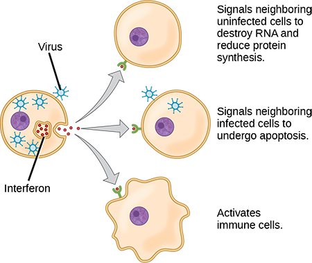
| Alpha interferons (Type I) | Produced by virally-infected host cells.
Induce AVP production in neighbouring cells to block viral RNA/DNA replication. Trigger apoptosis in infected neighbouring cells. Stimulate NK cells and macrophages. Increases MHC I expression. MHC is used by all cells to present non-self antigens to T cells (activating T cell specific immunity). |
| Beta interferons (Type I) | Produced by fibroblasts (a common stromal/mesenchymal/connective tissue cell).
Anti-inflammatory; released in preparation for healing after infection. |
| Gamma interferons (Type II) | Released by activated NK and T cells.
Stimulate macrophage activity. Increases MHC II expression in APCs (macrophages and dendritic cells). MHC II is used in presenting non-self antigens to T cells (activating T cells specific immunity). |
Cytokines
Cytokines are signalling glycoproteins produced by most cells of the body – macrophages, B cells, T cells, mast cells, endothelial cells, fibroblasts and other stromal (mesenchymal/connective tissue) cells. Some cytokines participate in innate (non-specific) defense; others in adaptive (specific) immune responses. Four main categories:
| Interferons | As described above. Main roles; interfere with viral replication, activate NK cells, T cells, and macrophages, acts as endogenous pyrogens, slow inflammation in preparation for healing. |
| Chemokines | Induce chemotaxis – directed migration and recruitment of WBCs to infection or injury sites. |
| Lymphokines | Produced by T lymphocytes to: (a) attract macrophages; and (b) stimulate B lymphocytes. |
| Interleukins | Produced by helper T cells. Functions:
(a) activate macrophages. (b) stimulate fever (act as endogenous pyrogens); (c) stimulate T and B cell differentiation; (d) stimulate hematopoietic cells to produce more WBCs. |
Media Attributions
- Private: interferons © Mary Ann Clark, Matthew Douglas, Jung Choi is licensed under a CC BY (Attribution) license
