Chapter 2 Innate and Adaptive Immunity: From Cell Defense to Tissue Repair
Section 10: Blood Composition and Blood Disorders
Zoë Soon
Blood Composition
Blood is fluid connective tissue with a liquid extracellular matrix called plasma. The formed elements are the cellular components: RBCs, WBCs, and platelets. If blood is allowed to settle by weight:
- Plasma (~55% by volume) rises to the top;
- Buffy coat (thin white layer of leukocytes (WBCs) and platelets, <1% forms in the middle); and
- Erythrocytes (~41-45%) form the bottom dark red layer.
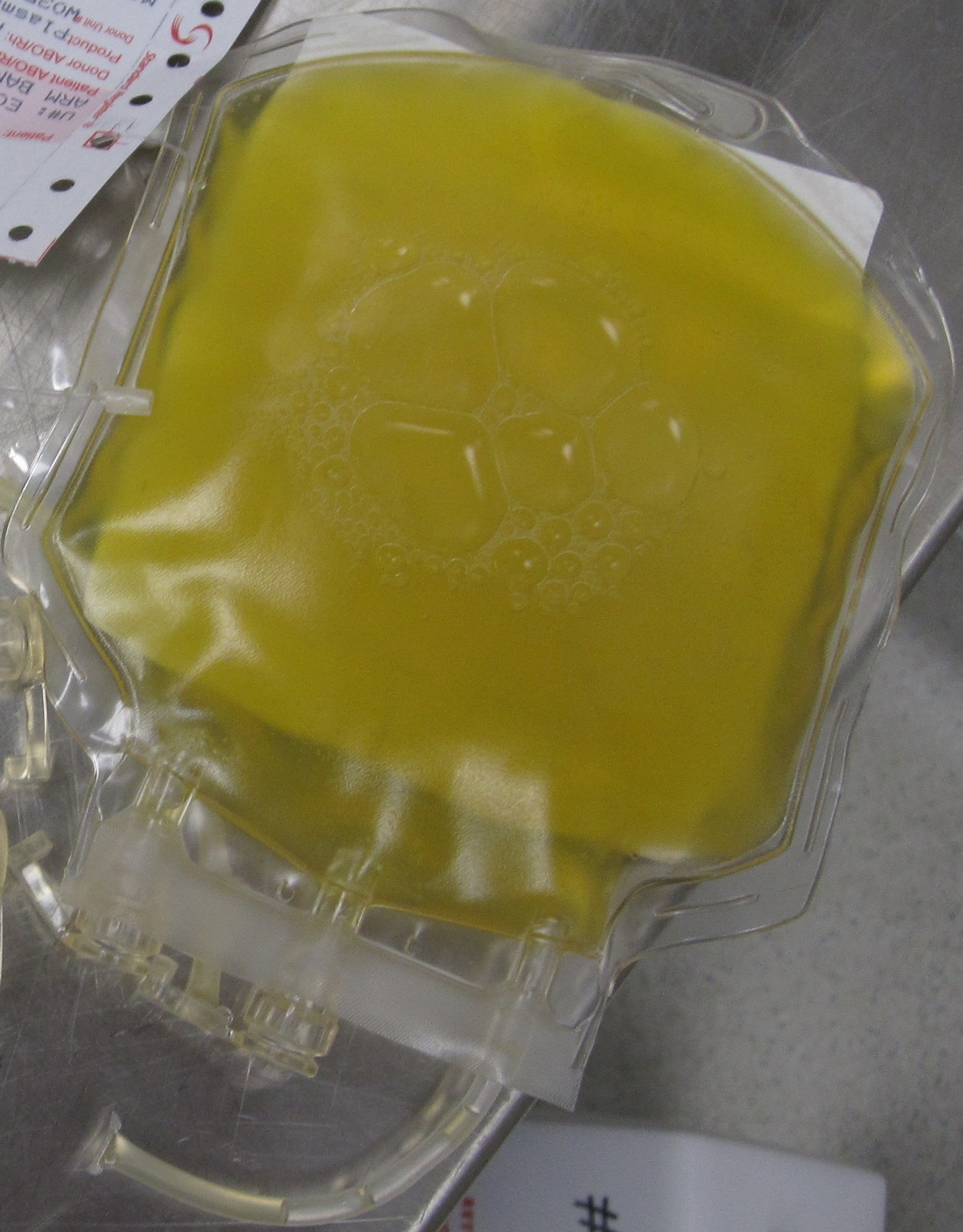
Plasma is a yellowish fluid and contains water, electrolytes (Na+, K+, Ca2+, Mg2+, Cl–, HCO3–, HPO4–, SO42-), dissolved gases, amino acids, fatty acids, glycerides, cholesterol, glucose, hormones, organic wastes (urea, uric acid, creatinine, bilirubin) and proteins.
It is likely not surprising that interstitial fluid (the fluid between tissue cells of the body) is very similar in composition to plasma as an equilibrium does exist particularly for small water soluble ions and molecules. In comparison with plasma, interstitial fluid contains similar concentrations of water, electrolytes, dissolved gases, amino acids and hormones. However, plasma contains many dissolved proteins, whereas interstitial fluid does not.
Plasma Proteins
| Albumin (60% of plasma protein) | Produced in the liver. Most abundant plasma protein.
Transports lipid-soluble substances (fatty acids, steroid hormones). Responsible for most of the blood’s osmotic force. |
| Globulins (35% of plasma proteins) | Include antibodies (immunoglobulins) produced by B cells, and liver-produced transport globulins that carry vitamins, lipids, metal ions, and hormones (e.g., sex hormone-binding globulins, thyroxine-binding globulin, and cortisol-binding globulin). |
| C-reactive proteins (CRP) | Produced by the liver; bind to dead cells and bacteria to activate the complement system.
CRP serum levels correlate with the extent of tissue damage and inflammation. |
| Fibrinogen & prothrombin (4%) | Produced in the liver; involved in blood clotting.
Require Vitamin K during synthesis. |
| Complement proteins | Produced by the liver; act as opsonins, form MAC complexes, and stimulate mast cell activity. |
| Lipoproteins | Produced in the liver, transport triglycerides and cholesterol in the blood (e.g., high- and low- density lipoproteins, HDLs and LDLs). |
Hormones
Hormones are important signalling molecules that travel the bloodstream and regulate complex biological processes, growth, development, maturation, and behaviour. Hormones are classified into 5 main categories based on molecular structure:
- Lipid (e.g. prostaglandin, thromboxane)
- Steroid (cortisol, estrogen and testosterone)
- Amino acid (e.g. epinephrine)
- Peptide (e.g. insulin)
- Gas (e.g. nitric oxide, NO)
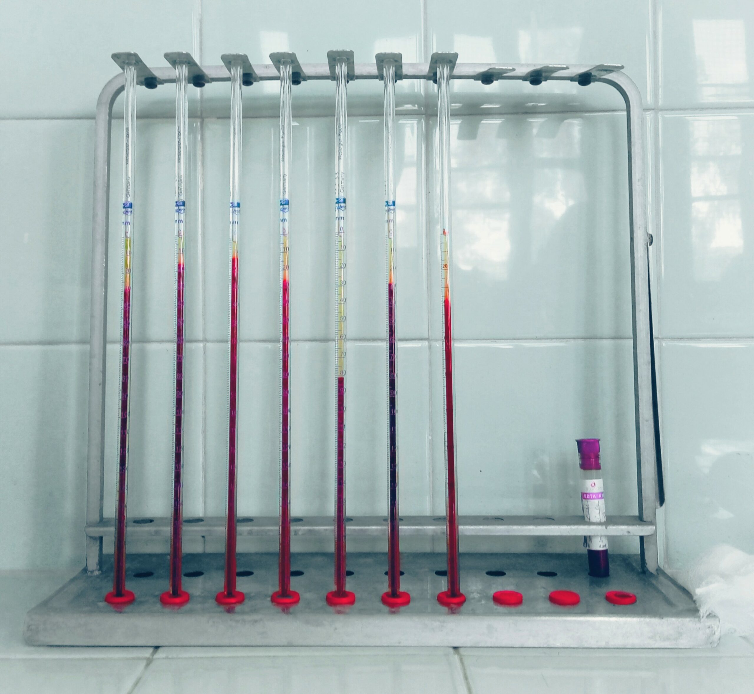
Erythrocyte Sedimentation Rate (ESR)
The ESR measures how quickly RBCs settle to the bottom of a blood tube. During injury or infection, plasma proteins: (C-reactive protein (CRP), prothrombin, complement proteins) increase causing RBCs to aggregate and settle faster. ESR therefore correlates with the level of inflammation in the body.
- Normal ESR: adult males ~12 mm/hr; adult females ~18 mm/hr.
- Serious infections, autoimmune diseases, and chronic illnesses can raise ESR to ≥100 mm/hr.
Blood Disorders
| Anemia | Reduced oxygen-carrying capacity of blood (an- = without, -emia = blood).
Due to low RBC count, low hemoglobin, or abnormal RBC shape. Risk factors include: blood loss, systemic lupus erythematosus, autoimmune diseases that affect intestinal absorption of food (e.g. Crohn disease, ulcerative colitis), blood cancers (e.g. lymphoma), chemotherapy and long term infections (e.g. HIV, osteomyelitis, hepatitis). |
| Iron-deficiency anemia | Most common in biological females (lower iron reserves).
Contributing factors: vegetarian die, poor nutrition, menstruation, frequent blood donation. Prevention involves healthy diet that includes iron-rich foods such as: eggs, meat, leafy green vegetables. |
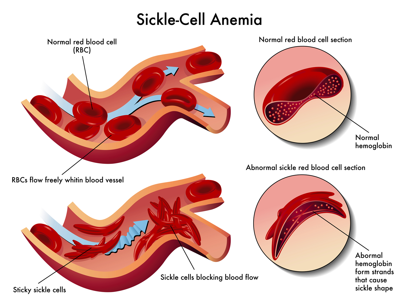
| Sickle Cell Anemia | Autosomal recessive – mutations in both copies of the hemoglobin beta-chain gene (chromosome 11).
Results in misfolded hemoglobin and rigid, sickle-shaped RBCs with shortened lifespan. Causes blockages and downstream hypoxia throughout the body. Blood transfusions and the donation of bone marrow stem cells is used to treat this disease. |
| Alpha/Beta Thalassemia | Autosomal recessive – mutations in alpha and beta hemoglobin chain genes.
Signs and symptoms range from mild to severe. |
| Pernicious anemia | Rare autoimmune disease; auto-antibodies inhibit Intrinsic Factor (required for vitamin B12 intestinal absorption). RBC production requires B vitamins, folate, amino acids and iron.
Without treatment, B12 deficiency causes anemia. |
| Hemorrhagic anemia | Due to excess blood loss. |
Polycythemia and the Dangers of Blood-Doping
Polycythemia: Excessive RBC production – thickens blood, increases clotting risk, strains the heart. Some athletes misuse EPO injections (blood doping) to artificially boost RBC count and oxygen delivery. Raises the risk of heart attacks and strokes.
* What is Serum?
Serum is the fluid plasma component of blood, with all of the clotting factors, platelets, and cells removed. Serum is obtained by allowing blood to clot and then centrifuging to separate the blood by weight. The top liquid portion (that has not coagulated) is serum. Serum contains water, electrolytes and soluble proteins (e.g. antibodies).
Diagnostic tests have been developed for some cancers, autoimmune reactions, and other diseases that analyze the presence or level of various serum biomarkers.
Media Attributions
- Private: FreshFrozenPlasma © DiverDave is licensed under a CC BY-SA (Attribution ShareAlike) license
- Private: Erythrocyte_sedimentation_rate_(ESR) © Rouibi Dhia Eddine Nadjm is licensed under a CC BY-SA (Attribution ShareAlike) license
- Private: Sickle Cell Anemia © Diana Grib is licensed under a CC BY-SA (Attribution ShareAlike) license

.jpg)
2.jpg)