Appendices: Introduction to Histology for first-time learners
Histology – Introduction to Staining
Willie Wu and Jennifer Kong
Learning Objectives
By the end of this section, you will be able to:
- Explain why staining is an essential step in preparing tissues for light microscopy.
- Describe the basic principle behind H&E staining and identify what cellular components it highlights.
- Identify the primary purpose of common special stains (Trichrome, Elastic, Reticulin) and the structures they colorize.
- Choose an appropriate stain to highlight a specific tissue component based on its function.
Why do We Stain?
Imagine a perfectly clear, glass-like sculpture. Shining a light through it would reveal little of its intricate detail. This is the challenge of histology; thin sections of tissue are mostly transparent and colorless. To solve this, we use stains, which are chemical dyes that bind to specific cellular components, imparting color and contrast to this invisible world. Staining transforms a featureless slide into a detailed map, allowing us to navigate and interpret the complex landscape of tissues. It is the fundamental tool that makes microscopic analysis possible.
H&E (Hematoxylin and Eosin)
The undisputed cornerstone of histological staining is the H&E (Hematoxylin and Eosin) method. It is the first stain applied to virtually every tissue sample and provides the critical baseline view of cellular architecture. Its power lies in its simplicity and reliability, offering a general overview that highlights the most fundamental structures.
The principle behind H&E is based on the acid-base affinities of biological molecules. Hematoxylin itself is a natural dye that, when combined with a metal ion mordant (commonly aluminum), forms a complex that binds strongly to acidic (negatively charged) tissue components. It is powerfully attracted to and stains acidic (negatively charged) structures a deep bluish-purple. These structures are described as basophilic (“base-loving”). The most clinically significant basophilic structures are those rich in nucleic acids: the DNA within the nucleus and the RNA on ribosomes in the rough endoplasmic reticulum. Thus, the most important takeaway from any H&E image is that blue-purple dots represent cell nuclei, providing an instant way to locate and count cells.
Conversely, Eosin is an acidic dye carrying a negative charge, which binds to and stains basic (positively charged) structures a vibrant pink or reddish-orange. These structures are termed acidophilic or eosinophilic. This category encompasses most common proteins in the cytoplasm of cells, as well as the majority of proteins found in the extracellular matrix, most notably the collagen fibers of connective tissue.
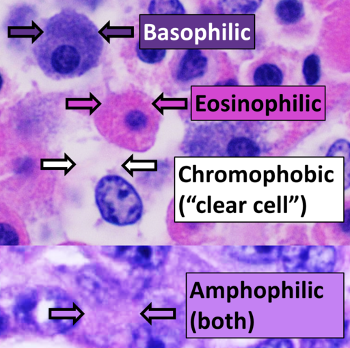
Therefore, when you look at an H&E-stained slide, your brain quickly learns to read its color-coded language: blue indicates the nuclei, while pink reveals the cytoplasm and connective tissue. This contrast creates a beautifully clear and informative picture of tissue organization.
Special Stains: Targeted Tools for Specific Questions
While H&E provides the essential map, it sometimes lacks the specificity to answer deeper diagnostic questions. This is where special stains come into play. These are specialized techniques that act like highlighter pens, selectively targeting and illuminating specific molecules that H&E shows but does not distinguish clearly.
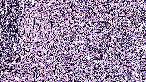
The Periodic Acid-Schiff (PAS) reaction is a vital chemical stain that targets carbohydrates. It beautifully highlights substances like glycogen (a glucose storage molecule in liver and muscle), the sugary glycocalyx on the surface of intestinal cells, and mucins (the gel-forming secretions of goblet cells) in a distinctive magenta hue. It is indispensable for identifying mucus production and assessing basement membranes (as shown below).

Masson’s Trichrome is another commonly used stain, known for its ability to clearly differentiate between different tissue types (see example below). It uses a three-dye technique to stain cytoplasm and muscle fibers bright red, while collagen fibers appear blue or green, depending on the protocol. This contrast is crucial for pathologists to identify and quantify fibrosis, which is the harmful scarring of organs that occurs in chronic diseases of the liver, kidney, and heart.

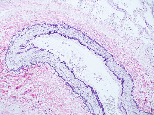
For assessing the elastic properties of tissues, the Elastic Stain (e.g., Verhoeff-Van Gieson) is used. It specifically targets elastic fibers, staining them a deep blue-black or purple-black against a light background. This is essential for evaluating the health of blood vessels, skin, and lungs, where the integrity of elastic fibers is paramount. A great example will be staining arteries with elastic stain, as shown below.
Silver Impregnation Stains (e.g., Reticulin stain) are used to visualize the finest of connective tissue fibers. They deposit silver onto reticular fibers (a type of thin collagen), making them stand out in sharp black against a pale yellow background. This stain is used to visualize the delicate architectural scaffolding of organs like the liver, spleen, and lymph nodes, providing a sensitive measure of tissue structure.
It is important to note that the stains mentioned here are just a few of the most common examples from the toolkit. There is a diverse group of other dyes and chemical reactions, each with a highly specific purpose. Pathologists can choose from dozens of specialized stains to detect unique features like microorganisms, specific minerals, or abnormal protein deposits, allowing for incredibly targeted diagnostic investigation.
Key Takeaways
- Staining is necessary to create contrast and allow visualization of transparent tissue components.
- H&E is the most commonly used staining method: staining basophilic (acidic) structures like nuclei blue and acidophilic (basic) structures like cytoplasm and collagen pink.
- PAS: Highlights carbohydrates (magenta), like mucus, glycogen, and basement membranes.
- Masson’s Trichrome: Differentiates muscle (red) from collagen (blue/green), crucial for assessing fibrosis.
- Elastic Stain: Visualizes elastic fibers (blue-black) in blood vessels and lungs.
- Reticulin Stain: Reveals the delicate network of reticular fibers (black).
Media Attributions
- 512px-Eosinophilic,_basophilic,_chromophobic_and_amphophilic_staining © Mikael Häggström is licensed under a CC BY (Attribution) license
- 512px-Renal_corpuscle © Ed Uthman is licensed under a CC BY-SA (Attribution ShareAlike) license
- 512px-Masson’s_trichrome_staining_on_rat’s_trachea © 22Kartika is licensed under a CC BY-SA (Attribution ShareAlike) license
- 512px-Normal_lung;_Artery_-_Elastic_Stain_(3626657933) © Yale Rosen is licensed under a CC BY-SA (Attribution ShareAlike) license
- 512px-Connective_Tissue_Reticular_(40885193495) © Berkshire Community College Bioscience Image Library is licensed under a CC0 (Creative Commons Zero) license

.jpg)
.jpg)