Atherosclerosis and Angina
Gross Anatomy and Histology of Normal Aorta
Tetiana Povshedna
Learning Objectives
By the end of this chapter, you will be able to:
- Compare and contrast the main histological features of tissues that can be identified via H&E and elastic/trichrome stains.
- Identify three main layers (tunics) in the wall of the aorta and their main components.
- Relate the structural features of aorta to its function.
Arteries are blood vessels that transport and distribute oxygenated blood from the heart to all the organs in the body (with one exception – pulmonary arteries transport deoxygenated blood from the heart to the lungs for gas exchange as a part of pulmonary circle). The aorta, which originates from the aortic valve in the left ventricle, is the largest artery in the human body. Arteries are responsible for regulating blood pressure, which morphologically translates into a thick smooth muscle layer that facilitates contraction and dilatation in response to physiological stimuli.
Aorta is a unique example of an elastic artery and contains a lot of elastic fibers within its wall that allow for a large variation of the lumen size.
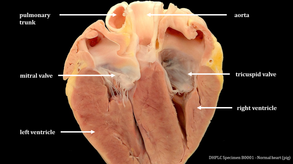
One the gross anatomy specimen that features ascending aorta, identify:
- smooth luminal surface, which is required for uninterrupted blood flow;
- wide diameter (around 20-30 mm in ascending aorta), necessary for allowing large volumes of oxygenated blood to be conducted from the heart into the vascular tree;
- thick walls (2-3 mm in the ascending aorta), which reflect high blood pressure in the aorta;
- color: aorta appears yellow/white due to a high amount of connective tissue fibers (collagen, elastin).
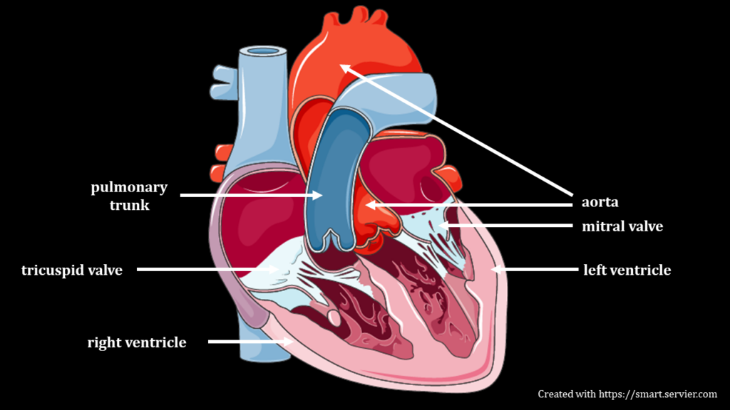
Please note: the classic depiction of normal heart anatomy usually examines the heart chambers and vessels from the anterior side. In this gross anatomy specimen, however, the heart chambers and vessels are examined from the posterior side. Thus, the left ventricle is on the left, and the aorta is in front of the pulmonary trunk.
Histology of the Aorta
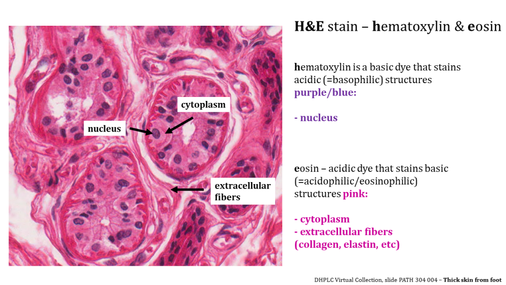
Before examining the histological sections of a normal human aorta, it is important to consider commonly used histological stains and their interpretation.
Generally, the stains are divided into non-specific, the ones that use dyes that stain all the cells within tissue in a similar manner, and special – the ones that use dyes that selectively bind to specific tissue/cell components.
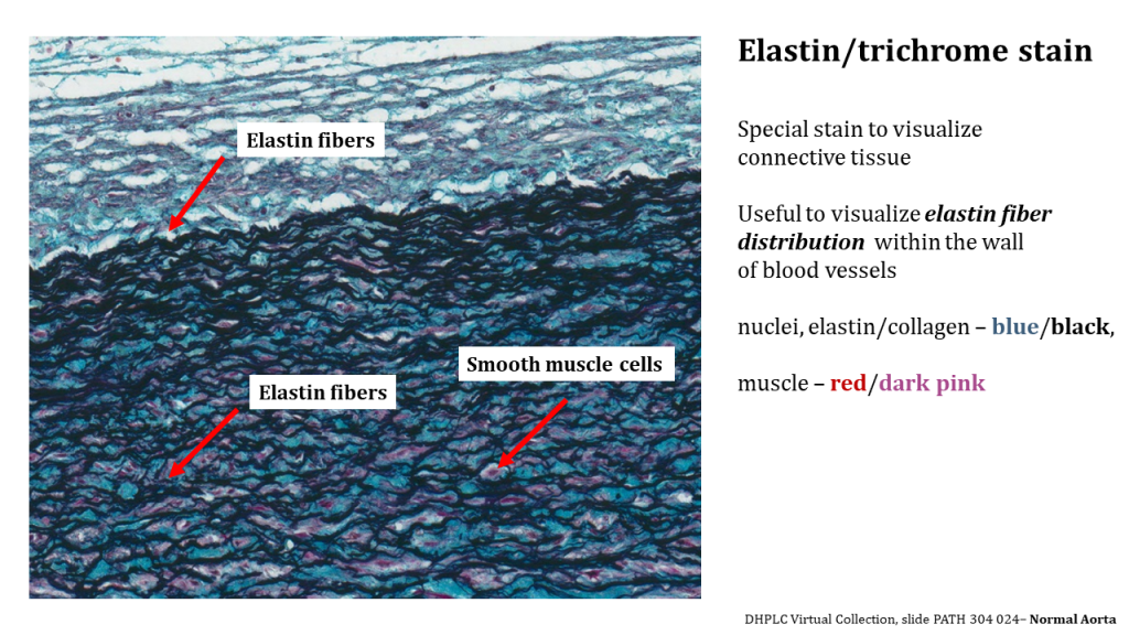
This section will examine the normal histology of the human aorta using histology slides stained with hematoxylin & eosin (H&E, non-specific) and elastin/trichrome (special) stains.
Please refer to the sections How Are the Specimens Obtained and Prepared? and Video Lessons on Histology to learn more about the stains and histology slide preparation.
H&E Stain Interpretation
To better understand changes that occur in the blood vessel wall during the development of atherosclerosis, it’s critical to understand the normal histology of large blood vessels.
Even though the morphology of blood vessels of different sizes and types (arteries, veins, capillaries) exhibits functional adaptations that reflect the location of a blood vessel and local blood pressure, the main features of the blood vessel wall remain the same across various types.
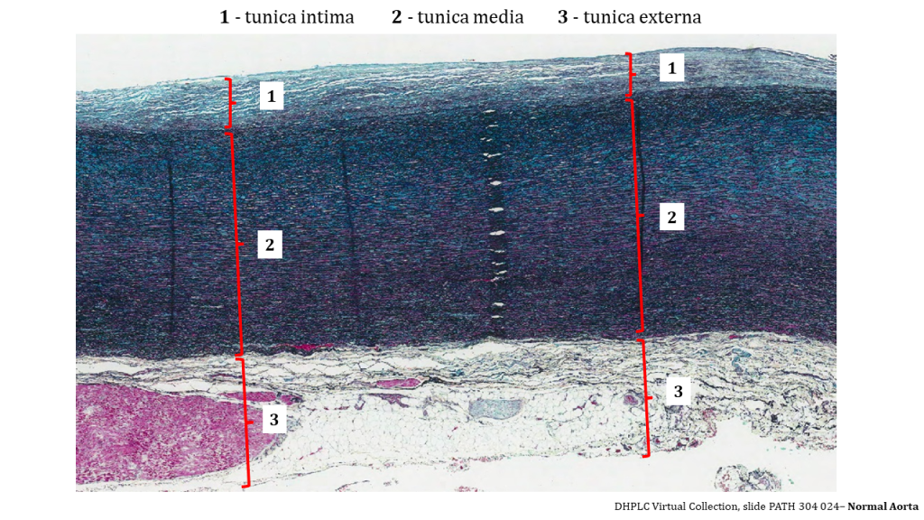
The wall of the aorta consists of 3 layers (tunics):
1. Tunica intima – thin innermost layer that includes:
a) Endothelium – a single layer of epithelial cells that forms the luminal surface of the vessel and is in direct contact with blood. Endothelium within the wall of blood vessels is continuous with the endocardium – the inner lining of the heart;
b) Loose connective tissue – a thin layer of connective tissue located under endothelium
2. Tunica media – the thickest layer in the aorta, includes:
a) Smooth muscle cells that allow for contraction and relaxation;
b) Elastin fibers that further expand the ability of aorta to dilate and contract; in aorta, numerous elastin fibers are located within tunica media, and some within tunica intima;
c) Connective tissue fibers and nerves.
3. Tunica adventitia -the outermost layer that includes:
a) Connective tissue for structural support and protection;
b) Nerve fascicles (nervi vasorum) and blood vessels (vasa vasorum) that supply and innervate the wall of the aorta
As an elastic artery, aorta contains a large amount of elastic fibers within the tunica media. This amount of elastin allows aorta to expand and recoil in response to blood pressure fluctuations (similarly to how an elastic band behaves) and conduct large volumes of blood that’s ejected from the heart under high pressure.
In addition to being present within tunica media, elastin fibers also form two structures on the border between tunics:
a) Internal elastic lamina – on the border between tunica intima and tunica media;
b) External elastic lamina – on the border between tunica media and tunica adventitia
Both elastic laminas are thick bands of elastin fibers that appear wavy on the histological slide, which reflects the properties of elastin in a relaxed state. Elastic laminas are more easily identified in smaller arteries, where elastin fibers are located only within the laminas. In the aorta, which has numerous elastin fibers within tunica media, elastic laminas would be the fibers on the border between the tunics.
Normal histology of aorta. Created and presented by Tetiana Povshedna. Histology slides are DHPLC specimens PATH 304-023 (H&E) and PATH 304-024 (elastin/trichome). Illustrations were created under license with Biorender.com.
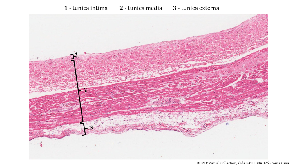
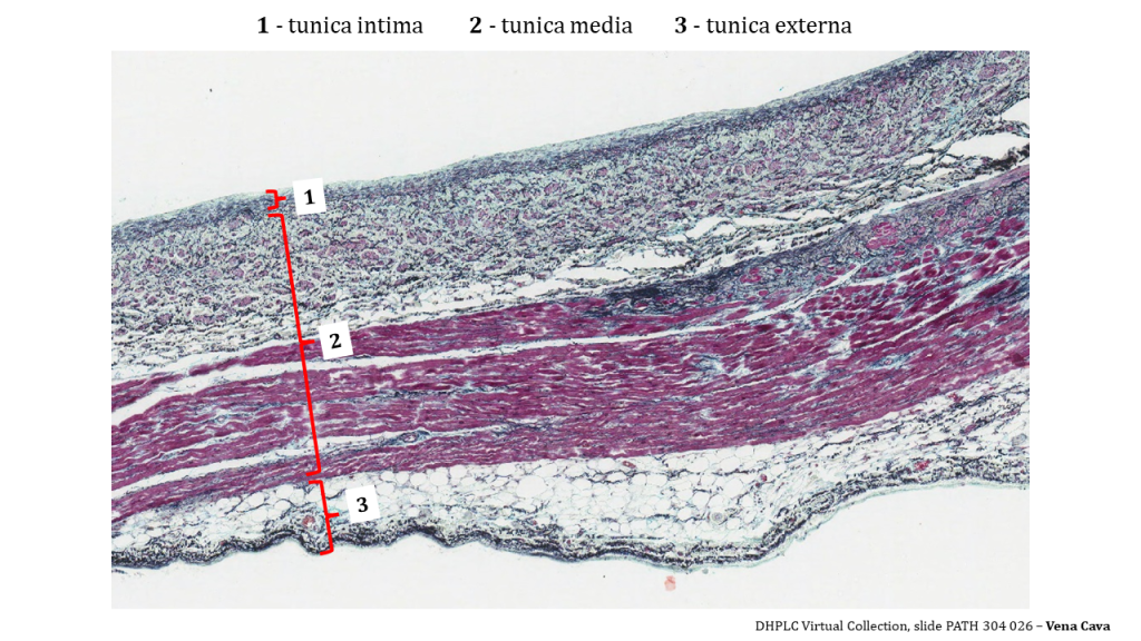
Compare and Contrast the Histological Appearance of 3 Tunics in Various Blood Vessel Types
Please note the variation in the thickness of tunica media and the amount of elastic fibers between the two large vessels – aorta and vena cava.
These morphological adaptations reflect blood vessel location and blood pressure levels – both thickness of tunica media and amount of elastin fibers decrease in veins and venules compared to arteries. Note that tunica media is the thickest layer in aorta, but not in vena cava. Tunica externa of the vena cava is the thickest layer that also contains some smooth muscle fibers.
Aorta
Vena Cava
Section Review
Blood vessels across the vascular tree share a similar histological structure – their wall consists of three layers (also known as tunics). The thickness and characteristics of these layers (tunica intima, tunica media, tunica externa) vary depending on the blood vessel type (artery vs vein) and location (closer or further from the heart). Aorta, a major vessel that carries oxygenated blood from the heart and distributes it to organs and tissues, is an elastic artery that contains numerous elastic fibers across the layers, with the majority of them accumulated within the middle muscular layer (tunica media). This histological feature allows aorta to expand and recoil in response to blood pressure fluctuations, and carry large amounts of oxygenated blood from the heart.
Review Questions
1. Fill in the blanks. Aorta is the largest artery in the human body that carries _____ blood from the heart to tissues and organs.
2. Finish the following sentence. The wall of human aorta consists of three layers (also known as tunics):
Select all that apply.
- Tunica interna
- Tunica media
- Tunica intermedialis
- Tunica extra
3. Generally, blood vessels of various types and sizes share a common histological structure.
- True
- False
4. Fill in the blanks.
Tunica _____ is the thickest layer in aorta, while tunica _____ is the thickest layer in vena cava.
5. Finish the following sentence. Large amount of elastic fibers in the tunica media of aorta allows for:
Select all that apply.
- Dilation and contraction in response to blood pressure and fluctuations
- Variability of lumen size depending on physiological conditions
- Transport the large volumes of oxygenated blood outside the heart
- Blood pressure regulation
- Pulse variability
Answer Key
- Oxygenated
- Tunica interna, tunica externa, tunica media
- True
- Media, tunica, externa
- Dilation and contraction in response to blood pressure fluctuations, variability of lumen size depending on physiological conditions, transport of large volumes of oxygenated blood outside of heart
Media Attributions
- Normal-heart-anatomy-figure2 © Tetiana Povshedna is licensed under a CC BY (Attribution) license
- Heart anatomy © DHPLC is licensed under a All Rights Reserved license
- HE Eosin adapted by Tetiana Povshedna is licensed under a All Rights Reserved license
- Elastin stain interpretation © DHPLC adapted by Tetiana Povshedna is licensed under a All Rights Reserved license
- Aorta Elastin © DHPLC adapted by Tatiana Povshedna is licensed under a All Rights Reserved license
- Vena Cava H&E © DHPLC adapted by Tatiana Povshedna is licensed under a All Rights Reserved license
- Vena Cava Elastin © DHPL adapted by Tetiana Povshedna is licensed under a All Rights Reserved license
(also, conducting artery) artery with abundant elastic fibers located closer to the heart, which maintains the pressure gradient and conducts blood to smaller branches
interior of a tubular structure such as a blood vessel or a portion of the alimentary canal through which blood, chyme, or other substances travel
part of the aorta that originates from the heart; total length - around 5 cm
large arterial vessel that carries blood ejected from the right ventricle; divides into the left and right pulmonary arteries
A blue basic dye that is used to stain tissues. It turns acidic elements such as RNA and DNA in the tissue blue.
Eosin is a pink acidic dye that stains basic elements such as most proteins in the tissue pink or red
blood vessel that conducts blood away from the heart; may be a conducting or distributing vessel
blood vessel that conducts blood toward the heart
smallest of blood vessels where physical exchange occurs between the blood and tissue cells surrounded by interstitial fluid
Tissue that lines vessels of the lymphatic and cardiovascular system, made up of a simple squamous epithelium.
innermost layer of the heart lining the heart chambers and heart valves; composed of endothelium reinforced with a thin layer of connective tissue that binds to the myocardium
a type of connective tissue that consists of fibroblasts, a large amount of extracellular matrix (also called ground substance), and space connective tissue fibers (collagen, elastin). Loose connective tissue often contains cappillaries, arterioles, and venules that supply epithelium on top of it
Fibers made of the protein elastin that increase the elasticity of the dermis.
small nerve fibers found in arteries and veins that trigger contraction of the smooth muscle in their walls
small blood vessels located within the walls or tunics of larger vessels that supply nourishment to and remove wastes from the cells of the vessels
middle layer or tunic of a vessel (except capillaries)
(also, tunica adventitia) outermost layer or tunic of a vessel (except capillaries)
large vein that brings deoxygenated blood from the organs in the body back to the heart
(also, tunica interna) innermost lining or tunic of a vessel
