Atherosclerosis and Angina
Structure and Function of Blood Vessels
J. Gordon Betts; Kelly A. Young; James A. Wise; Eddie Johnson; Brandon Poe; Dean H. Kruse; Oksana Korol; Jody E. Johnson; Mark Womble; and Peter DeSaix
Learning Objectives
By the end of this section, you will be able to:
- Compare and contrast the three tunics that make up the walls of most blood vessels.
- Distinguish between elastic arteries, muscular arteries, and arterioles on the basis of structure, location, and function.
- Explain the structure and function of arteries vs veins.
Blood is carried through the body via blood vessels. An artery is a blood vessel that carries blood away from the heart, where it branches into ever-smaller vessels. Eventually, the smallest arteries, vessels called arterioles, further branch into tiny capillaries, where nutrients and wastes are exchanged, and then combine with other vessels that exit capillaries to form venules, small blood vessels that carry blood to a vein, a larger blood vessel that returns blood to the heart.
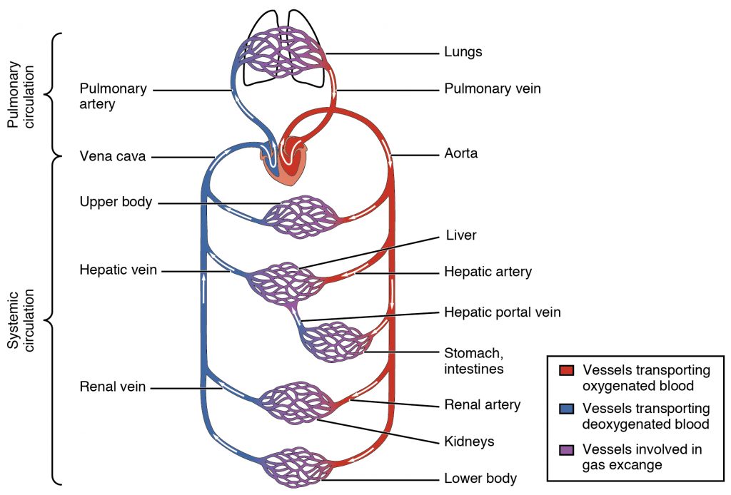
Arteries and veins transport blood in two distinct circuits: the systemic circuit and the pulmonary circuit. Systemic arteries provide blood rich in oxygen to the body’s tissues. The blood returned to the heart through systemic veins has less oxygen, since much of the oxygen carried by the arteries has been delivered to the cells. In contrast, in the pulmonary circuit, arteries carry blood low in oxygen exclusively to the lungs for gas exchange. Pulmonary veins then return freshly oxygenated blood from the lungs to the heart to be pumped back out into systemic circulation. Although arteries and veins differ structurally and functionally, they share certain features.
Shared Structures
Different types of blood vessels vary slightly in their structures, but they share the same general features. Arteries and arterioles have thicker walls than veins and venules because they are closer to the heart and receive blood that is surging at a far greater pressure). Each type of vessel has a lumen—a hollow passageway through which blood flows. Arteries have smaller lumens than veins, a characteristic that helps to maintain the pressure of blood moving through the system. Together, their thicker walls and smaller diameters give arterial lumens a more rounded appearance in cross-section than the lumens of veins.
In comparison to arteries, veins and venules withstand a much lower blood pressure, which is morphologically reflected in thinner walls and larger lumens. These adaptations allow for large volumes of blood to flow with less vessel resistance. Additionally, some veins in the limbs contain valves that assist the unidirectional flow, which is challenging in the extremities due to lower pressure and gravity.
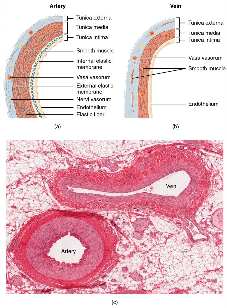
Larger arteries and veins contain small blood vessels within their walls known as the vasa vasorum—literally “vessels of the vessel”. Vasa vasorum are located in the outer layers of the vessel (Figure 2) and provide nutrients and allow for waste exchange for the cells and tissues composing the blood vessel wall. There are also minute nerves (known as nervi vasorum within the walls of both types of vessels that control the contraction and dilation of smooth muscle.
Both arteries and veins have the same three distinct tissue layers, called tunics. From the most interior layer to the outer, these tunics are the tunica intima, the tunica media, and the tunica externa.
Comparison of tunics within arteries and veins
| Arteries | Veins | |
|---|---|---|
| General appearance | Thick walls with small lumens, appear rounded | Thin walls with large lumens, appear flattened |
| Tunica intima | Endothelium appears wavy due to constriction of smooth muscle. Internal elastic membrane present in larger vessels. | Endothelium appears smooth. Internal elastic membrane absent. |
| Tunica media | Normally the thickest layer in arteries. Smooth muscle cells and elastic fibers predominate (the proportions of these vary with distance from the heart). External elastic membrane present in larger vessels. | Normally thinner than the tunica externa. Smooth muscle cells and collagenous fibers predominate. Nervi vasorum and vasa vasorum present. External elastic membrane absent. |
| Tunica externa | Normally thinner than the tunica media in all but the largest arteries. Collagenous and elastic fibers. Nervi vasorum and vasa vasorum present. | Normally the thickest layer in veins. Collagenous and smooth fibers predominate. Some smooth muscle fibers. Nervi vasorum and vasa vasorum present. |
Tunica Intima
The tunica intima (also called the tunica interna) is composed of epithelial (endothelium) and connective tissue layers.
- endothelium is the specialized simple squamous epithelium, makes up the lining of the blood vessel lumen and is continuous throughout the vascular system, including the heart. The permeable basement membrane is located underneath endothelium and provides strength while maintaining flexibility.
- thin layer of connective tissue that contains elastic and collagen fibers.
In larger arteries, there is also a thick, distinct layer of elastic fibers known as the internal elastic membrane (also called the internal elastic lamina) at the boundary with the tunica media. Like the other components of the tunica intima, the internal elastic membrane provides structure while allowing the vessel to stretch.
Under the microscope, the lumen and the entire tunica intima of a vein will appear smooth, whereas those of an artery will normally appear wavy because of the partial constriction of the smooth muscle in the tunica media, the next layer of blood vessel walls.
Tunica Media
The tunica media is the thickest layer in arteries and consists of smooth muscle layers and connective tissue that is made up of elastic fibers, which appear wavy in histological slides.
Contraction and relaxation of the circular muscles decrease and increase the diameter of the vessel lumen, respectively. Specifically in arteries, vasoconstriction decreases blood flow as the smooth muscle in the walls of the tunica media contracts, making the lumen narrower and increasing blood pressure. Similarly, vasodilation increases blood flow as the smooth muscle relaxes, allowing the lumen to widen and blood pressure to drop. Both vasoconstriction and vasodilation are regulated in part by nervi vasorum.
Separating the tunica media from the outer tunica externa in larger arteries is the external elastic membrane (also called the external elastic lamina). This structure is not usually seen in smaller arteries, nor is it seen in veins.
Tunica Externa
The outer tunic, the tunica externa (also called the tunica adventitia), is a substantial sheath of connective tissue composed primarily of collagenous fibers and some elastic fibers. The tunica externa in veins also contains groups of smooth muscle fibers. This is normally the thickest tunic in veins and may be thicker than the tunica media in some larger arteries. The outer layers of the tunica externa are not distinct but rather blend with the surrounding connective tissue outside the vessel, helping to hold the vessel in relative position. If you are able to palpate some of the superficial veins on your upper limbs and try to move them, you will find that the tunica externa prevents this. If the tunica externa did not hold the vessel in place, any movement would likely result in disruption of blood flow.
Arteries
An artery is a blood vessel that conducts blood away from the heart. All arteries have relatively thick walls that can withstand the high pressure of blood ejected from the heart. However, those close to the heart have the thickest walls, containing a high percentage of elastic fibers in all three of their tunics. This type of artery is known as an elastic artery).. Vessels larger than 10 mm in diameter are typically elastic. Their abundant elastic fibers allow them to expand, as blood pumped from the ventricles passes through them, and then to recoil after the surge has passed. The elastic recoil of the vascular wall helps to maintain the pressure gradient that drives the blood through the arterial system. An elastic artery is also known as a conducting artery, because the large diameter of the lumen enables it to accept a large volume of blood from the heart and conduct it to smaller branches.
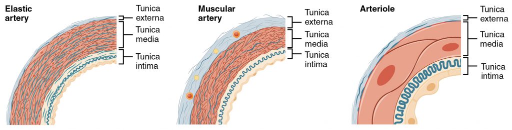
Farther from the heart, where the surge of blood has dampened, the percentage of elastic fibers in an artery’s tunica intima decreases and the amount of smooth muscle in its tunica media increases. The artery at this point is described as a muscular artery. The diameter of muscular arteries typically ranges from 0.1 mm to 10 mm. Their thick tunica media allows muscular arteries to play a leading role in vasoconstriction. In contrast, their decreased quantity of elastic fibers limits their ability to expand. Fortunately, because the blood pressure has eased by the time it reaches these more distant vessels, elasticity has become less important.
Notice that although the distinctions between elastic and muscular arteries are important, there is no “line of demarcation” where an elastic artery suddenly becomes muscular. Rather, there is a gradual transition as the vascular tree repeatedly branches. In turn, muscular arteries branch to distribute blood to the vast network of arterioles. For this reason, a muscular artery is also known as a distributing artery.
Arterioles
An arteriole is a very small artery that leads to a capillary. Arterioles have the same three tunics as the larger vessels, but the thickness of each is greatly diminished. The critical endothelial lining of the tunica intima is intact. The tunica media is restricted to one or two smooth muscle cell layers in thickness. The tunica externa remains but is very thin.
With a lumen averaging 30 micrometers or less in diameter, arterioles are critical in slowing down—or resisting—blood flow and, thus, causing a substantial drop in blood pressure. Because of this, you may see them referred to as resistance vessels.
Capillaries
A capillary is a microscopic channel that supplies blood to the tissues themselves, a process called perfusion. Exchange of gases and other substances occurs in the capillaries between the blood and the surrounding cells and their tissue fluid (interstitial fluid). The diameter of a capillary lumen ranges from 5–10 micrometers; the smallest are just barely wide enough for an erythrocyte to squeeze through. Flow through capillaries is often described as microcirculation.
The wall of a capillary consists of the endothelial layer surrounded by a basement membrane with occasional smooth muscle fibers. There is some variation in wall structure: In a large capillary, several endothelial cells bordering each other may line the lumen; in a small capillary, there may be only a single cell layer that wraps around to contact itself.
Venules
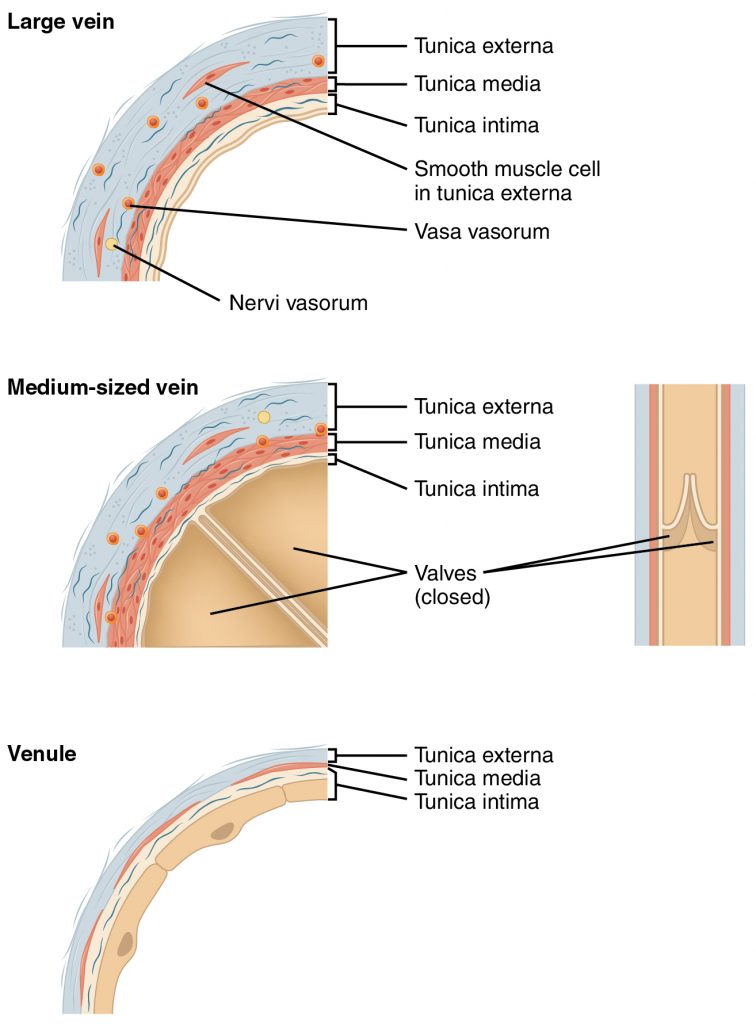
A venule is an extremely small vein, generally 8–100 micrometers in diameter. Multiple venules join to form veins. The walls of venules consist of endothelium, a thin middle layer with a few muscle cells and elastic fibers, plus an outer layer of connective tissue fibers that constitute a very thin tunica externa. Venules as well as capillaries are the primary sites of emigration or diapedesis, in which the white blood cells adhere to the endothelial lining of the vessels and then squeeze through adjacent cells to enter the tissue fluid.
Veins
A vein is a blood vessel that conducts blood toward the heart. Compared to arteries, veins are thin-walled vessels with large and irregular lumens. Because they are low-pressure vessels, larger veins are commonly equipped with valves that promote the unidirectional flow of blood toward the heart and prevent backflow toward the capillaries caused by the inherent low blood pressure in veins as well as the pull of gravity.
Comparison of Arteries and Veins
| Arteries | Veins | |
| Direction of blood flow | Conducts blood away from the heard | Conducts blood toward the heard |
| General appearance | Rounded | Irregular, often collapsed |
| Pressure | High | Low |
| Wall thickness | Thick | Thin |
| Relative oxygen concentration | Higher in systemic arteries
Lower in pulmonary arteries |
Lower in systemic vein
Higher in pulmonary veins |
| Valves | Not present | Present most commonly in limbs and in veins inferior to the heart |
Vascular Surgeons and Technicians Vascular surgery is a specialty in which the physician deals primarily with diseases of the vascular portion of the cardiovascular system. This includes repair and replacement of diseased or damaged vessels, removal of plaque from vessels, minimally invasive procedures including the insertion of venous catheters, and traditional surgery. Following completion of medical school, the physician generally completes a 5-year surgical residency followed by an additional 1 to 2 years of vascular specialty training. In the United States, most vascular surgeons are members of the Society of Vascular Surgery.
Vascular technicians are specialists in imaging technologies that provide information on the health of the vascular system. They may also assist physicians in treating disorders involving the arteries and veins. This profession often overlaps with cardiovascular technology, which would also include treatments involving the heart. Although recognized by the American Medical Association, there are currently no licensing requirements for vascular technicians, and licensing is voluntary. Vascular technicians typically have an Associate’s degree or certificate, involving 18 months to 2 years of training. The United States Bureau of Labor projects this profession to grow by 29 percent from 2010 to 2020.
Visit this site to learn more about vascular surgery.
Visit this site to learn more about vascular technicians.
Section Review
Blood pumped by the heart flows through a series of vessels known as arteries, arterioles, capillaries, venules, and veins before returning to the heart. Arteries transport blood away from the heart and branch into smaller vessels, forming arterioles. Arterioles distribute blood to capillary beds, the sites of exchange with the body tissues. Capillaries lead back to small vessels known as venules that flow into the larger veins and eventually back to the heart.
The arterial system is a relatively high-pressure system, so arteries have thick walls that appear round in cross-section. The venous system is a lower-pressure system, containing veins that have larger lumens and thinner walls. They often appear flattened. Arteries, arterioles, venules, and veins are composed of three tunics known as the tunica intima, tunica media, and tunica externa. Capillaries have only a tunica intima layer. The tunica intima is a thin layer composed of a simple squamous epithelium known as endothelium and a small amount of connective tissue. The tunica media is a thicker area composed of variable amounts of smooth muscle and connective tissue. It is the thickest layer in all but the largest arteries. The tunica externa is primarily a layer of connective tissue, although in veins, it also contains some smooth muscle. Blood flow through vessels can be dramatically influenced by vasoconstriction and vasodilation in their walls.
Review Questions
1. The endothelium is found in the:
- Tunica intima
- Tunica media
- Tunica adventitia
- Lumen
2. Closer to the heart, arteries would be expected to have a higher percentage of:
- Endothelium
- Smooth muscle fibres
- Elastin fibers
- Collagen fibers
3. Which of the following best describes veins?
- Thick walled, small lumens, low pressure, lack valves
- Thin walled, large lumens, low pressure, have valves
- Thin walled, small lumens, high pressure, have valves
- Thick walled, large lumens, high pressure, lack valves
Answer Key
- Tunica intima
- Elastin fibers
- Thin walled, large lumens, low pressure, have valves
Adaption
This chapter is adapted from the following text:
Structure and function of blood vessels in Anatomy and Physiology by OSCRiceUniversity is licensed under a Creative Commons Attribution 4.0 International License
Media Attributions
- 2101_Blood_Flow_Through_the_Heart © OSCRiceUniversity is licensed under a CC BY (Attribution) license
- 2102_Comparison_of_Artery_and_Vein © OSCRiceUniversity is licensed under a CC BY (Attribution) license
- 2103_Muscular_and_Elastic_Artery_Arteriole © OSCRiceUniversity is licensed under a CC BY (Attribution) license
- 2106_Large_Medium_Vein_Venule © OSCRiveUniversity is licensed under a CC BY (Attribution) license
blood vessel that conducts blood away from the heart; may be a conducting or distributing vessel
(also, resistance vessel) very small artery that leads to a capillary
small vessel leading from the capillaries to veins
blood vessel that conducts blood toward the heart
interior of a tubular structure such as a blood vessel or a portion of the alimentary canal through which blood, chyme, or other substances travel
small blood vessels located within the walls or tunics of larger vessels that supply nourishment to and remove wastes from the cells of the vessels
small nerve fibers found in arteries and veins that trigger contraction of the smooth muscle in their walls
(also, tunica interna) innermost lining or tunic of a vessel
middle layer or tunic of a vessel (except capillaries)
(also, tunica adventitia) outermost layer or tunic of a vessel (except capillaries)
Tissue that lines vessels of the lymphatic and cardiovascular system, made up of a simple squamous epithelium.
Tissue that consists of a single layer of flat scale-like cells; promotes diffusion and filtration across surface.
membrane composed of elastic fibers that separates the tunica intima from the tunica media; seen in larger arteries
constriction of the smooth muscle of a blood vessel, resulting in a decreased vascular diameter
relaxation of the smooth muscle in the wall of a blood vessel, resulting in an increased vascular diameter
membrane composed of elastic fibers that separates the tunica media from the tunica externa; seen in larger arteries
(also, conducting artery) artery with abundant elastic fibers located closer to the heart, which maintains the pressure gradient and conducts blood to smaller branches
(also, distributing artery) artery with abundant smooth muscle in the tunica media that branches to distribute blood to the arteriole network
distribution of blood into the capillaries so the tissues can be supplied
blood flow through the capillaries
