Blood, Anemia, Leukemia, and Blood Tests
Normal Histology of Blood
Shing Tat Theodore Lam and William Wang
Learning Objectives
By the end of this section, you will be able to
- Rank the relative sizes of red blood cells, platelets, and granulocytes in the peripheral blood.
- Relate the appearance of red blood cells and neutrophils to their morphology and composition.
- Identify the three distinct features in the bone marrow core: trabecular bone, blood cells, adipocytes.
- Define and estimate cellularity based on one’s age.
- State the difference between blasts and cytes.
- State key features of the blood cells in the bone marrow: stem cells, blasts, reticulocytes.
The diversity of normal blood cell physiology and anatomy is reflected through microscope images of histological samples from multiple different areas of the human body. This section will introduce said diversity with the assistance of histological images from three regions of interest (ROIs).
ROI 1 – Peripheral Blood Smear
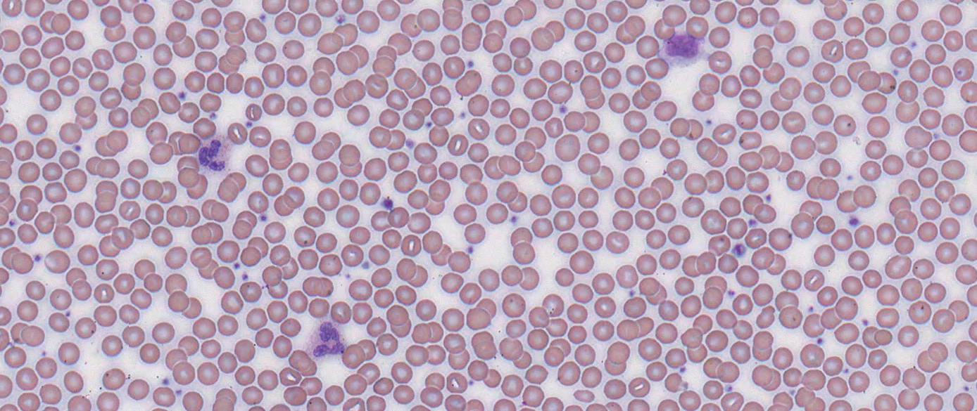
The image above is derived from a thin peripheral blood smear with normal histology. Contrasting the white background is an overwhelming majority of red blood cells. Mainly responsible for gas exchange, they appear as round red donuts with a white center. This is a reflection of their natural shape: the biconcave disk. Occasionally, the background would be interrupted by purple specks that are much smaller than the red blood cells. Those are platelets, a major component of hemostasis and coagulation. The purple cells that are much larger than the red blood cells are neutrophils, which are characterized by their light purple cytoplasm and dark purple, multi-segmented nuclei. Amongst the leukocytes, cells part of the immune system, neutrophils are present in the blood with the highest concentration, far outnumbering their fellow granulocytes (eosinophils and basophils).
ROI 2- Bone Marrow Core
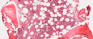
Bone marrow biopsies can yield normal histological images of the bone marrow core just like the one above. The trabecular bone is visualized as large, smooth, red patches interwoven amongst the rest of the tissue. In between the trabecular bone is a myriad of light and dark red dots of different sizes and intensities. These are the bone marrow blood stem cells and blasts (young blood cells in earlier stages of development). Dotting these cells are adipocytes (fat storing cells), which appear as large, round patches of emptiness. As one ages, the percentage of cells (also called cellularity) in the bone marrow core decreases and the amount of adipocytes increases. Changes in cellularity can be indicative of many different blood disorders.
ROI 3 – Bone Marrow Smear
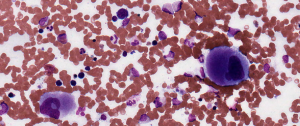
In addition to biopsies, smears of bone marrow aspirate (example above) can also provide insight into the normal cellular ecosystem of the bone marrow. Most cells in the bone marrow are blasts (immature cells) as opposed to cytes (mature cells). The small, dark purple cells are the stem cells and the blasts of various leukocytic lineages. They are characterized by their high nucleus/cytoplasm (N/C) ratio as well as their smaller size compared to their mature counterparts. Between the stem cells and the blasts, the blasts are usually larger with a lower N/C ratio. Reticulocytes, immature red blood cells, are also present in abundance in the bone marrow. They are multi-lobular, irregularly shaped red cells that are larger than their mature counterparts. Under a microscope, the most distinctive feature of reticulocytes is the residual cytoplasmic RNA. This gives some reticulocytes dark granules that clearly separates them from the clear center of healthy, mature red blood cells.
Watch the following video lessons of author Theodore Lam looking at normal and peripheral blood smear and bone marrow biopsy.
Video lesson was created and recorded by Shing Tat Theodore Lam, licensed under All Rights Reserved.
Video lesson was created and recorded by Shing Tat Theodore Lam, licensed under All Rights Reserved
Video lesson was created and narrated by Shing Tat Theodore Lam. Visuals were created by by Sarah Pinault, licensed under All Rights Reserved
Section Review
Circulating blood – or peripheral blood -consists predominantly is full of RBCs, which are easily identified by its biconcave (donut) shape disk with a pale centre. The next most common cell are the larger leukocytes which is easily identified by the dominant purple nucleus with varying amounts of cytoplasm. Periodically, a very small dense purple speck – lacking recognizable organelle or nucleus – are platelets.
Mature blood cells (formed elements) are made in the bone marrow in a healthy adult. An aspirate or bone marrow core biopsy will reveal a mixture of fat cells (adipocytes) and hemopoietic stem cells in varying levels of differentiation, known as blasts. There would be few mature blood cells in marrow as they would normally leave the bone and enter the peripheral circulation. However, early red blood cells (reticulocytes) can be seen but they lack the characteristic biconcave disk with pale centre
Review Questions
Media Attributions
- Peripheral Blood Smear Histology © DHPLC is licensed under a All Rights Reserved license
- Normal Bone Marrow Core © DHPLC is licensed under a All Rights Reserved license
- Bone Blood Smear Histology © DHPLC is licensed under a All Rights Reserved license
