Acute Kidney Injury
Gross Anatomy of the Kidney
J. Gordon Betts; Kelly A. Young; James A. Wise; Eddie Johnson; Brandon Poe; Dean H. Kruse; Oksana Korol; Jody E. Johnson; Mark Womble; and Peter DeSaix
Learning Objectives
By the end of this section, you will be able to:
- Describe the external structure of the kidney, including its location, support structures, and covering.
- Identify the major internal divisions and structures of the kidney.
- Identify the major blood vessels associated with the kidney and trace the path of blood through the kidney.
- Name structures found in the cortex and medulla.
- Describe the physiological characteristics of the cortex and medulla.
- Identify the ureters, urinary bladder, and urethra, as well as their location, structure, histology, and function.
- Compare and contrast male and female urethras.
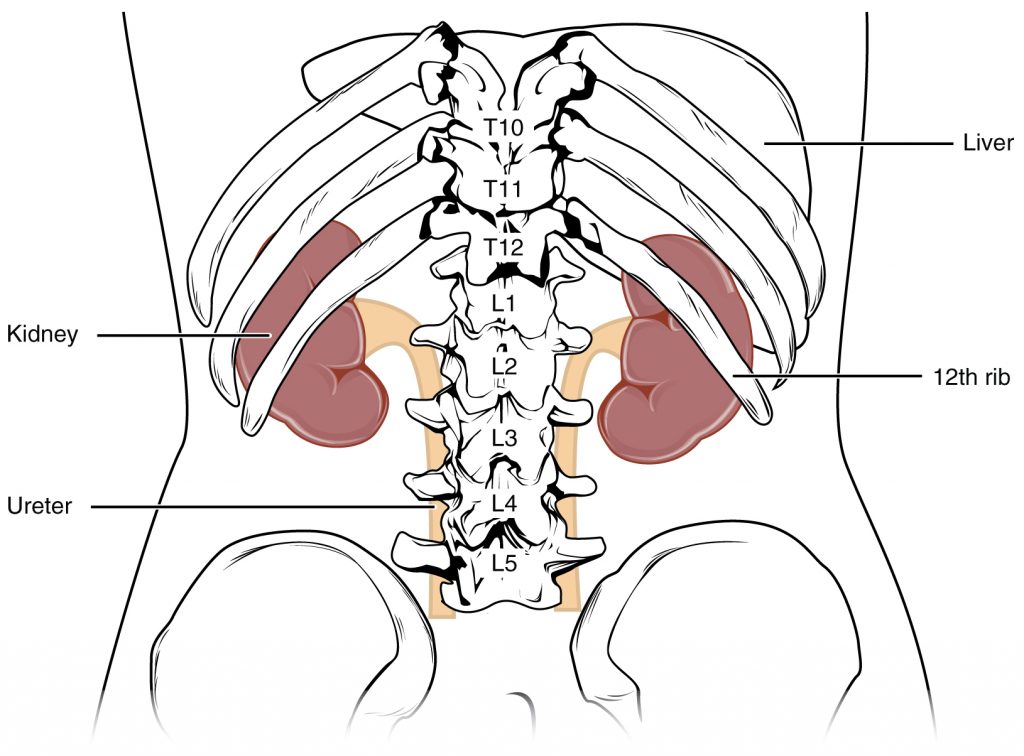
The kidneys lie on either side of the spine against the back of the abdominal wall, well protected by muscle, fat, and ribs. They are roughly the size of your fist. The kidneys require a lot of blood flow, receiving about 25 percent of the blood pumped by the heart at rest.
External Anatomy
The right kidney is located slightly lower than the left, due to displacement by the liver. Upper portions of the kidneys are somewhat protected by the eleventh and twelfth ribs and are also surrounded and protected by a connective tissue capsule and surrounding fat.
Atop each kidney is an adrenal gland. The adrenal cortex, or outer layer of the adrenal gland, influences renal function by producing the hormone aldosterone to stimulate sodium reabsorption.
Internal Anatomy
Inside the kidney there is an outer region called the renal cortex and an inner region called the medulla, as shown in the figure below. The renal columns are connective tissue extensions that pass from the cortex through the medulla to separate the renal pyramids (triangular segments formed of tissue from the medulla) and renal papillae. The papillae are bundles of collecting ducts that transport urine made by the functional tissue of the kidney to the calyces of the kidney for excretion.
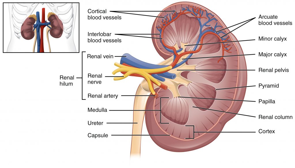
Renal Hilum
The renal hilum is the entry and exit site for structures servicing the kidneys: blood vessels (artery and vein), nerves, lymphatics, and ureters.
Nephrons and Vessels
The renal arteries branch into smaller and smaller vessels, eventually forming afferent arterioles, as seen in the figure below. The afferent arterioles feed the capillaries in the glomeruli that are vital to the function of the kidney.
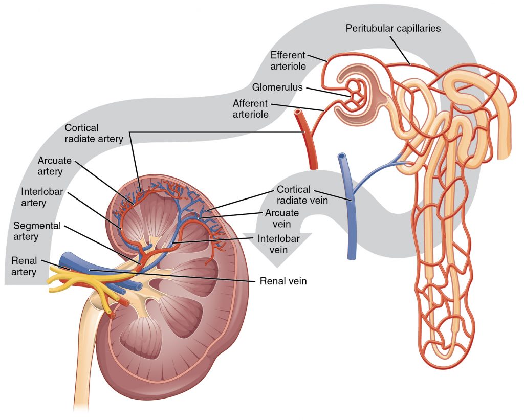
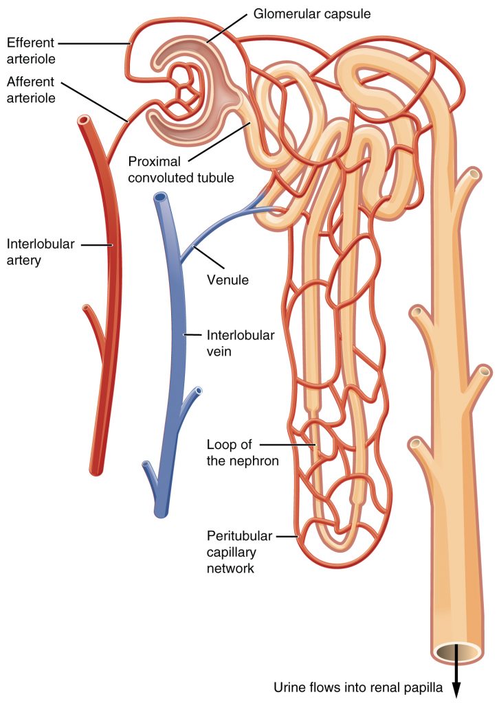
Nephrons are the “functional units” of the kidney; they cleanse the blood and balance water and electrolytes, like sodium and potassium. The diagram below shows the blood flow and urine formation in the nephron. The afferent arterioles form the capillaries of the glomeruli (sing. glomerulus). The glomerulus is surrounded by Bowman’s capsule, which forms a space around the glomerulus called Bowman’s space. The nephron is composed of tubules that are continuous with Bowman’s space. The glomerulus and Bowman’s capsule together form the renal corpuscle. These glomerular capillaries filter the blood based on particle size. After passing through the renal corpuscle, the capillaries form a second arteriole, the efferent arteriole. These will next form the peritubular capillary network around the more distal portions of the nephron tubule before returning to the venous system. As the glomerular filtrate passes through the nephron, these peritubular capillary networks recover most of the solutes, nutrients, and water, and return them to the circulation.
Ureters
As urine is formed, it drains into the calyces of the kidney, which merge to form the funnel-shaped renal pelvis in the hilum of each kidney. The renal pelvis narrows to become the ureter of each kidney. As the ureters approach the bladder, they turn and enter the bladder at an angle. This creates a one-way valve that allows urine into the bladder but prevents reflux of urine from the bladder back into the ureter.
Bladder
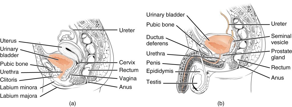
The urinary bladder collects urine from the ureters and lies in front of the uterus in females, behind the pubic bone and in front of the rectum. In males, the anatomy is similar, minus the uterus, and with the addition of the prostate below the bladder, surrounding the urethra.
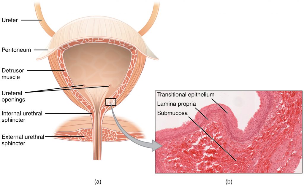
The urethra transports urine from the bladder to the outside of the body for disposal. The urethra is the only part of the urinary system that shows any significant anatomic difference between males and females; all other urine transport structures are identical.
The urethra in both males and females begins inferior and central to the two ureteral openings.
Female Urethra
The main difference between the from the male urethra is the female urethra’s short length of about 4 cm. This makes it less of a barrier to fecal bacteria than the longer male urethra and the best explanation for the greater incidence of urinary tract infections (UTIs) in women.
Male Urethra
The male urethra passes through the prostate gland immediately inferior to the bladder before passing below the pubic symphysis. The length of the male urethra varies but averages 20 cm in length.
Section Review
The structure of the kidney is divided into two main regions—the peripheral rim of cortex and the central medulla. The two kidneys receive about 25 percent of cardiac output (amount of blood pumped by the heart). They are protected by fat and overlying ribs and muscle. Ureters, blood vessels, lymph vessels, and nerves enter and leave at the renal hilum. There are about 1.3 million nephrons per kidney and these are the “functional units” that perform the important physiologic functions. The glomerulus (a specialized capillary bed) filters blood and the filtrate is captured by Bowman’s capsule. Most water and solutes are recovered by a second capillary bed that surrounds the tubules of the nephron. This filtrate is processed into urine by the renal tubules and finally flows into collecting ducts that drain into the minor calyces, which merge to form major calyces. The urine then proceeds to the renal pelvis and finally the ureters to the bladder. Urine is stored in the bladder until urination. The male and female urethras differ in their length and the presence of a prostate in males.
Review Questions
1. Finish the following sentence. The renal pyramids are separated from each other by extensions of the renal cortex called:
- Renal medulla
- Minor calyces
- Medullary cortices
- Renal columns
2. Finish the following sentence. The primary structure found within the medulla is the:
- Loop of Henle
- Minor calyces
- Portal System
- Ureter
3. Finish the following sentence. The right kidney is slightly lower because:
- It is displaced by the liver
- It is displaced by the heart
- It is slightly smaller
- It needs protection of the lower ribs
4. Fill in the blanks.
The sturdy _____ and conformable _____ provide physical protection to the kidney.
5. What is NOT part of the renal hilum?
- Bladder
- Renal Artery
- Renal Vein
- Ureter
- Lymphatics
- Nerves
6. Fill in the blanks.
Females are more likely to develop bladder infections than males because _____ must travel farther in the _____ urethra, making it less likely for infections.
Answer Key
- Renal columns
- Loop of Henle
- It is displaced by the liver
- ribs, fat pads
- Bladder
- bacteria, male
Adaption
This chapter is adapted from the following text:
Gross Anatomy of the Kidney in Anatomy and Physiology by OSCRiceUniversity is licensed under a Creative Commons Attribution 4.0 International License
Media Attributions
- 2608_Kidney_Position_in_Abdomen © OSCRiceUniversity is licensed under a CC BY (Attribution) license
- 2610_The_Kidney © OSCRiceUniversity is licensed under a CC BY (Attribution) license
- Blood Flow in the Kidney © OSCRiceUniversity is licensed under a CC BY (Attribution) license
- 2611_Blood_Flow_in_the_Nephron © OSCRiceUniversity is licensed under a CC BY (Attribution) license
- 2605_The_Bladder © OSCRiceUniversity is licensed under a CC BY (Attribution) license
- Female_and_Male_Urethra © OSCRiceUniversity is licensed under a CC BY (Attribution) license
outer part of kidney containing all of the nephrons; some nephrons have loops of Henle extending into the medulla
inner region of kidney containing the renal pyramids
extensions of the renal cortex into the renal medulla; separates the renal pyramids; contains blood vessels and connective tissues
six to eight cone-shaped tissues in the medulla of the kidney containing collecting ducts and the loops of Henle of some nephrons
medullary area of the renal pyramids where collecting ducts empty urine into the minor calyces
cup-like structures receiving urine from the collecting ducts where it passes on to the renal pelvis and ureter
medial area of the kidney through which the renal artery, renal vein, ureters, lymphatics, and nerves pass
functional units of the kidney that carry out filtration and modification to produce urine; consist of renal corpuscles, proximal and distal convoluted tubules, and descending and ascending loops of Henle; drain into collecting ducts
tuft or cluster of capillaries surrounded by Bowman’s capsule; filters the blood based on molecular size
cup-shaped sack lined by a simple squamous epithelium (parietal surface) and specialized cells called podocytes (visceral surface) that participate in the filtration process; receives the filtrate which then passes on to the PCTs
consists of the glomerulus and Bowman’s capsule
arteriole carrying blood from the glomerulus to the capillary beds around the convoluted tubules and loop of Henle; portion of the portal system
second capillary bed of the renal portal system; surround the proximal and distal convoluted tubules; associated with the vasa recta
transports urine from the bladder to the outside environment
