Heart Failure
Video Lessons of Normal Heart Function, Anatomy, and Histology
Jennifer Kong
Learning Objectives
By the end of this section, you will be able to:
- Identify the main chambers and great vessels of the normal heart.
- Describe the path blood normally takes from when it enters the right atrium to when it leaves the aorta.
- Demonstrate knowledge of histology by identifying normal myofibrils on a tissue stained with H&E.
Normal Cardiac Cycle – Interactive Video
Normal Heart Structure and Function by Jennifer Kong, licensed under Creative Commons Attribution-NonCommercial 4.0 International License
Normal Cardiac Anatomy
Of course, looking at the heart in a multicolour textbook image is very different than identifying structures in a real heart. The image below demonstrates a frontal section of a normal pig heart (i.e. heart is sliced into front & back). Thus, both the right and left heart structures are visible at the same time.
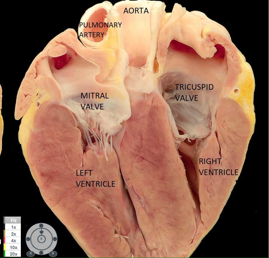
Normal Frontal Section of Pig Heart by Jennifer Kong, licensed under All Rights Reserved
Normal Cardiac Histology
Before viewing this video, recall your knowledge of H&E staining of tissue preparations from “How are specimens obtained and prepared.” Key points to remember are:
- Protein-rich areas will stain pink/purple. The stronger the stain in the cytoplasm, the more protein is present.
- Nuclei will stain purplish blue. The bigger the nuclei, the more nuclear activity is present (e.g. protein synthesis).
- Cells that fulfill a single function will create tissues with little variety in cell appearance.
Critical Thinking and Histology Exercises
Answer these questions for yourself before viewing the next video:
- Consider the function of the heart wall – especially the ventricles.
- What is the purpose of the ventricular tissue? Will there be a need for more than one type of cell type?
- Will a cardiomyocyte (ie the typical cell in the ventricle) have a large nucleus? A small nucleus? Why or why not?
- Will a cardiomyocyte have a large or small cytoplasm? What would be “filling up” that cytoplasm?
- Would there be much space between cardiomyocytes? HINT: think about the near-instantaneous communication between cardiomyocytes during electrical conduction.
- Why would the wall to the atria be thinner than the ventricular wall?
- If the slice of heart tissue cut through a coronary artery, nestled between the atrium and ventricle, what shape would it be?
Histology of Normal Heart Tissue by Jonathan Bush, licensed under All Rights Reserved
Section Review
- Deoxygenated blood (visible as blue/purple colour) enters the right atrium from the superior and inferior vena cavae. This blood passes through the tricsupid valve into the right ventricle. Upon ventricular systole, this blood is ejected through the pulmonary valve into the pulmonary arteries which goes to the lungs. The blood is now oxygenated and can be seen as it makes the blood the colour red. Oxygenated blood returns to the heart via the pulmonary veins, entering the left atrium and into the left ventricle via the mitral (or bicuspid) valve. During ventricular systole, the left ventricle contracts and pushes the blood through the aortic valve into the aorta which delivers the oxygenated blood to the heart via the coronary arteries and the rest of the body.
- The left ventricular wall is thicker than the right because more pumping force is needed to reach the rest of the body (compared to the lungs which is the destination of the right ventricle). Similarly, the atrial walls are thinner than the ventricular walls because their pumping force just needs to reach the respective neighbouring ventricle.
- Ventricular tissue is predominantly made of cardiomyocytes which allow for contraction. These myocytes have large protein-filled cytoplasms which are comprised of contractile units (i.e. the proteins actin, myosin, troponin, and tropomyosin).
- Once mature, cardiomyocytes tend to have small nuclei as they do very little protein synthesis and are not replicating.
- Because the contractile cardiomycoytes need near-instantaneous communication between cells, cardiomyocytes are joined by gap junctions with no appreciable space between cells.
Review Questions
Blue _____ blood enters the _____ atria and ventricle during diastolic filling. Atrial systole occurs, completely filling the ventricles and ends with the closing of the _____ valve in the right heart. At the start of ventricular systole, pressure builds up in the right ventricle, exerting pressure on both the atrioventricular _____ valve and the semilunar _____ valve. The atrioventricular valve can not open in response to this pressure due to the strong _____ which are holding the valve leaflets closed. Thus, the right ventricle produces enough pressure to open the _____valve thus ejecting the blood to the lungs. At the lungs, the blood becomes oxygenated becoming _____ in colour. This oxygenated blood returns to the left atrium via the “pulmonary veins”. During diastolic filling, the _____ valve is open as the left atria and ventricles fill. During ventricular systole, the _____ valve can’t open due to the strong _____ holding it closed. As such, the _____valve is opened during ventricular systole, thus allowing oxygenated blood to be ejected to the rest of the body.2. Cardiac cells have large nuclei and little cytoplasm because of the intense amount of protein synthesis that is involved.
- True
- False
3. Which is the correct statement? The walls of the right ventricle are thicker than those of the right atria
- The right ventricle wall is thicker than that of the left ventricle
- The aortic valve separates the right atria and ventricle
- The tricuspid valve separates the left atria and ventricle
Answer Key
- Blue deoxygenated blood enters the right atria and ventricle during diastolic filling. Atrial systole occurs, completely filling the ventricles and ends with the closing of the tricuspid valve in the right heart. At the start of ventricular systole, pressure builds up in the right ventricle, exerting pressure on both the atrioventricular tricuspid valve and the semilunar pulmonary valve. The atrioventricular valve can not open in response to this pressure due to the strong chordae tendinae which are holding the valve leaflets closed. Thus, the right ventricle produces enough pressure to open the pulmonary valve thus ejecting the blood to the lungs. At the lungs, the blood becomes oxygenated becoming red in colour. This oxygenated blood returns to the left atrium via the “pulmonary veins”. During diastolic filling, the mitral/bicuspid valve is open as the left atria and ventricles fill. During ventricular systole, the “mitral/bicuspid” valve can’t open due to the strong chordae tendinae holding it closed. As such, the aortic valve is opened during ventricular systole, thus allowing oxygenated blood to be ejected to the rest of the body.
- False
- The walls of the right ventricle are thicker than those of the right atria
Media Attributions
- annotate normal heart and aorta
