Pneumonia and Pulmonary Edema
Pulmonary Circulation, Organs and Structures of the Respiratory Zone
J. Gordon Betts; Kelly A. Young; Mark Womble; Brandon Poe; Oksana Korol; Dean H. Kruse; Peter DeSaix; James A. Wise; Jody E. Johnson; and Eddie Johnson
Learning Objectives
By the end of this section, you will be able to:
- List the structures that make up the respiratory zone.
- Describe how the respiratory system processes oxygen and CO2.
- Trace the path of oxygenated and deoxygenated blood as it arrives and leaves the alveoli.
- Identify the vessels through which blood travels within the pulmonary circuit, beginning from the right ventricle of the heart and ending at the left atrium.
Respiratory Zone
In contrast to the conducting zone, the respiratory zone includes structures that are directly involved in gas exchange. The respiratory zone begins where the terminal bronchioles join a respiratory bronchiole, the smallest type of bronchiole, which then leads to an alveolar duct, opening into a cluster of alveoli.
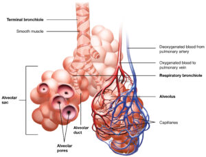
Alveoli
Alveolar duct is a tube composed of smooth muscle and connective tissue, which opens into a cluster of alveoli. An alveolus is one of the many small, grape-like sacs that are attached to the alveolar ducts.
An alveolar sac is a cluster of many individual alveoli that are responsible for gas exchange. An alveolus is approximately 200 μm in diameter with elastic walls that allow the alveolus to stretch during air intake, which greatly increases the surface area available for gas exchange. The image below shows that alveoli are connected to their neighbors by alveolar pores, which help maintain equal air pressure throughout the alveoli and lung.
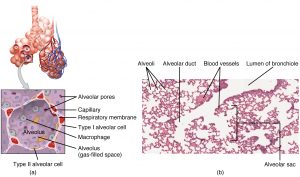
The alveolar wall consists of three major cell types: type I alveolar cells, type II alveolar cells, and alveolar macrophages. A type I alveolar cell is a squamous epithelial cell of the alveoli, which constitute up to 97 percent of the alveolar surface area. These cells are about 25 nm thick and are highly permeable to gases. A type II alveolar cell is interspersed among the type I cells and secretes pulmonary surfactant, a substance composed of phospholipids and proteins that reduces the surface tension of the alveoli. Roaming around the alveolar wall is the alveolar macrophage, a phagocytic cell of the immune system that removes debris and pathogens that have reached the alveoli.
The simple squamous epithelium formed by type I alveolar cells is attached to a thin, elastic basement membrane. This epithelium is extremely thin and borders the endothelial membrane of capillaries. Taken together, the alveoli and capillary membranes form a respiratory membrane that is approximately 0.5 μm (micrometers) thick. The respiratory membrane allows gases to cross by simple diffusion, allowing oxygen to be picked up by the blood for transport and CO2 to be released into the air of the alveoli.
Pulmonary Circulation
Recall that blood returning from the systemic circuit enters the right atrium via the superior and inferior venae cavae and the coronary sinus, which drains the blood supply of the heart muscle. These vessels will be described more fully later in this section. This blood is relatively low in oxygen and relatively high in carbon dioxide, since much of the oxygen has been extracted for use by the tissues and the waste gas carbon dioxide was picked up to be transported to the lungs for elimination. From the right atrium, blood moves into the right ventricle, which pumps it to the lungs for gas exchange. This system of vessels is referred to as the pulmonary circuit.
The single vessel exiting the right ventricle is the pulmonary trunk. At the base of the pulmonary trunk is the pulmonary semilunar valve, which prevents backflow of blood into the right ventricle during ventricular diastole. As the pulmonary trunk reaches the superior surface of the heart, it curves posteriorly and rapidly bifurcates (divides) into two branches, a left and a right pulmonary artery. To prevent confusion between these vessels, it is important to refer to the vessel exiting the heart as the pulmonary trunk, rather than also calling it a pulmonary artery. The pulmonary arteries in turn branch many times within the lung, forming a series of smaller arteries and arterioles that eventually lead to the pulmonary capillaries. The pulmonary capillaries surround lung structures known as alveoli that are the sites of oxygen and carbon dioxide exchange.
Once gas exchange is completed, oxygenated blood flows from the pulmonary capillaries into a series of pulmonary venules that eventually lead to a series of larger pulmonary veins. Four pulmonary veins, two on the left and two on the right, return blood to the left atrium. At this point, the pulmonary circuit is complete (see the diagram below) defines the major arteries and veins of the pulmonary circuit discussed in the text.
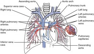
| Pulmonary Arteries and Veins | |
|---|---|
| Vessel | Description |
| Pulmonary trunk | Single large vessel exiting the right ventricle that divides to form the right and left pulmonary arteries |
| Pulmonary arteries | Left and right vessels that form from the pulmonary trunk and lead to smaller arterioles and eventually to the pulmonary capillaries |
| Pulmonary veins | Two sets of paired vessels—one pair on each side—that are formed from the small venules, leading away from the pulmonary capillaries to flow into the left atrium |
Section Review
The lining of the conducting zone is composed mostly of pseudostratified ciliated columnar epithelium with goblet cells. The mucus traps pathogens and debris, whereas beating cilia move the mucus superiorly toward the throat, where it is swallowed. As the bronchioles become smaller and smaller, and nearer the alveoli, the epithelium thins and is simple squamous epithelium in the alveoli. The endothelium of the surrounding capillaries, together with the alveolar epithelium, forms the respiratory membrane. This is a blood-air barrier through which gas exchange occurs by simple diffusion. The right ventricle pumps oxygen-depleted blood into the pulmonary trunk and right and left pulmonary arteries, which carry it to the right and left lungs for gas exchange. Oxygen-rich blood is transported by pulmonary veins to the left atrium after gas exchange occurs across the respiratory membrane inside the capillaries of the lung. The left ventricle pumps this blood into the aorta.
Review Questions
1. Fill in the blanks.The _____ ventricle of the heart pumps oxygen-depleted blood to the lungs via the pulmonary _____ which will continue to branch until they become pulmonary capillaries which envelop the _____ where gas exhange occurs. The vessels that leave now carry oxygenated blood via the pulmonary _____ back to the _____ atrium.
2. What is the role of alveolar macrophages?
- To secrete pulmonary surfactant
- To secrete antimicrobial proteins
- To remove pathogens and debris
- To facilitate gas exchange
3. What would be the consequence of the respiratory membrane suddenly getting thicker (e.g. during inflammation)?
- Reduced gas exchange
- Reduced blood flow to the alveoli
- Reduced blood flow away from the alveoli
- Improved gas exchange
Answer Key
- Right, arteries/trunk, alveoli, veins, left
- To remove pathogens and debris
- Reduced gas exchange
Adaptation
This chapter is adapted from the following text:
Pulmonary Circulatory Pathway and Organs and Structures of the Respiratory Zone and Pulmonary Circulation in Anatomy and Physiology by OSCRiceUniversity is licensed under a Creative Commons Attribution 4.0 International License
References
Bizzintino J, Lee WM, Laing IA, Vang F, Pappas T, Zhang G, Martin AC, Khoo SK, Cox DW, Geelhoed GC, et al. Association between human rhinovirus C and severity of acute asthma in children. Eur Respir J [Internet]. 2010 [cited 2013 Mar 22]; 37(5):1037–1042. Available from: http://erj.ersjournals.com/gca?submit=Go&gca=erj%3B37%2F5%2F1037&allch=
Kumar V, Ramzi S, Robbins SL. Robbins Basic Pathology. 7th ed. Philadelphia (PA): Elsevier Ltd; 2005.
Martin RJ, Kraft M, Chu HW, Berns, EA, Cassell GH. A link between chronic asthma and chronic infection.
Allergy Clin Immunol [Internet]. 2001 [cited 2013 Mar 22]; 107(4):595-601. Available from: http://erj.ersjournals.com/gca?submit=Go&gca=erj%3B37%2F5%2F1037&allch=
Media Attributions
- 2309_The_Respiratory_Zone © OSCRiceUniversity is licensed under a CC BY (Attribution) license
- 2310_Structures_of_the_Respiratory_Zone © OSCRiceUniversity is licensed under a CC BY (Attribution) license
- 2119_Pulmonary_Circuit © OSCRiceUniversity is licensed under a CC BY (Attribution) license
