Cirrhosis
The Normal Liver and Gallbladder
J. Gordon Betts; Kelly A. Young; James A. Wise; Eddie Johnson; Brandon Poe; Dean H. Kruse; Oksana Korol; Jody E. Johnson; Mark Womble; and Peter DeSaix
Learning Objectives
By the end of this section, you will be able to:
- State the main digestive roles of the liver and gallbladder.
- Identify three main features of liver histology that are critical to its function.
- Discuss the composition and function of bile.
Chemical digestion in the small intestine relies on the activities of three accessory digestive organs: the liver, pancreas, and gallbladder. The digestive role of the liver is to produce bile and export it to the duodenum. The gallbladder primarily stores, concentrates, and releases bile.
The Liver
The liver is the largest gland in the body, weighing about three pounds in an adult. It is also one of the most important organs. In addition to being an accessory digestive organ, it plays a number of roles in metabolism and regulation of blood plasma composition. The liver lies inferior to the diaphragm in the right upper quadrant of the abdominal cavity and receives protection from the surrounding ribs.
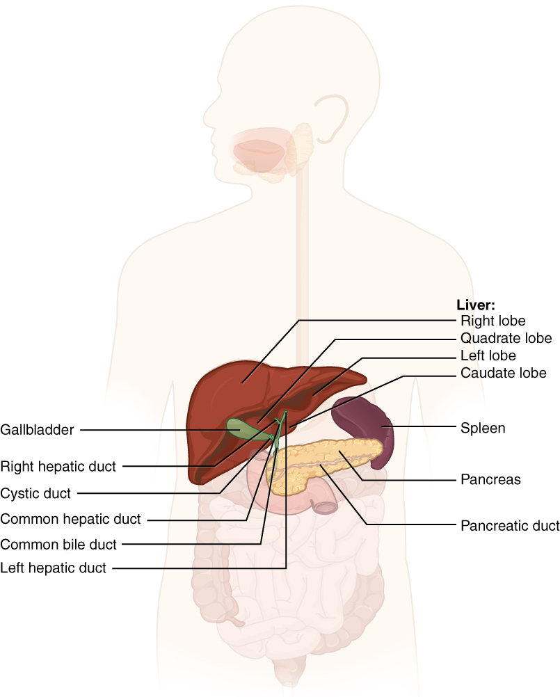
The liver is divided into two primary lobes: a large right lobe and a much smaller left lobe. In the right lobe, some anatomists also identify an inferior quadrate lobe and a posterior caudate lobe, which are defined by internal features. The liver is connected to the abdominal wall and diaphragm by five peritoneal folds referred to as ligaments. These are the falciform ligament, the coronary ligament, two lateral ligaments, and the ligamentum teres hepatis. The falciform ligament and ligamentum teres hepatis are actually remnants of the umbilical vein, and separate the right and left lobes anteriorly. The lesser omentum tethers the liver to the lesser curvature of the stomach.
The porta hepatis (“gate/door to the liver”) is where the hepatic artery and hepatic portal vein enter the liver. These two vessels, along with the common hepatic duct, run behind the lateral border of the lesser omentum on the way to their destinations. As shown in the figure below, the hepatic artery delivers oxygenated blood from the heart to the liver. The hepatic portal vein delivers approximately 80% of total blood flow to the liver; portal vein blood is partially deoxygenated, but contains nutrients absorbed from the small intestine and actually supplies more oxygen to the liver than do the much smaller hepatic arteries. In addition to nutrients, drugs and toxins are also absorbed. After processing the bloodborne nutrients and toxins, the liver releases nutrients needed by other cells back into the blood, which drains into the central vein and then through the hepatic vein to the inferior vena cava. With this hepatic portal circulation, all blood from the alimentary canal passes through the liver. This largely explains why the liver is the most common site for the metastasis of cancers that originate in the alimentary canal.
Histology
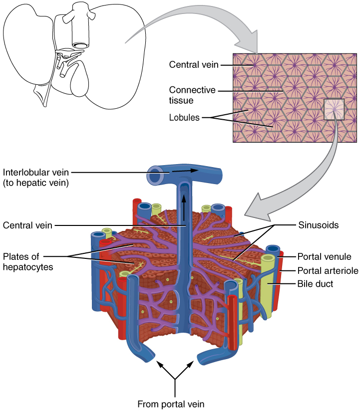
The liver has three main components: hepatocytes, bile canaliculi, and hepatic sinusoids. A hepatocyte is the liver’s main cell type, accounting for around 80 percent of the liver’s volume. These cells play a role in a wide variety of secretory, metabolic, and endocrine functions. Plates of hepatocytes called hepatic laminae radiate outward from the central vein in each hepatic lobule.
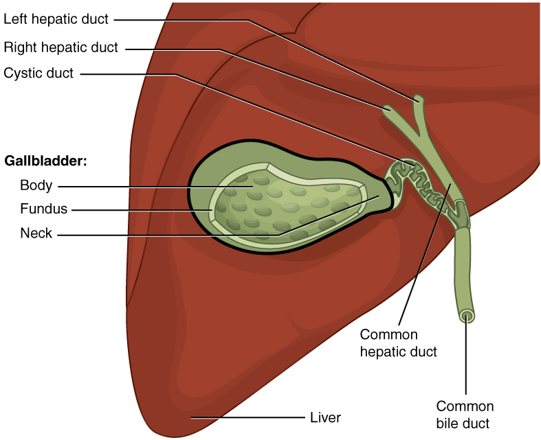
Between adjacent hepatocytes, grooves in the cell membranes provide room for each bile canaliculus (plural = canaliculi). These small ducts transport the bile that is produced and secreted by hepatocytes. From here, bile flows first into bile ductules and then into bile ducts. The bile ducts unite to form the larger right and left hepatic ducts, which themselves merge and exit the liver as the common hepatic duct. This duct then joins with the cystic duct from the gallbladder, forming the common bile duct through which bile flows into the duodenum of the small intestine.
Hepatic sinusoids are an open, porous blood space lined with simple squamous endothelial cells that takes the form of discontinuous fenestrated capillaries. Sinusoids are formed by the merging of nutrient-rich hepatic portal veins and oxygen-rich hepatic arteries and therefore carry mixed blood containing nutrients from the alimentary canal as well as oxygen. Sinusoids are instrumental in that they infiltrate each hepatic laminae delivering their contents to the hepatocytes. From their central position, hepatocytes absorb and process the nutrients, toxins, and waste materials carried by the blood. Materials such as bilirubin are processed and excreted into the bile canaliculi. Other materials including proteins, lipids, and carbohydrates are processed and secreted into the sinusoids at levels that maintain specific levels within the blood. Hepatocytes have the ability to store excess lipid, carbohydrates and amino acids until the blood plasma levels become depleted. Maintaining blood plasma composition is very important as each of these blood plasma components are required by cells of the entire body. The liver therefore has an important role in ensuring maintenance, survival, and homeostasis of all of the tissues and organs.
Once the hepatic sinusoids have passed by the hepatocytes within each laminae, the sinusoids merge and send blood through a central vein. Blood then flows through a hepatic vein into the inferior vena cava (IVC). This means that blood and bile flow in opposite directions. The hepatic sinusoids also contain star-shaped reticuloendothelial cells (Kupffer cells), phagocytes that remove dead red and white blood cells, bacteria, and other foreign material that enter the sinusoids. The portal triad is a distinctive arrangement around the perimeter of hepatic lobules, consisting of three basic structures: a bile duct, a hepatic artery branch, and a hepatic portal vein branch.
Bile
In order to underscore the importance of the bile that is produced by hepatocytes. It is important to remember that the food that is eaten does contain fat which is a valuable energy source and building block for the body. Recall that lipids are hydrophobic, that is, they do not dissolve in water. Thus, before they can be digested in the watery environment of the small intestine, large lipid globules must be broken down into smaller lipid globules, a process called emulsification. Bile is a mixture secreted by the liver to accomplish the emulsification of lipids in the small intestine.
Hepatocytes secrete about one liter of bile each day. A yellow-brown or yellow-green alkaline solution (pH 7.6 to 8.6), bile is a mixture of water, bile salts, bile pigments, phospholipids (such as lecithin), electrolytes, cholesterol, and triglycerides. The components most critical to emulsification are bile salts and phospholipids, both of which have a nonpolar (hydrophobic) region as well as a polar (hydrophilic) region. The hydrophobic region interacts with the large lipid molecules, whereas the hydrophilic region interacts with the watery chyme in the intestine. This results in the large lipid globules being pulled apart into many tiny lipid fragments of about 1 µm in diameter. This change dramatically increases the surface area available for lipid-digesting enzyme activity. Emulsification is similar to way dish soap can disperse oil slicks or fats that can be seen on the top of dishwater.
Bile salts act as emulsifying agents, so they are also important for the intestine’s ability to digest and then absorb lipids. While most constituents of bile are eliminated in feces, bile salts are reclaimed by the enterohepatic circulation. Once bile salts reach the ileum, they are absorbed and returned to the liver in the hepatic portal blood. The hepatocytes absorb and then excrete the bile salts as part of the newly formed bile. Thus, this precious resource is recycled.
Bilirubin, the main bile pigment, is a waste product produced when the spleen removes old or damaged red blood cells from the circulation. These breakdown products, including proteins, iron, and toxic bilirubin, are transported to the liver via the splenic vein of the hepatic portal system. In the liver, proteins and iron are recycled, whereas bilirubin is processed and then excreted in the bile. It accounts for the green color of bile. Bilirubin is eventually transformed by intestinal bacteria into stercobilin, a brown pigment that gives your stool its characteristic colour! In some disease states, bile does not enter the intestine, resulting in white (‘acholic’) stool with a high fat content, since virtually no fats are able to be broken down or absorbed in the absence of bile.
Hepatocytes work non-stop, but bile production increases when fatty chyme enters the duodenum and stimulates the secretion of the gut hormone secretin. Between meals, bile is produced but conserved. The valve-like hepatopancreatic ampulla closes, allowing bile to divert to the gallbladder, where it is concentrated and stored until the next meal.
Watch this video on liver to see the structure of the liver and how this structure supports the functions of the liver, including the processing of nutrients, toxins, and wastes. At rest, about 1500 mL of blood per minute flow through the liver. What percentage of this blood flow comes from the hepatic portal system?
The Gallbladder
The gallbladder is 8–10 cm (~3–4 in) long and is nested in a shallow area on the posterior aspect of the right lobe of the liver. This muscular sac stores, concentrates, and, when stimulated, propels the bile into the duodenum via the common bile duct. It is divided into three regions. The fundus is the widest portion and tapers medially into the body, which in turn narrows to become the neck. The neck angles slightly superiorly as it approaches the hepatic duct. The cystic duct is 1–2 cm (less than 1 in) long and turns inferiorly as it bridges the neck and hepatic duct.
The simple columnar epithelium of the gallbladder mucosa is organized in rugae, similar to those of the stomach. There is no submucosa in the gallbladder wall. The wall’s middle, muscular coat is made of smooth muscle fibers. When these fibers contract, the gallbladder’s contents are ejected through the cystic duct and into the bile duct. Visceral peritoneum reflected from the liver capsule holds the gallbladder against the liver and forms the outer coat of the gallbladder. The gallbladder’s mucosa absorbs water and ions from bile, concentrating it by up to 10-fold.
Section Review
Chemical digestion in the small intestine cannot occur without the help of the liver and pancreas. The liver produces bile and delivers it to the common hepatic duct. Bile contains bile salts and phospholipids, which emulsify large lipid globules into tiny lipid droplets, a necessary step in lipid digestion and absorption. The gallbladder stores and concentrates bile, releasing it when it is needed by the small intestine.
The pancreas produces the enzyme- and bicarbonate-rich pancreatic juice and delivers it to the duodenum of the small intestine through ducts. Pancreatic juice buffers the acidic gastric juice in chyme, inactivates pepsin from the stomach, and enables the optimal functioning of digestive enzymes in the small intestine.
Review Questions
1. What percentage of blood flow in the liver comes from the hepatic portal system?
- 33%
- 50%
- 80%
2. Which of these statements about bile is true?
- About 500 mL is secreted daily.
- Its main function is the denaturation of proteins.
- It is synthesized in the gallbladder.
- Bile salts are recycled.
3. Fill in the blanks with the following words.
| central vein hepatic artery |
portal vein sinusoids hepatocytes |
In a liver lobule, the _____ are the main cell type of the liver. They process, store, and release nutrients into the blood. Radiating out from the _____, they are tightly packed around the hepatic _____, allowing the hepatocytes easy access to the blood flowing through. Blood enters the lobule from divisions of the smaller _____ and larger _____ of the portal triad. As the blood flows towards the central vein, the blood bathes the hepatocytes.
Answer Key
- 80%
- Bile salts are recycled.
- hepatocytes, central vein, sinusoids, hepatic artery, portal vein
Adaption
This chapter is adapted from the following text:
Accessory organs of the digestive in Anatomy and Physiology by OSCRiceUniversity is licensed under a Creative Commons Attribution 4.0 International License
Media Attributions
- 2422_Accessory_Organs © OSCRiceUniversity is licensed under a CC BY (Attribution) license
- 2423_Microscopic_Anatomy_of_Liver © OSCRiceUniversity is licensed under a CC BY (Attribution) license
- 2425_Gallbladder © OSCRiceUniversity is licensed under a CC BY (Attribution) license
largest gland in the body whose main digestive function is the production of bile
organ with both exocrine and endocrine functions located posterior to the stomach that is important for digestion and the regulation of blood glucose
alkaline solution produced by the liver and important for the emulsification of lipids
“gateway to the liver” where the hepatic artery and hepatic portal vein enter the liver
artery that supplies oxygenated blood to the liver
vein that supplies deoxygenated nutrient-rich blood to the liver
duct formed by the merger of the two hepatic ducts
vein that receives blood from hepatic sinusoids
vein that drains into the inferior vena cava
major functional cells of the liver
blood capillaries between rows of hepatocytes that receive blood from the hepatic portal vein and the branches of the hepatic artery
hexagonal-shaped structure composed of hepatocytes that radiate outward from a central vein
small ducts between hepatocytes that collects bile
structure formed by the union of the common hepatic duct and the gallbladder’s cystic duct
main bile pigment, which is responsible for the brown color of feces
(also, Kupffer cell) phagocyte in hepatic sinusoids that filters out material from venous blood from the alimentary canal
bile duct, hepatic artery branch, and hepatic portal vein branch
recycling mechanism that conserves bile salts
accessory digestive organ that stores and concentrates bile
duct through which bile drains and enters the gallbladder
secretion of the pancreas containing digestive enzymes and bicarbonate
