Appendices: Introduction to Histology for first-time learners
A New Learner’s Guide to Epithelial Tissue: Lining Epithelia
Willie Wu and Athena Li
Learning Objectives
By the end of this section, you will be able to:
- Describe the key characteristics that define all epithelial tissues.
- Classify epithelium based on the number of cell layers and cell shape.
- Match different types of epithelium to their location and function in the body.
- Explain the structure and function of glandular epithelium.
- Identify common epithelial features like microvilli and cilia.
As we briefly discussed in the introduction, there are four types of basic tissue: epithelial, connective, muscle, and nervous. Epithelial tissue is the most varied in shape as it has to accomplish many types of function. They form the covering of all body surfaces – both external and internal. Epithelial tissue line external body (e.g. skin), internal cavities (e.g. inside blood vessels), and hollow organs (e.g. the stomach). They are also the major tissue in glands that secrete substances (e.g. oil gland on hair follicles, pancreatic duct that deliver enzymes from pancreas into GI tract). They perform a variety of functions and that is why they have different layers of cells (depending on the function) and specialized shapes to help it achieve its function.
An analogy for why there are different layers & shapes is to consider the different kind of skins/rinds on fruit. The skin/rind on a grape is a different thickness to a watermelon, probably due to the fact that grapes usually are suspended in the air whereas watermelon grows on the ground and the fact that the watermelon weights more than 1000x that of a grape! Similarly, think of the surface of the rinds of oranges vs apples vs bananas. They all serve the purpose of wrapping and protecting their fruit but their outer surface has different shapes and textures.
Characteristics of Epithelium
All epithelial tissue share the following four key features:
- Cellularity: They are composed almost entirely of tightly packed cells, with very little extracellular space between them. This creates a strong, continuous sheet.
- Polarity: Every epithelial cell has an apical surface (the “top” that faces a space or the outside world) and a basal surface (the “bottom” that is attached to the underlying tissue). These surfaces have different functions and different structures.
- Attachment: The basal surface sits on a thin, non-living, non-cellular layer called the basement membrane. This membrane is like the sticky mortar that holds the epithelial “tiles” firmly to the underlying connective tissue “floor.”
- Avascularity: Epithelium itself contains no blood vessels. Cells receive nutrients and oxygen by diffusion from blood vessels in the underlying connective tissue. This is why a shallow paper cut does not cause bleeding, since you have only sliced through avascular epithelium.
Classification of Epithelium: A Two-Name System
We classify epithelia by two simple characteristics: the number of cell layers and the shape of the cells at the apical surface.
| Epithelium Type | Description | Location | Function |
| Simple Squamous | A single layer of flat tiles | Lung air sacs (alveoli), lining of blood vessels (endothelium) | Allows rapid diffusion and filtration |
| Simple Cuboidal | A single layer of cubes | Kidney tubules, ducts of glands, surface of ovaries | Secretion and absorption |
| Simple Columnar | A single layer of tall columns | Lining of stomach, intestines, and gallbladder | Secretion (of mucus, enzymes) and absorption |
| Pseudostratified Columnar | A single layer of columns of differing heights | Lining of the trachea and most of the upper respiratory tract | Secretion and propulsion of mucus |
| Stratified Squamous | Multiple layers, apical cells are flat | Non-keratinized: mouth, esophagus
Keratinized: epidermis of skin |
Protection against abrasion and pathogens |
| Stratified Cuboidal | Multiple layers, apical cells are round | Ducts of exocrine glands (e.g. sweat glands, mammary glands, salivary glands) | Protection, limited secretion and absorption |
| Transitional (Urothelium) | Stratified epithelium with rounded, “plump” apical cells that can flatten | Lines the urinary bladder, ureters, and part of the urethra | Stretches to allow distension of organs |
By Layers
- Simple: A single layer of cells. Found in areas where the primary function is absorption, secretion, or filtration (e.g., lungs, blood vessels, kidney tubules).
- Stratified: Multiple layers of cells. Found in areas subject to wear and tear, where the primary function is protection (e.g., skin, mouth, esophagus).
- Pseudostratified: Looks layered because the nuclei are at different heights, but every cell touches the basement membrane. It is, in fact, a simple epithelium. A classic trick of histology!
Simple Squamous – Lung Alveoli
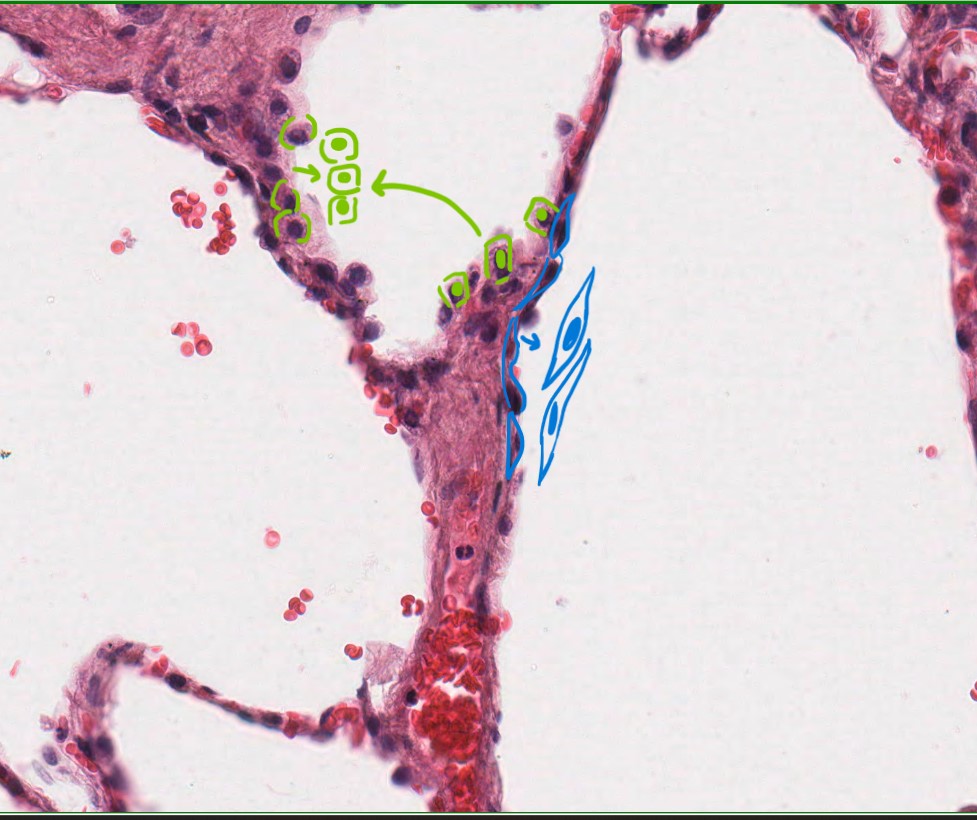
The image below shows the simple squamous epithelium lining the alveoli and allowing efficient gas exchange between air and blood. Because the cells are so thin, oxygen and carbon dioxide can easily pass through. The same type of epithelium also lines blood vessels and body cavities, where it helps regulate the passage of materials across the membrane.
Stratified Squamous – Foot Skin
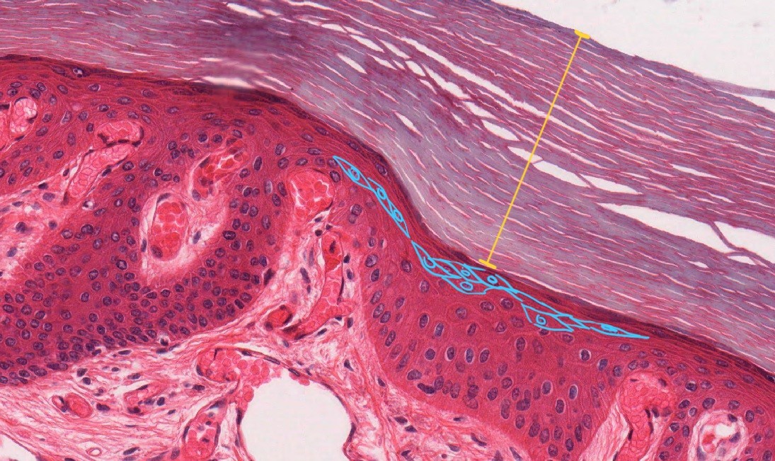
Stratified squamous epithelium is made up of multiple layers of cells, which makes it ideal for protecting the underlying tissues from physical and environmental damage. In areas like the skin of the foot as shown in the image below, this type of epithelium provides a tough and durable barrier against friction, microbes, and other external pathogens. The outermost cells are constantly shed and replaced to help maintain a strong protective surface.
Keep in mind that the epithelial tissue (whether one layer thick or more) sits on top of the basement membrane with other tissues below. So, at first glance, EVERYTHING looks stratified. However, with practice, you will be able to identify the basement membrane, which is the boundary between the basal surface of the epithelial tissue and a the deeper tissue (e.g. connective, muscle). You will also notice that the pattern of cell shapes change as you go from epithelial tissue into deeper structures.
Introduction to Alive vs Dead Cells
We often don’t realize how many dead cells cover our bodies. As mentioned in the Metastatic Cancer: Melanoma chapter, our entire outermost skin consists of dead cells. So when looking at histological specimens, how can we tell if the tissue we are looking at is made up of living or dead cells? The simplest way to determine this is by looking for the presence of a nucleus. All living cells must have a functional nucleus to manage cellular functions, start the synthesis of products, and for cellular reproduction. If a cell has clearly defined, intact nucleus, typically stained a deep purple color with hematoxylin and eosin (H&E) stain, it is considered alive. In contrast, dead cells may still absorb the H&E stain due to the remaining proteins, but they will lack a visible nucleus. This distinction is evident in the H&E stained thick foot image, where the yellow line highlights the layer of dead skin cells.
Interestingly, the lung alveoli image also contains “dead” cells – red blood cells (RBCs). These appear as round, red discs with pale centers. As outlined in the Blood, Anemia, Leukemia, and Blood Tests chapter, RBCs lack nuclei, which is why they show no dark purple staining. Because they cannot reproduce or synthesize new products, RBCs are technically not considered alive, which is something that can be surprising given that blood is often thought o as the very essence of life.
By Shape
- Squamous: Cells are flat and scale-like. The nucleus is often flattened. Ideal for rapid diffusion.
- Cuboidal: Cells are box-like, as tall as they are wide. The nucleus is round and central. Often involved in secretion and absorption.
- Columnar: Cells are tall and column-like, taller than they are wide. The nucleus is usually elongated and located near the base. Often involved in secretion and absorption.
When looking at shapes of epithelial cells, we always focus on the apical surface layer, which is the topmost layer exposed to the external environment or internal space. In stratified epithelium, it is important to remember that not all layers will have the same cell shape. This is because the apical layer serves a distinct function, such as protection or secretion, while the primary role of the basal layer is cell regeneration. Basal cells continuously divide to replace the cells above them, which creates the layered structure of the typical stratified epithelium. Therefore, only the shape of the apical layer is used to classify the tissue types (e.g. stratified squamous, stratified cuboidal).
Squamous Cells – Think SQUAshed Like a Fried Egg!
In the lung alveoli image, the squamous cells are marked in blue, while the cuboidal cells are marked in green. Squamous cells can also be recognized based on their “eye-like” or “fried egg” appearance. The blue outlined cells are as if you are looking at a fried egg on its side. If we were viewing the tissue from above rather than in cross section, the squamous layer would look like a crowded pan of fried eggs. The cells would appear irregular in their shapes and sizes, with dominant nuclei scattered without any obvious pattern. This irregular arrangement, while seemingly disorganized, serves as an advantage that maximizes the surface area and makes squamous epithelium high efficient for diffusion.
As mentioned earlier, the shape of epithelial cells often reflect their function. Because squamous cells are very flat and thin (like a fried egg), they allow rapid and easy diffusion of gases or substances between two compartments. The alveolar squamous cells form the barrier between inhaled air (seen in the open lumen) and the blood in nearby capillaries. The RBCs are visible as round red discs with no stained nuclei, which confirms their identity and reminding us of their unique anucleate nature.
Cuboidal Cells – Think as Beads/Pearls
In cuboidal lining epithelia, the cells themselves may vary slightly in their shapes, but a key identifying feature is their round, centrally located nuclei. These nuclei are typically evenly spaced and uniform in size, which creates a distinctive pattern. A helpful analogy is that the nuclei in a cuboidal epithelial layer resemble a string of pearls, looking round, orderly, and neatly aligned. This characteristic arrangement can make cuboidal epithelium easy to recognize in histological sections, especially when compared to the irregular appearance of squamous cells.
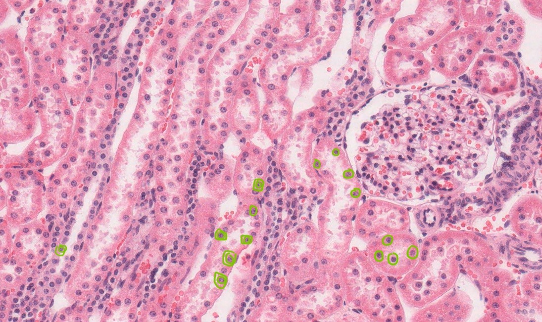
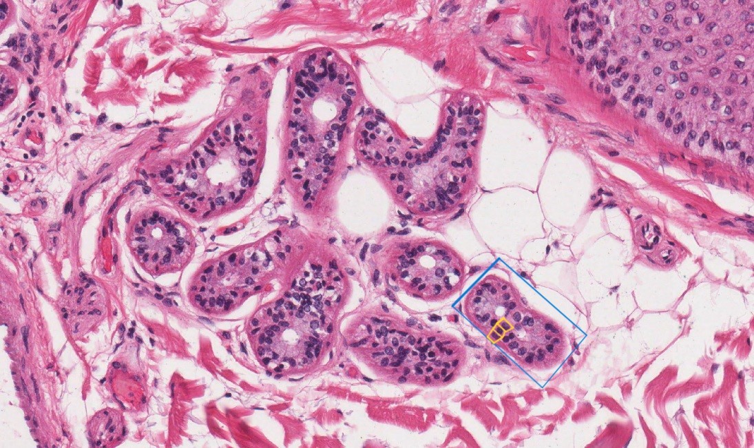
The two images (kidney and sweat glands) shown above illustrate examples of simple and stratified cuboidal cells, respectively. In the kidney image , the nuclei (highlighted in green) appear large and round, even though the surrounding cells may not be perfectly cuboidal in shape. So why do cuboidal cells typically have such prominent, round nuclei?
Let’s return to the relationship between structure and function. Cuboidal epithelial cells are commonly found in glandular tissues, such as sweat glands and in kidney tubules, where their primary role is to synthesize and secrete substances into ducts or tubule lumens. To support this active metabolic function, these cells require a large, active nucleus to manage high levels of protein and enzyme production. Additionally, the cuboidal shape provides the cell with enough cytoplasmic volume to house the organelles needed for secretion. Because these cells often surround the lumen of a duct, their structures are well-suited for both production and directional release of substances.
Columnar Cells
Columnar epithelial cells are easily recognized by their tall, rectangular shapes and their elongated, oval nuclei, which are typically located near the basal surface (the side of the cell closest to the basement membrane). These nuclei are usually aligned in a neat row, which gives columnar epithelium a very organized and structured appearance under the microscope.
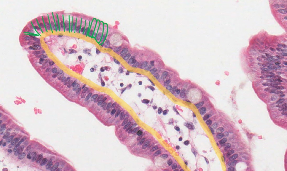
The shape of a columnar cell directly reflects its specialized function. The presence of a large, elongated nucleus indicates a high level of nuclear activity, such as the synthesis of proteins and other cellular products. The tall cytoplasmic space between the nucleus and the apical surface suggests that these cells house abundant organelles and machinery needed to process and transport these products. Additionally, the apical surface of columnar cells often features specialized structures such as cilia or microvilli (highlighted in green boxes), which are adaptations that help movement or absorption, depending on the tissue’s function. This structural complexity allows columnar cells to play a key role in secretion, absorption, and transport in organs such as the intestines and respiratory tract.
There are two major functions for columnar cells: the secretion of product and the absorption of substances from the apical surface. These well known functions of columnar cells help explain why they are commonly found in the epithelial lining of the gastrointestinal (GI) tract, among other locations.
In the GI tract, different regions have specialized needs. For example, mucus-secreting cells are abundant throughout, making columnar cells ideal due to their large cytoplasmic volume and robust organelle content for product synthesis and secretion. In the small intestine, the primary role of the epithelium is to absorb nutrients. While squamous epithelium is efficient for diffusion due to its thinness, columnar cells can actually provide even greater surface area for absorption—thanks to specialized apical structures called microvilli.
Microvilli, highlighted in green boxes in the duodenum image above, significantly increase the absorptive surface area. These projections require an internal cytoskeletal framework to maintain their structure, which is why they are commonly found on columnar cells (the elongated shape of these cells provides enough depth to support and anchor these features). Despite being taller than squamous cells, simple columnar epithelium still maintains a single-cell thickness, ensuring efficient absorption across the epithelium. In contrast, the urethra shows stratified columnar epithelium, which appears in regions like the kidney tubules, where a harsher chemical environment demands additional protective layers. Here, the priority is not absorption but protection, and multiple layers of columnar cells help maintain epithelial integrity.
In summary, the body maximizes nutrient absorption by lining the intestines with simple columnar cells equipped with microvilli. The elongated shape of columnar cells is especially suited to supporting specialized apical features, such as microvilli for absorption or cilia for movement, which makes them highly versatile for both secretion and absorption across various organ systems.
Transitional Epithelium – Umbrella Cells
As the name suggests, transitional epithelium is a type of epithelial tissue specialized for rapid transitions in shape and structure. Its most well-known location is the urinary bladder, which undergoes frequent and dramatic changes in volume throughout the day. Rather than increasing or decreasing the number of cells, transitional epithelium adapts by stretching and recoiling. This unique flexibility allows the tissue to maintain a protective barrier while expanding to accommodate urine and contracting once the bladder empties. Its ability to stretch without tearing is a key feature that distinguishes it from other epithelial types.
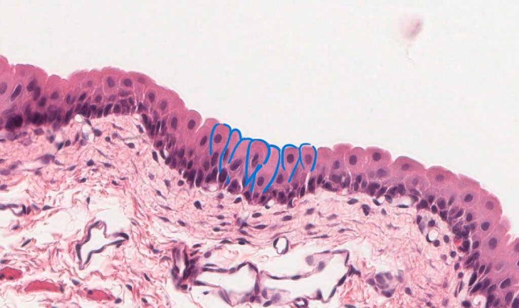
In the image above, transitional epithelium is recognizable by the presence of its distinctive “umbrella cells” (or raindrop-shaped cells) at the apical surface. These large, dome-shaped cells are a hallmark of transitional tissue and play a key role in allowing the epithelium to stretch and recoil without losing its integrity. Because transitional epithelium lines most of the urinary tract, including the bladder, it must be capable of adapting to rapid changes in volume throughout the day.
A helpful analogy is to think of transitional epithelium like a marshmallow. In its relaxed state, a marshmallow stands tall and resembles a columnar shape. When pressure is applied, similar to the bladder filling with urine, it compresses, flattening into a cuboidal or even squamous-like shape. However, once the pressure is released, the marshmallow returns to its original form. This analogy illustrates how transitional epithelium can change shape and height in response to stretching, without altering cell number or overall mass, ensuring both flexibility and protection in dynamic environments like the bladder.
Pseudostratified Columnar Epithelium
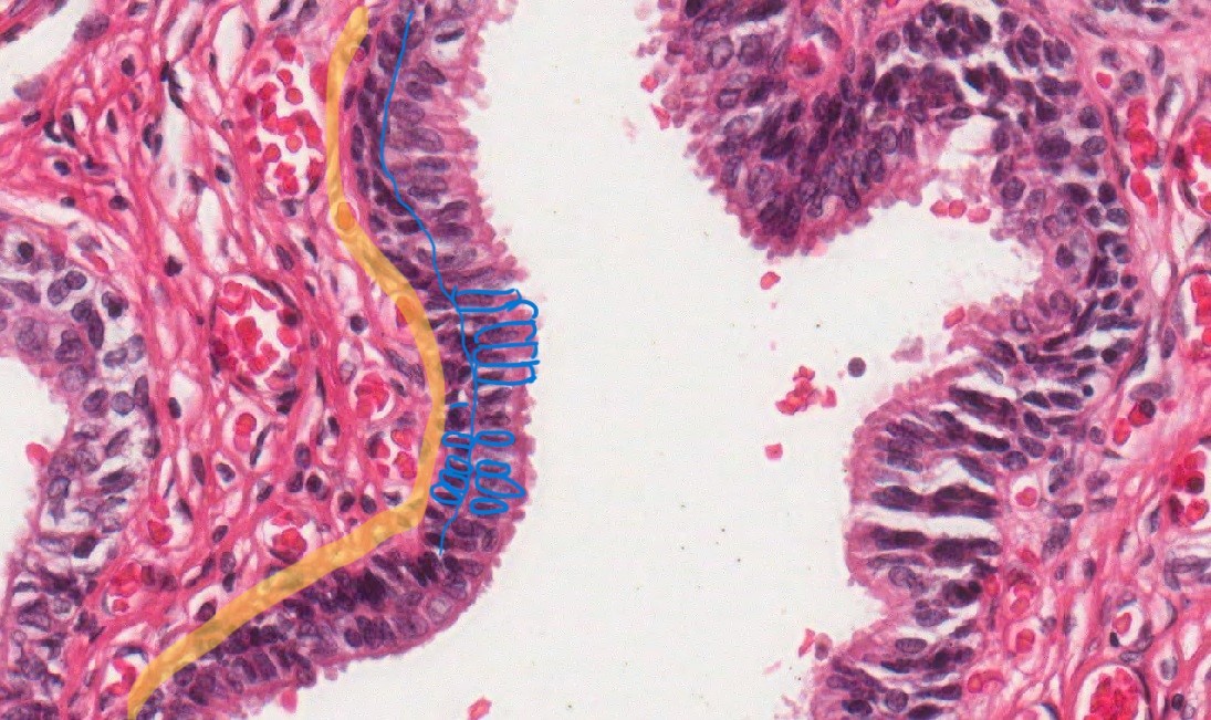
Pseudostratified columnar epithelium is found almost exclusively in the respiratory and reproductive systems, where it is typically ciliated. The presence of cilia is functionally important—it enables the movement of substances along the epithelial surface, such as clearing mucus from the airways or transporting ova through the reproductive tract.
This epithelial type can be challenging to identify, especially for beginners, because at first glance, it closely resembles stratified columnar epithelium (see the image comparison below). The key distinguishing feature is that in pseudostratified epithelium, all cells contact the basement membrane, even though the nuclei appear at different heights, giving a false impression of multiple layers. In contrast, true stratified columnar epithelium consists of stacked layers, with only the basal cells anchored to the basement membrane.
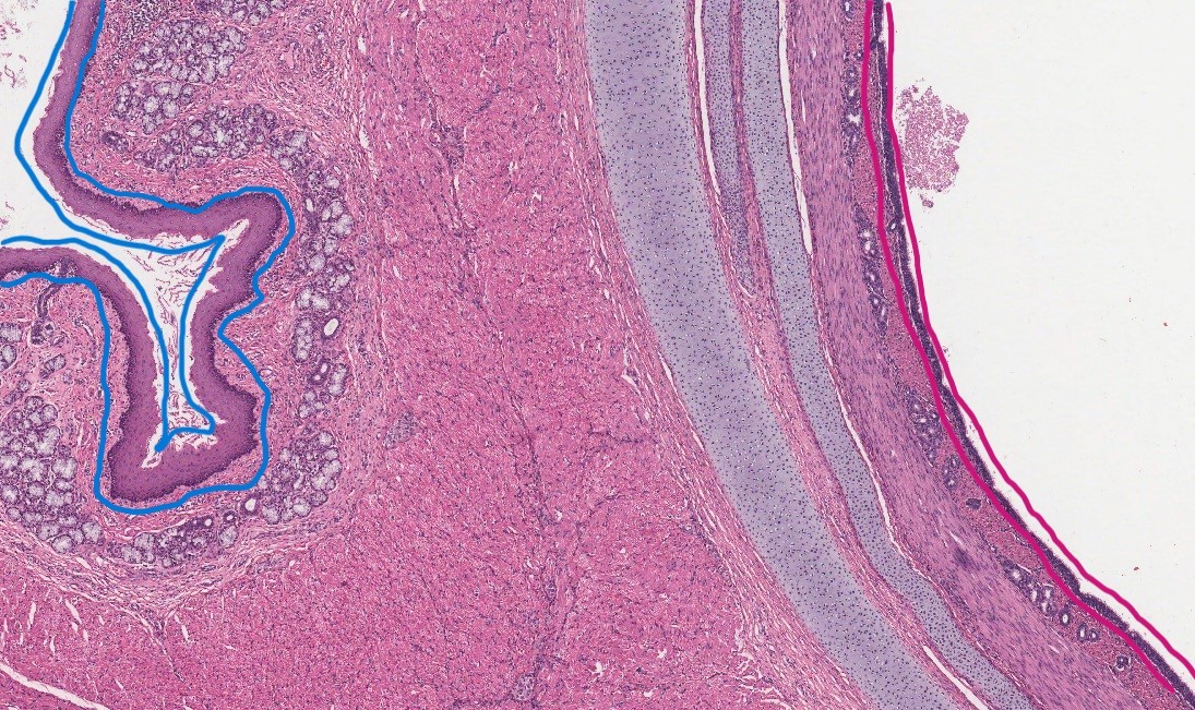
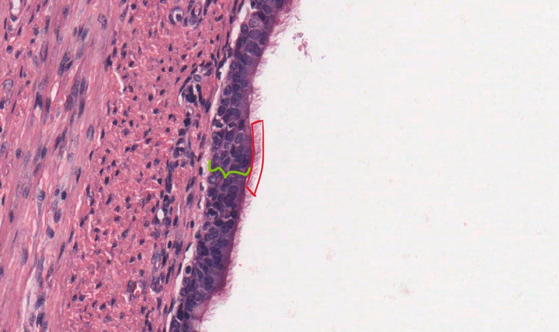
When comparing pseudostratified epithelium to a true stratified epithelial lining, the difference in layer thickness becomes abundantly clear. In trachea, the nuclei in the pseudostratified layer (highlighted by the green line) appear staggered at various heights, unlike the more uniform, neatly stacked nuclei seen in stratified cuboidal and stratified columnar epithelia in sweat glands and urethra, respectively. Despite the seemingly disorganized appearance, if one traces each nucleus to its respective cell body, it becomes clear that every cell in pseudostratified epithelium touches the basement membrane. This confirms that it is technically a simple epithelium, not a true stratified layer—hence the name “pseudostratified”, meaning “falsely layered.”
But this raises an important question: Why use pseudostratified epithelium at all, if stratified layers offer more protection? One possible advantage lies in the presence of differently sized columnar cells, which may offer a built-in reserve. If a taller, exposed columnar cell is damaged—such as from inhaled irritants in the airway—a shorter adjacent cell can quickly step in to maintain function. This may offer a faster protective response than waiting for the mitotic generation of a new full-length columnar cell. In this way, pseudostratified epithelium balances efficiency, flexibility, and protection, making it especially well-suited to the respiratory tract, where ciliary motion and rapid repair are essential.
Media Attributions
- morphology guide squamous © Athena Li is licensed under a CC BY-NC-ND (Attribution NonCommercial NoDerivatives) license
- morphology guide stratified squamous © Athena Li is licensed under a CC BY-NC-ND (Attribution NonCommercial NoDerivatives) license
- morphology guide cuboidal © Athena Li is licensed under a CC BY-NC-ND (Attribution NonCommercial NoDerivatives) license
- morphology guide stratified cuboidal © Athena Li is licensed under a CC BY-NC-ND (Attribution NonCommercial NoDerivatives) license
- morphology guide columnar © Athena Li is licensed under a CC BY-NC-ND (Attribution NonCommercial NoDerivatives) license
- morphology guide stratified columnar © Athena Li is licensed under a CC BY-NC-ND (Attribution NonCommercial NoDerivatives) license
- morphology guide transitional © Athena Li is licensed under a CC BY-NC-ND (Attribution NonCommercial NoDerivatives) license
- morphology guide pseudostratified and stratified © Athena Li is licensed under a CC BY-NC-ND (Attribution NonCommercial NoDerivatives) license
- morphology guide pseudostratified © Athena Li is licensed under a CC BY-NC-ND (Attribution NonCommercial NoDerivatives) license
