Appendices: Introduction to Histology for first-time learners
A New Learner’s Guide to Epithelial Tissue: Glandular Epithelia
Willie Wu and Athena Li
Learning Objectives
By the end of this section, you will be able to:
-
Define what a gland is and distinguish between endocrine and exocrine glands based on the presence or absence of a duct.
- Compare and contrast the function and secretion method of endocrine and exocrine glands.
- Classify exocrine glands based on their duct structure and the shape of their secretory units.
- Classify endocrine glands based on their four primary morphological patterns.
- Identify the key histological features of one major example for each exocrine and endocrine structural class.
Introduction to Glands
In the previous section, we learned about the lining epithelia and their classification. Now, let’s talk about glands. If you think about your body as a busy city, then glands are the factories, water treatment plants, and postal services all organized into one. These specialized structures, which mostly develop from your sheets of epithelial tissue, have one main role of making and secreting products. This could be anything from sweat and saliva to hormones that control and affect your metabolism. The field of studying these important structures is called adenology.
The first and most important thing to understand is that all glands fall into one of two big categories. It all comes down to how they deliver their products!
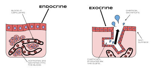
- Endocrine Glands are the body’s broadcast system. They are “ductless,” meaning they don’t have pipes to carry their secretions. Instead, they release their chemical messages, called hormones, directly into your bloodstream. From there, the hormones can travel anywhere and affect distant target organs. Think of it like a radio station broadcasting a signal! Anyone with a receiver can pick it up.
- Exocrine Glands are the local delivery services. They always have a duct (a little pipe) that carries their product to a specific location, like your skin surface or the inside of your stomach. Their effects are local, not systemic.
This difference in delivery method also means they look completely different under a microscope. Endocrine glands often seem simpler, like clusters of cells surrounded by a huge network of tiny blood vessels (capillaries), ready to soak up those hormones. Exocrine glands are all about the ducts, which makes their structure more complex and varied.
| Features | Endocrine Glands | Exocrine Glands |
| Ducts | No ducts (“ductless” glands) | Have ducts that carry secretions to a surface |
| Method of Secretion | Secrete hormones directly into the bloodstream or surrounding tissue fluid | Secrete products (enzymes, mucus, sweat, etc.) through a duct to a surface |
| Structure | Often appear as cords or clusters (islets) of secretory cells surrounded by a rich network of capillaries | Structurally more complex, consisting of a secretory unit and a branching duct system |
| Examples | Thyroid gland, pituitary gland, Islets of Langerhans in the pancreas | Salivary glands, sweat glands, mammary glands, pancreas (acinar cells) |
For the purpose of this appendix, we will focus on exocrine glands and their ducts as they are a tricky structure to identify, as a first time learner.
Exocrine Glands
Since exocrine glands have such interesting structures, we classify them based on their architecture, shown in the diagram below. We look at two things:
- The duct: Does it branch like a tree, or is it a single, simple tube?
- The secretory portion: What is the shape of the factory where the product is actually made?

Simple Tubular Glands
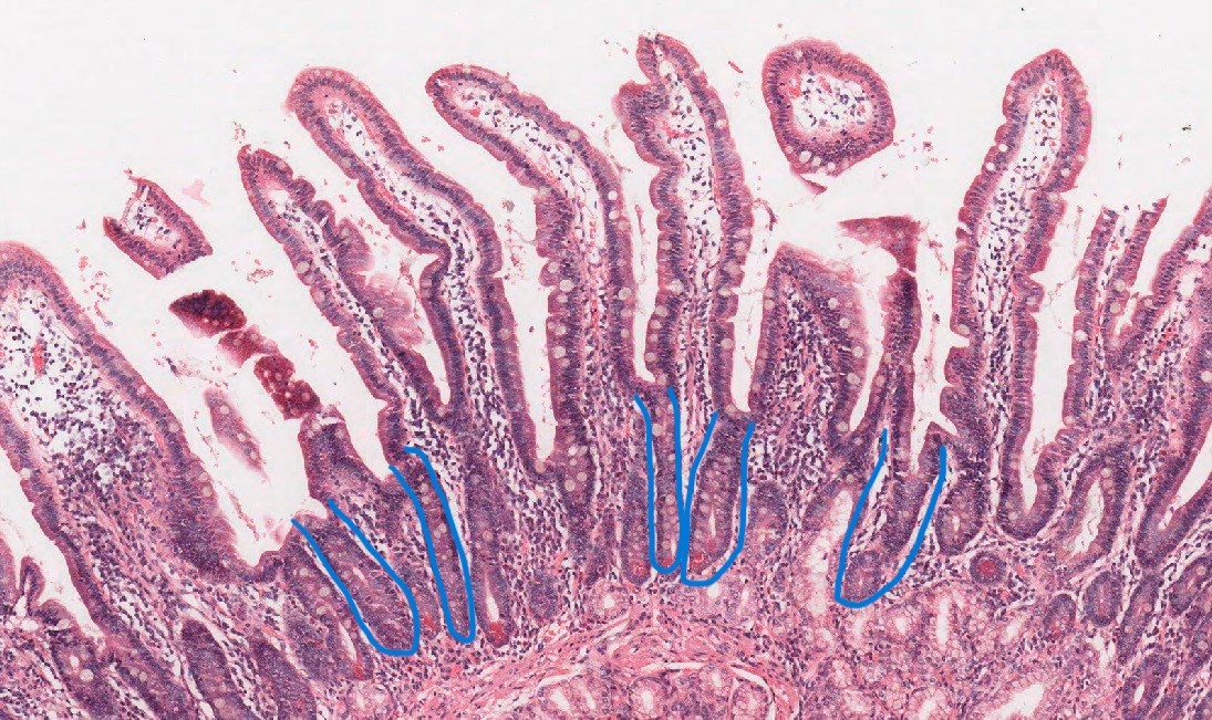
Imagine a test tube that is sunk into the tissue. This is essentially a simple tubular gland. It’s a straight, unbranched tube. In your intestines, you’ll find these as Crypts of Lieberkühn. They’re simple little pockets that secrete mucus and house cells that are crucial for repair and absorption. What you see is what you get with these—no fancy twists or branches.
Simple Branched Tubular Glands
Now, imagine that same test tube, but at the bottom, it splits into several smaller tubes. This is the branched tubular design. They still have just one duct to the surface, but the “factory floor” has multiple chambers. This is a clever way to increase production space. Your stomach is lined with these glands, as shown below. They branch out to house different cells that produce acid, enzymes, and protective mucus, all funneling into the stomach to help digest your food.
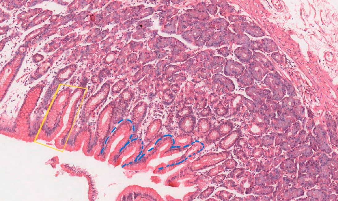
It is worthing mentioning that the yellow box shows an interesting sample which could be mistaken for two simple tubular glands, but this would be due to the cut of this stomach section.
The possible cut made is visualized below:
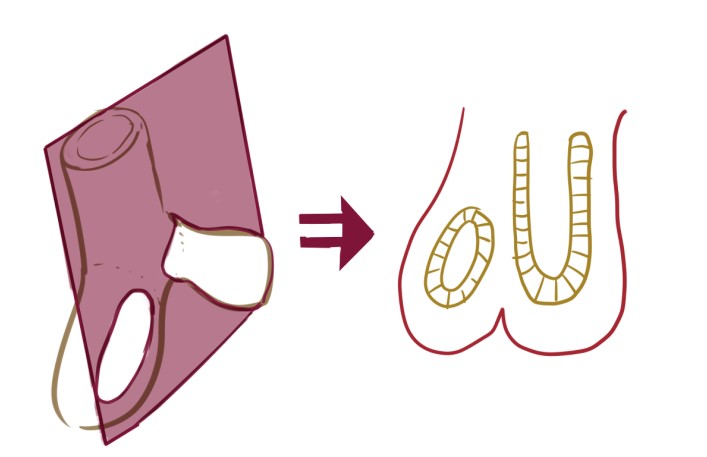
It takes time and practice to recognize simple tubular branched glands compared to simple. When you have a section with lumens of different circumferences, around prominent tubular shaped lumens – yet the secretory cuboidal/columnar cell looks the same around each lumen space, this is highly suggestive of branched tubular gland (shown in the yellow box).
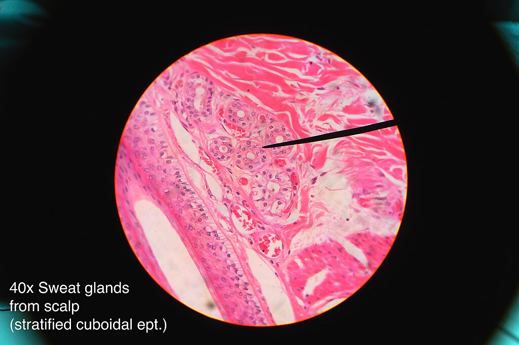
Simple Coiled Tubular Glands
What if you needed a very long tube but didn’t have much space? You would coil it up, right? That is exactly what your eccrine sweat glands do. Their secretory portion is wound into a tight little ball deep in your skin, connected to a straight duct that runs to the surface. This clever design packs a huge amount of tube into a tiny space, allowing you to produce a lot of sweat very efficiently to cool down.
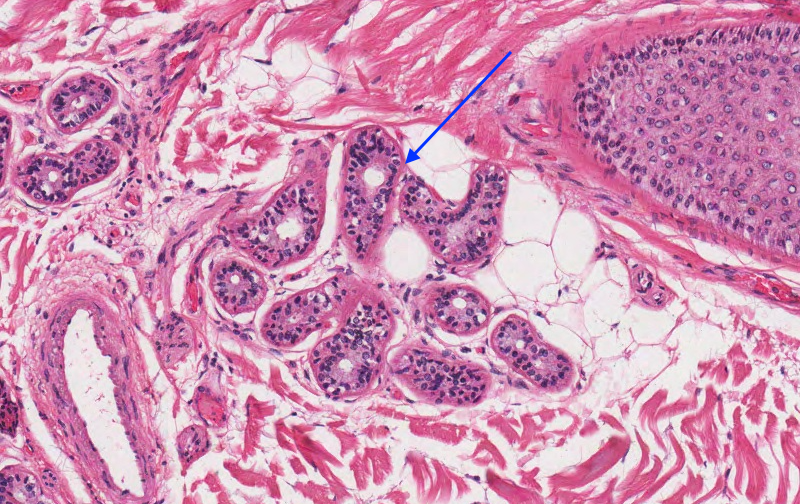
Here is a helpful analogy: imagine a garden hose coiled on the ground. If you look at the coil from above, it will look like a lot of circles which may (or may not) lie neatly on top of each other. Or, if the hose is in a tangled mass, the sectional plane would shows cut sections of small round to oval profiles of varying sizes. If you are lucky, the plane might go along the length of the hose allowing for a dominant tube to be seen with lots of coils nearby.
Simple Acinar & Branched Acinar Glands
Sometimes, a tube might not always be the best shape. If you need a small, round sac to produce and store a product, you form an acinus (which means “berry” in Latin).
- A simple acinar gland is just a single, berry-like sac connected to a duct. You can find tiny versions of these in your urethra.
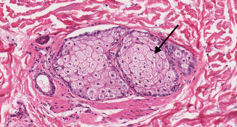
- A simple branched acinar gland is a cluster of these berries (acini) all connected to the same single duct. The best example is your sebaceous (oil) glands. Multiple little sacs filled with oily cells all drain into one short duct that leads to a hair follicle, keeping your skin and hair lubricated.
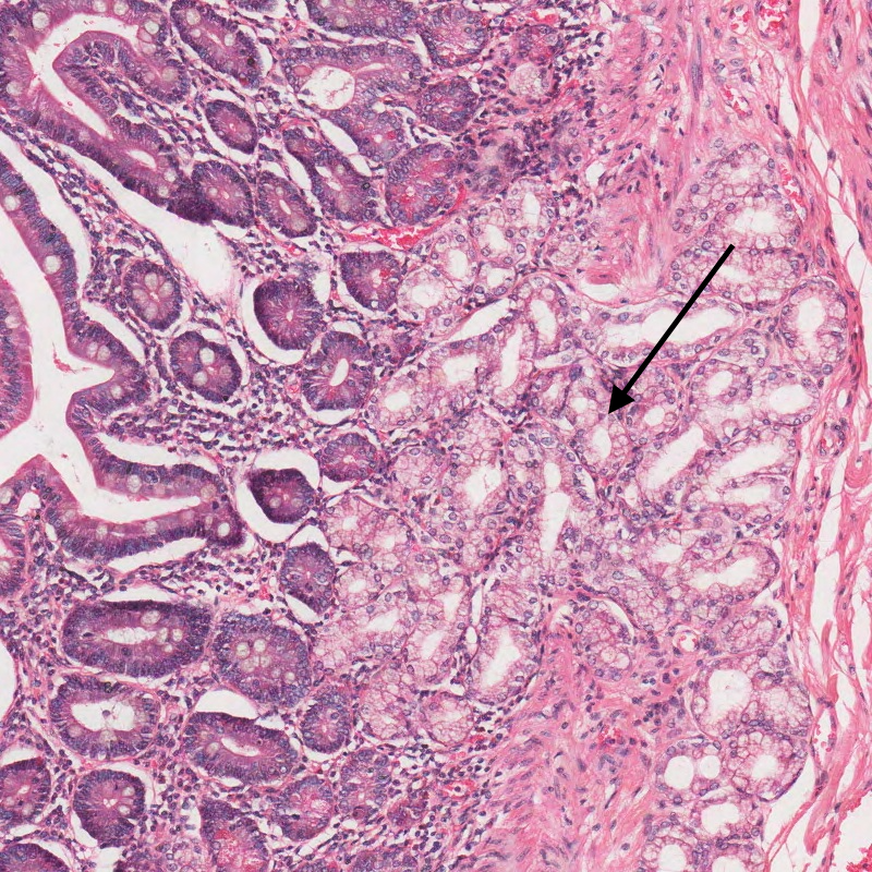
Compound Tubular Glands
Imagine a tree with many small branches, and at the end of each tiny twig, there is not a leaf but a straight, narrow tube. This is essentially the structure of a compound tubular gland. These glands feature an elaborate, branching duct system where even the smallest ducts end in tubular secretory units. It is like having a sophisticated pipeline network where the product is both manufactured and transported through an increasingly complex system of tubes.
A perfect anatomical example of this can be found in the Brunner’s glands of the duodenum. These small glands are located in the submucosa of the first part of the small intestine and secrete an alkaline mucus rich in bicarbonate. This secretion helps neutralize the acidic chyme arriving from the stomach, protecting the intestinal lining and creating an optimal environment for enzymatic digestion. The compound tubular architecture allows numerous tubular secretory units to operate simultaneously, all feeding into a branched duct system that efficiently channels the mucus into the intestinal lumen.
Compound Acinar Glands
Now picture that same branching tree, but instead of tubes at the end of the twigs, imagine clusters of berries. That’s the structure of compound acinar glands. These glands feature an extensive branching duct system that terminates in numerous spherical, berry-like sacs called acini. Each acinus is a tiny factory where secretions are synthesized and promptly released into the duct system for delivery.
The exocrine pancreas is a textbook example of a compound acinar gland. Each pancreatic acinus consists of pyramidal-shaped serous cells that produce digestive enzymes. These enzymes are secreted into an intricate duct system, beginning with intercalated ducts, moving into intralobular and interlobular ducts, and eventually merging into the main pancreatic duct. This complex arrangement allows the pancreas to safely and efficiently transport large quantities of digestive enzymes to the small intestine.

Compound Tubuloacinar Glands
Some glands need to produce different types of secretions, and for this purpose, evolution created the compound tubuloacinar design—one of the most sophisticated glandular architectures. These glands have a branching duct system that ends in a mix of tubular and acinar secretory units. Think of it as a hybrid production facility, with both tube-like factories and berry-shaped factories, all linked to a shared delivery network.
The parotid salivary gland, the largest salivary gland, located just in front of your ears, is a classic example of this structural design. Although it is composed almost entirely of serous acini, its secretory architecture still classifies it as tubuloacinar due to the tubular components involved in early duct formation and branching. The serous cells produce a watery, enzyme-rich saliva packed with amylase, crucial for breaking down starches in the mouth. This secretion drains through an intricately branched duct system, ensuring rapid delivery into the oral cavity, especially when eating or smelling food.
Endocrine Glands
Now that we have solid understanding of exocrine glands, let’s switch gears and take a look at endocrine glands! There are three main structural classes based on the glandular morphology.
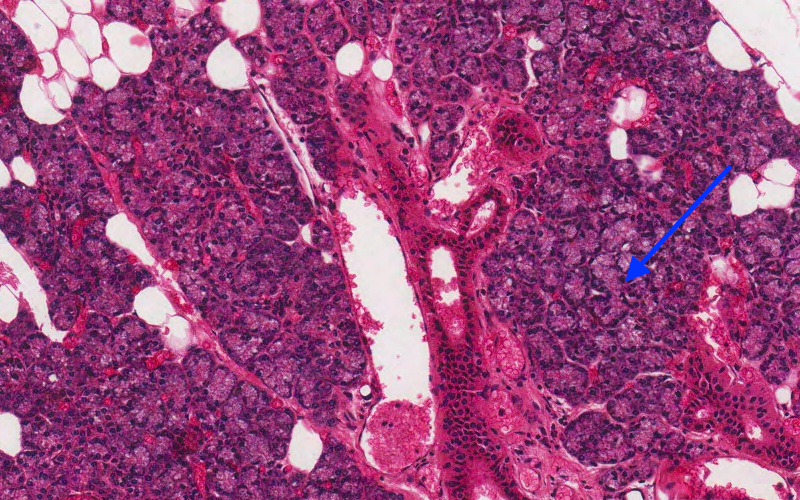
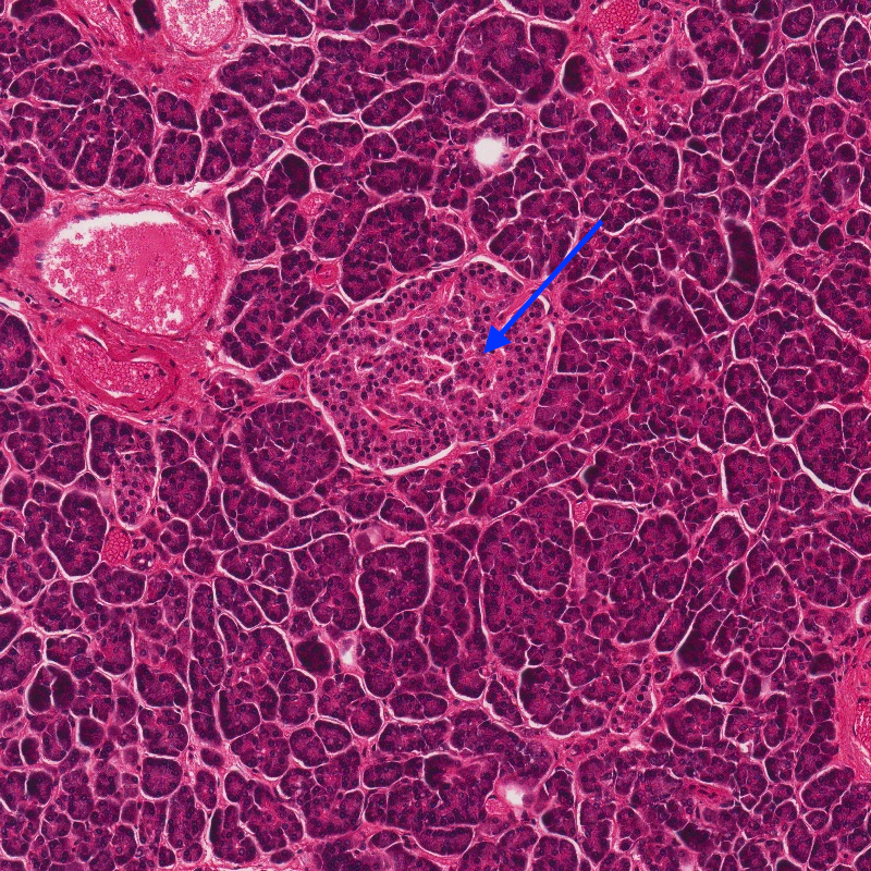
Insular Pattern
The insular pattern is characterized by discrete, rounded clusters or “islands” of endocrine cells embedded within a supporting framework of connective tissue. These cellular islands are not isolated, but are thoroughly infiltrated by an extensive and delicate network of fenestrated capillaries, which creates an immense surface area for the efficient transfer of hormones from the secretory cells into the circulatory system. This structure functions like a series of specialized manufacturing plants, each with direct access to a highway system for immediate product distribution. The prime example of this pattern is the pancreatic islets (islets of Langerhans), which appear as pale-staining, spherical clusters scattered like islands in a sea of darker exocrine tissue. Within each islet, different cell types, such as insulin-producing beta cells and glucagon-producing alpha cells, are intermingled, all contributing to the precise regulation of blood glucose.
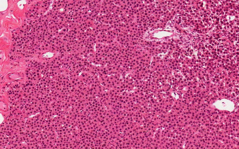
Trabecular Pattern
In the trabecular pattern, endocrine cells are organized into long, branching, and often interconnecting cords or ribbons that are typically only one or two cells thick. These linear cords are separated by parallel rows of fenestrated capillaries, ensuring that every single secretory cell is in close proximity to the blood supply. This architectural design prioritizes rapid and direct secretion, as hormones can be released and absorbed almost instantly without the need for a storage phase. The parathyroid glands are a classic example of this pattern, with dense, winding cords of chief cells responsible for producing parathyroid hormone. This structure is perfectly suited for the gland’s role in minute-to-minute calcium homeostasis that requires swift hormone release into the circulation. Similarly, the adrenal cortex also shows a cord-like arrangement in its zones, with long, straight columns of steroid-producing cells aligned alongside extensive capillary networks, which help facilitate the immediate release of hormones like cortisol and aldosterone.
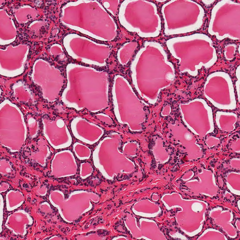
Follicular Pattern
The follicular pattern presents a unique and highly distinctive structure defined by spherical units called follicles. Each follicle consists of a single layer of secretory cells arranged in a circle, creating a central lumen that is filled with a stored precursor of the hormone known as colloid. This design is fundamentally different from other patterns, as it is built for storage rather than immediate release. The follicular cells synthesize a glycoprotein called thyroglobulin and secrete it into the colloid reservoir, where it is stored until needed. Upon stimulation, the cells reclaim the thyroglobulin, convert it into the active thyroid hormones (T3 and T4), and then secrete them into the capillaries on the follicle’s outer surface. The thyroid gland is a great example of this pattern, as it is composed entirely of countless follicles of varying sizes. The appearance of the follicles can even indicate the gland’s activity level: tall follicular cells and pale, scalloped colloid suggest active hormone production, while flat cells and dense, pink colloid indicate more of a resting state.
Key Takeaways
- Exocrine glands use ducts to transport their secretions to an epithelial surface.
- Endocrine glands are ductless and release their hormones directly into the bloodstream.
- The structural classification of exocrine glands depends on their duct branching pattern and the shape of their secretory unit.
- The structural classification of endocrine glands is based on their cellular arrangement, such as insular, follicular, or trabecular.
- A key functional difference is that exocrine secretions, like enzymes, have a local effect, while endocrine hormones have a systemic effect.
- The pancreas is a mixed gland, as it contains both exocrine acini that secrete digestive enzymes and endocrine islets that secrete hormones.
Media Attributions
- 512px-Endocrine_vs._Exocrine.svg © Mntrue15 is licensed under a CC BY-SA (Attribution ShareAlike) license
- 1024px-Types_Arrangements_of_Glands_1 © Holly Fischer is licensed under a CC BY (Attribution) license
- glands © Athena Li is licensed under a CC BY-NC-ND (Attribution NonCommercial NoDerivatives) license
- branched tubular gland © Athena Li is licensed under a CC BY-NC-ND (Attribution NonCommercial NoDerivatives) license
- 3D to 2D © Athena Li is licensed under a CC BY-NC-ND (Attribution NonCommercial NoDerivatives) license
- 10x Skin Coiled Tubular Sweat Glands © Willie Wu is licensed under a CC BY-NC-ND (Attribution NonCommercial NoDerivatives) license
- 1024px-Sweat_gland_histology_2014 © Athikhun.suw is licensed under a CC BY-SA (Attribution ShareAlike) license
- 10x Sebaceous Gland © Willie Wu is licensed under a CC BY-NC-ND (Attribution NonCommercial NoDerivatives) license
- 10x Brunner’s Submucosal Glands © Willie Wu is licensed under a CC BY-NC-ND (Attribution NonCommercial NoDerivatives) license
- 10x Pancreas Compound Acinar Glands © Willie Wu is licensed under a CC BY-NC-ND (Attribution NonCommercial NoDerivatives) license
- 10x Parotid Compound Tubular Glands © Willie Wu is licensed under a CC BY-NC-ND (Attribution NonCommercial NoDerivatives) license
- 10x Pancreas Endocrine Insular © Willie Wu is licensed under a CC BY-NC-ND (Attribution NonCommercial NoDerivatives) license
- 10x Parathyroid Trabecular Glands © Willie Wu is licensed under a CC BY-NC-ND (Attribution NonCommercial NoDerivatives) license
- 10x Thyroid Follicular Gland © Willie Wu is licensed under a CC BY-NC-ND (Attribution NonCommercial NoDerivatives) license
