The Back
Bones
Cervical Region 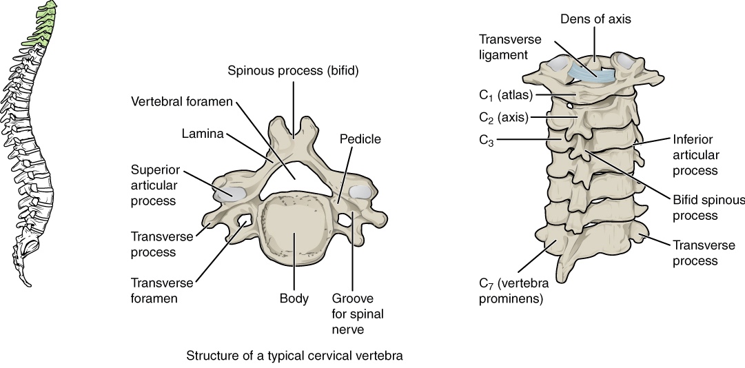
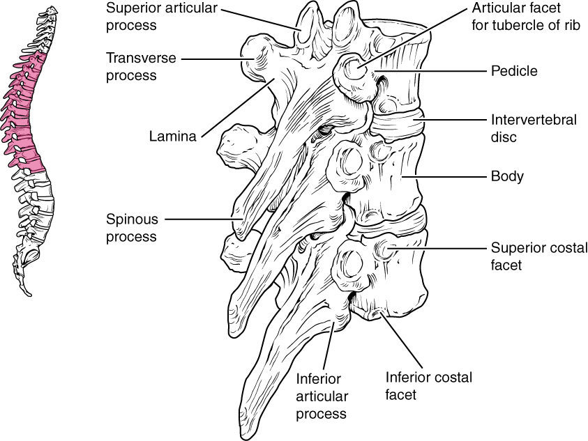
Thoracic Spine:
The thoracic spine consists of twelve vertebrae and labelled as T1-T12. These twelve vertebrae anchor the rib cage posteriorly and have special features which distinguish them from other vertebrae.
- The heart shaped vertebral bodies and the presence of demifacets on the sides of each vertebral body allow for articulation with the heads of the ribs.
- Costal facets on the transverse process, which articulate with the tubercles of the ribs. They are present on T1-T10 only.
- The spinous processes are long/slender and are slanted inferiorly.
The thoracic vertebrae supports the weight of head, neck, upper limbs and chest. It articulate with the ribs to allow changes in volume of thoracic cage. MORE DETAILS ABOUT THE THORACIC CAGE GO CHECK OUT THE THORACIC CAGE SECTION.
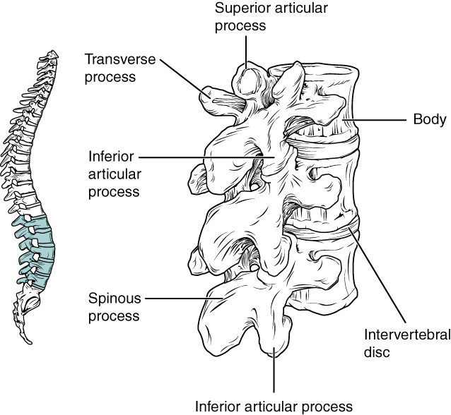
Lumbar Region
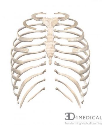
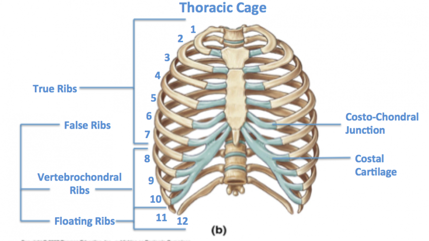
The thoracic cage consists of 12 different sets of ribs, the sternum, and the connecting costal cartilage; when these are all connected together they form a cage. These three sections (ribs, sternum, cartilage) are responsible for the protection vital organs such as the lungs and the heart.
Sternum:
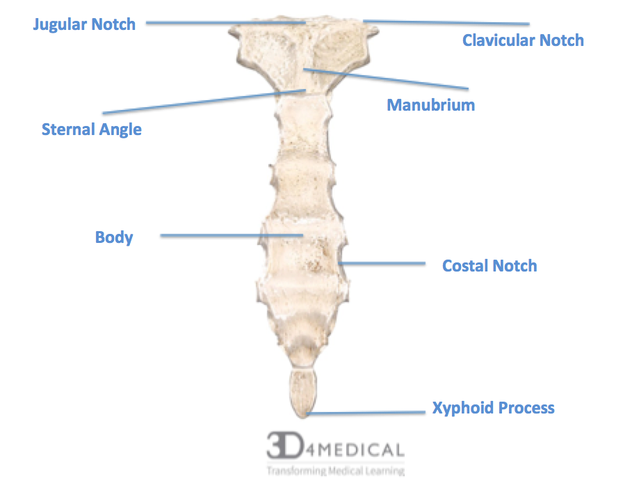
The sternum is a structure that anchors the anterior aspect of the thoracic cage. There are three major aspects to the sternum; the manubrium, the body, and the xyphoid process.
The manubrium is the wider, and most superior portion of the sternum. On the superior portion of the manubrium the jugular notch can be located between the two clavicles. On each side of the superior aspect of the manubrium there is a clavicular notch, These clavicular notches are where the sternum and clavicles articulate, creating the sternoclavicular joint (SC Joint).
The central, elongated portion of the sternum is called the body. The manubrium and the body are connected at a slight bend called the sternal angle. The body of the sternum articulates with ribs 2 through 7 at the costal notches.
The inferior tip of the sternum is the xiphoid process. This structure is cartilaginous early in life, but gradually becomes ossified during middle age. The xiphoid process does not articulate with any ribs.
Ribs:
Each rib is a curved flattened bone that contributes to the wall of the thorax. The ribs articulate posteriorly with the T1-T12 vertebrae, and are numbered 1-12 in accordance with the thoracic vertebrae. Ribs 1-10 attach via their costal cartilages to the sternum. With the first rib is hidden behind the clavicle, the second rib is most superior rib identified through palpations. This means that the second rib and the sternal angle are important landmarks for the identification and counting of lower ribs.
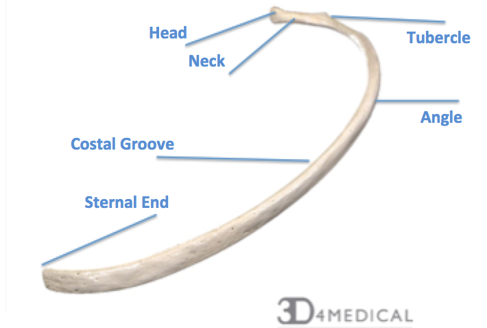
Typical ribs consist of 3 parts; a head, a body, and a neck:
- The head has two articular facets (separated by a wedge of bone); one connects to the corresponding vertebrae, the other to the vertebrae superior.
- The neck simply attaches the body to the head, and the only big landmark is the rough tubercle that attaches to the costal facet.
- The body, or the shaft, is flat and curved. On the inner part of the body, there is a grove for the neurovascular supply of the thorax (protecting them from external damage).
Thoracic Cage:
located on the posterior surface of a rib is the tubercle of the rib; this tubercle articulates with the facet located on the transverse process of the same numbered vertebrae. Just lateral to the tubercle is the angle of the rib, the angle of the rib is point where the rib has the greatest degree of curvature. In anatomical position, the angles align with the medial border of the scapula. A shallow costal groove is found on the inferior margin of each rib, where blood vessels and nerves for the intercostal muscles are protected.
Ribs do not extend all the way to the sternum, as costal cartilage bridges the gap between sternal end of the rib and the costal notches. The costal cartilage is made up of hyaline cartilage, which can extend several inches. The ribs then are classified into three groups based on their relationship to the sternum.
- True Ribs (Ribs 1-7) are ribs that have attachments directly to the sternum through costal cartilage.
*Underneath Rib 1 is the scalene tubercle that project along the medial border between two grooves that travel anteriorly for the subclavian vein and posteriorly for the subclavian artery.
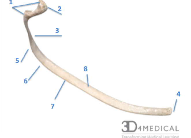
True Rib:
- The first three false ribs (Ribs 8-10) are Vertebrochondular ribs. meaning they do not directly attach to the sternum, as the costal cartilage attaches directly to the next superior rib. This means that the cartilage of rib 10 would attach to the cartilage of rib 9, and that the cartilage of rib 9 would attach to the cartilage of rib 8.
False Rib:
1.) Articulating Surfaces 2.)Head 3.)Neck 4.)Sternal End 5.) Tubercle 6.) Angle 7.) Costal Groove 8.)Body
Floating Ribs (Ribs 11 & 12), or vertebral ribs, are the final two false ribs that do not attach to the sternum at all. Their costal cartilages terminate within the musculature of the lateral abdominal wall.
These muscles have attachment sites on the ribs:
Serratus Anterior: Origin: Ribs 1-8
External Oblique: Origin: Ribs 5-12
Iliocostalis: Origin: Ribs 3-12
Rectus Abdominus: Insertion: Ribs 5-7
Latisimus Dorsi: Origin: Ribs 9-12
Pectoralis Major: Origin: Ribs 1-6
Pectoralis Minor: Origin: Ribs 3-5
Diaphragm: Ribs 6-12
Exteral intercostals; Origin is Inferior border of the ribs, and Insertion is the Superior border of the ribs)
Scalenes: Anterior (Insertion: Rib 1), Middle (Insertion: Rib 1 dorsal to Anterior), and Posterior (Insertion: Rib 2)
